Neuro - Special Senses Vision & Hearing Flashcards

(50 cards)

1
Q

What are the 2 photoreceptors of the eyeball?

A
  • Cones
  • Rods
2
Q

Where are the photoreceptors of the eye located?

A

Retina

3
Q

What vision are the cones responsible for?

A

Vision in the dalylight

4
Q

What vision are rods responsible for?

A

Dim light

5
Q

What is the term for the part of the retina on the nasal side (medial)?

A

Nasal hemiretina

6
Q

The nasal hemiretina receives stimulie from what visual field?

A

From the temporal visual field

7
Q

What is the term for the part of the retina on the temporal side?

A

Temporal hemiretina

8
Q

The temporal hemiretina receives stimuli from which visual field?

A

The nasal visual field

9
Q

If you damage your right temporal retina what could you not see?

A

Objects to the left of your right eye (obj. in the nasal visual field of the right eye)

10
Q

If you damage your left temporal retina what could you not see?

A

Obj. in the right side of your left eye (obj. in the nasal visual field of your left eye)

11
Q

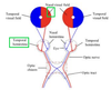
Does the nasal retina cross the optic chiasm to the opposite side?

A

YES

12
Q

Does the temporal retinal cross the optic chiasm to the opposite side?

A

NO

13
Q

What forms the optic nerve?

A

axons of ganglion cells of the retina

14
Q

Where does the optic nerve exit the orbit?

A

Optical canal

15
Q

Is the optic nerve considered a peripheral nerve or a tract?

A

Tract - b/c it’s surronded by cranial meninges and subarachnoid space

16
Q

Are the fibers of the optic n. myelinated or unmylinated?

A

Myelinated by oligodendrocytes (like CNS tracts)

17
Q

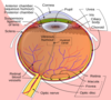
What is the optic disc?

A
  • Blind spot
  • point of exit of the optic n. from the eyeball
18
Q

Does the optic disc have photoreceptors?

A

NO - it’s a blind spot

19
Q

What part of the eye has the greatest acuity (sharpness)

A

Fovea Cetralis

20
Q

Does the fovea contain cones and rods?

A

ONLY cones

21
Q

Is the fovea lateral or medial to the optic n.?

22
Q

Where is the fovea located?

A

In the Macula Lutea

23
Q

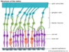
What is the visual pathway?

A

Visual Pathway

Light –> Rods & cones –> Bipolar cells –> Ganglion cells –> Optic Nerve –> Optic Chiasm –> Optic Tract –> LGB –> Optic Radiations –> Primary Visual Cortex

24
Q

Another pic of the visual pathway showing the end of it

A

Visual Pathway

Light –> Rods & cones –> Bipolar cells –> Ganglion cells –> Optic Nerve –> Optic Chiasm –> Optic Tract –> LGB –> Optic Radiations –> Primary Visual Cortex

25
What is a visual field?
All of what you see
26
How many visual fields are there for **both** eyes?
**ONE** visual field for both eyes
27
How many quadrants is the visual field divided in to? And what are they?
4 quadrants: * Upper Left * Lower Left * Upper Right * Lower Right
28
What is the function of the optic chiasm?
Allows images from one side of the **visual _field_** to go to the contralateral **visual _cortex_**
29
Do both nasal retina fibers and temporal retina fibers cross the optic chiasm?
NO - only **nasal** retina fibers
30
Is the internal carotid a. located lateral or medial to the optic chiasm?
Lateral
31
If there is an aneurysm in the ICA what will it do to the optic chiasm?
It can press on the lateral side of the optic chiasm - which would affect the **temporal retina fibers**
32
If the ICA presses on the lateral side of the optic chiasm and the temporal retina fibers are affected, what can you not see?
You can't see out of your nasal visual fields
33
Bilateral atherosclerosis and calcification of the ICA affects BOTH temporal retina fibers, what would not be able to see?
You would not be able to see out of your left and right nasal visual fields so you'd only be able to see peripherally
34
Where is the pituitary gland located in regards to the optic chiasm?
Deep underneather the optic chiasm
35
If there is a pituitary tumor present how will it affect the optic chiasm?
It will press on the middle of it
36
If a pituitary tumor is pressing on the middle of the optic chiasm, which retinal fibers are affected?
Nasal retinal fibers
37
If the nasal retina fibers of **BOTH EYES** are affected, what will you not be able to see?
You will not be able to see the temporal visual field of either eye So you lose peripheal vision and can only see centrally
38
Where do the optic tracs travel to?
* From the optic chiasm * To the pretectal area * To the LBG of the thalamus
39
If 90% of retinal afferent fibers go to the LGB of the thalamus, where do the remiander go?
Pretectal area - for the pupillary light reflex
40
Where do the optic radiation run?
From the LGB to the visual cortex in the occipital lobe
41
Why are optic radiation called "radiations"?
The fibers spead out through 3 lobes of the barin before reaching the visual cortex
42
What 3 lobes of the brain can you find the optic radiations?
* Parietal lobe * Temporal lobe * Occipital lobe
43
What is the part of the temporal lobe that has optic radiations called?
Meyer's loop - it relays in the lingual guyrus of the visual cortex
44
What is the orientation of Myer's loop
* In the anterior temporal lobe * Adjacent to the inferior horn of the lateral ventricle * Dips forward, down then back
45
Where is Broadmann are 17 (primary visual cortex) located?
Adjacent to the calcarine fissure in the occipital lobe
46
The **INFERIOR** retina fibers are stimulated by things seen in which visual field? And will end up in what part of the primary visual cortex?
Simulated by things seen in the **SUPERIOR** visual field and will end up on the contralateral lingual guyrus (below the calcarine fissure/lower part of the primary visual cortex)
47
The **SUPERIOR** retina fibers are stimulated by things seen in which visual field? And will end up in what part of the primary visual cortex?
Simulated by things seen in the **INFERIOR** visual field and will end up on the contralateral cuneus (above the calcarine fissure/upper part of the primary visual cortex)
48
Lower retina visual fields
L. upper visual field ---\> inferior retina fibers ---\> R. lingual gyrus R. upper visual field ---\> inferior retina fibers ---\> L. lingual gyrus
49
Upper retina visual field
L. lower visual field ---\> upper retina fibers ---\> R. cuneus gyrus R. lower visual field ---\> upper retina fibers ---\> L. cuneus gyrus \*\*\* NO Meyer's loop in upper retina visual field
50
Images from the fovea are represented where?
Larger area on the lateral aspects of the each primary cortex