Neuroanatomy Class Exam 1 Flashcards

(180 cards)

1
Q

Where does the CNS bend occur?

A

At the cephalic flexure

2
Q

Where is the Cephalic Flexure Visible?

A

It is visible at the junction between the brainstem and the diencephalon

3
Q

What are the divisions of the brainstem?

A

Midbrain, Medulla, and Pons

4
Q

What joins the two cerebral hemispheres?

A

The Corpus Callosum; this is a huge fiber bundle

5
Q

What develops into the ventricular system?

A

The cavity of the neural tube

6
Q

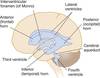
How does the system of ventricles connect?

A

The third ventricle opens into the lateral ventricle (of each hemisphere) through the inerventricular foramen. Posteriorly, the third ventricle is continuous with a narrow channel through the midbrain known as the cerebral aqueduct. This aqueduct connects with the fourth ventricle of the pons and medulla. The fourth ventricle is continuous with the central canal of the caudal medulla and the spinal cord.

7
Q

What is burried deep within the lateral sulcus?

A

The insula is hidden within this sulcus. It overlies the site where the telencephalon and the diencephalon fuse during embryological development.

8
Q

What is the circular sulcus?

A

The circular sulcus outlines the insula and marks its borders withe opercular areas of the cortex.

9
Q

What are the four divisions of the frontal lobe?

A

The Superior, middle, and inferior frontal gyri. The fourth major division is the

10
Q

What does the frontal lobe contain?

A

The frontal lobe mainly contains the motor areas.

11
Q

What are the subdivions of the parietal lobe?

A

1) Postcentral sulcus and gyrus
2) Superior Parietal Lobule
3) Supramarginal Gyrus
4) The Angular Gyrus
5) The Precuneus (only visible on the medial surface of a sagittal cut)

12
Q

What is the parital lobe mainly responsible for?

A

Integrates sensory information

13
Q

What are the subdivisions of the Temporal Lobe?

A

These include the superior, middle, and inferior temporal gyri as well as the occipitotemporal gyrus and the inferior temporal gyrus. The last two are only visible medially with a sagittal cut and viewed from slightly below the brain.

14
Q

What is the temporal lobe primarily responsible for?

A

It contains the auditory areas.

15
Q

What are the subdivisions of the Occipital Lobe?

A

These include the lateral occipital gyri, lingual gyrus, and the Cuneus.

16
Q

What is the occipital lobe mainly responsible for?

A

Primary responsiblity is vision

17
Q

What are the subdivisions of the Limbic Lobe?

A

These include the Cingulate sulcus and gyrus, the Uncus, and the Parahippocampal Gyrus.

18
Q

What is the Limbic Lobe primairly responsible for?

A

Primary responsiblity is emotion and memory processing.

19
Q

What does the Diendephalon include?

A

The thalamus and hypothalamus.

20
Q

What is the function of the thalamus and the hypothalamus?

A

The thalamus serves as a relay center for sensory information to the cortex. All sensory info goes through the thalamus except taste and smell. It also regulates consciousness and alertness.

The hypothalamus is responsible for autonomic actions, such as controlling body temperature, heart rate, and hunger.

21
Q

Where are most cranial nerves located?

A

Most cranial nerves are located in the brainstem

22
Q

What are the three functions of the parietal lobe?

A

1) Primary Somatosensory Cortex
2) Langauge Comprehension (inferior parietal lobule of one hemisphere paired with portiosns of the temporal lobe)
3) Spatial Orientation adn direction of attention

23
Q

What is the interthalamic adhesion?

A

The point of fusion for the two thalami

24
Q

What is one exception to sensory information that does not go through the thalamus?

A

Olfactory Information; all other information goes through the thalamus

25
What separates the thalamus from the hypothalamus?
The hypothalamic wall in the wall of the third ventricle.
26
What connects the hypothalamus to the pituitary gland?
The infundibular stalk
27
What is the tectum?
In the midbrain, a portion posterior to the cerebral aqueduct. Is it the roof of the cerebral aqueduct
28
Understand the strucure of the brainstem and surrounding structures
29
What forms part of the floor of the fourth ventricle?
The rhomboid foss, a depression in the brainstem. It is in the shape of a diamond.
30
Describe the open and closed portion of the medulla
The open medulla is the medulla at the level of the 4th ventricle, giving the impression of an open cavity. The closed medulla is caudal to the 4th ventricle.
31
What are the embryonic origins of the optic nerves?
Outgrowths of the Diencephalon
32
What is the Caudate Nucleus?
A subcortical structure that lies deep within the brain near the thalamus. Plays an important role in various higher neurological functions.
33
What is the function of an oligodendrocyte?
An oligodendrocyte myelinates axons of CNS neurons. They are also distinguishable because they form MULTIPLE sheets of myelin each on separate axons.
34
What is the function of microglia?
Microglia remove debris and infectous organisms
35
What are the derivatives of the neural crest?
The derivatives of the neural crest give rise to most of the peripheral nervous system (PNS) and to several non-neural cell types, including smooth muscle cells of the cardiovascular system, pigment cells in the skin, and craniofacial bones, cartilage, and connective tissue
36
The cerebellum is derived from what secondary brain vesicle?
The rhombencephalon gives rise to the metencephalon (and myelencephalon) and the metencephalon gives rise to the cerebellum.
37
Why does damage to a sensory area on the left cerebral cortex cause deficits on the right?
Second order sensory axons cross the midline on the way to the thalamus
38
What does the falx cerebri separate?
The two cerebral hemispheres
39
What does the tentorium cerebelli separate?
The Occipital Lobes and the Cerebellum
40
What is the cingulate gyrus?
An important part of the limbic system, the cingulate gyrus helps regulate emotions and pain. It is also involved in predicting and avoiding negative consequences
41
What is the Epidural Space?
Space between the bone of the skull and the dura. Site of epidural hematomas.
42
What does the interventricular foramen connect?
It connects the lateral ventricles and the third ventricle.
43
What do the median and lateral apertures accomplish?
The median aperture is an opening in the roof of the fourth ventricle; it drains CSF into the cisterna magna The lateral apertures drain CSF into the subarachnoid space. They are right below the median aperture.
44
Where are Choroid Plexus Cells NOT found?
In the anterior horns of the Lateral Ventricle
45
What are lower motor neurons?
Fibers that convey messages to skeletal muscles
46
What are primary afferent neurons?
Primary afferents are sensory neurons (axons or nerve fibers) in the peripheral nervous system that transduce information about mechanical, thermal, and chemical states of the body and transmit it to sites in the central nervous system.
47
Do the axons of lower motor neurons cross the midline?
No, the axons of lower motor neurons leaving the CNS do NOT cross the midline
48
What is the mode of travel for somatosensory pathways?
Most somatosensory pathways (the expections are taste and smell) cross the midline and pass through the thalamus which relays the information to the cerebral cortex.
49
From which side of the body does each half of the cerebellum receive information?
The ipsilateral side
50
What is the primary function of the cerebellum?
Interpret sensory information to coordinate movement
51
Do somatosensory pathways from the periphery to the cerebellum pass through the thalamus?
No, it goes directly to the cerebellum. Can involve as few as two neurons.
52
What is the corticospinal tract?
A collection of fibers that descend from cell bodies in motor areas of the cerebral cortex and terminate in the spinal cord. Commonly referred to as upper motor neurons.
53
How does the basal nuclei and cerebellum work to induce movement?
They have few or no outputs of their own, so they influence the motor areas of the cerebral cortex. When these two are damaged, the motor areas and lower motor neurons remain intact - movment may be slow or uncoordinated but there is no loss of strength
54
Do corticospinal axons cross the midline?
Just as the somatosensory pathways cross the midline between the periphery and the cerebral cortex, so to deos the corticospinal tract. Thus, damage to one half will cause deficits in the contralateral side.
55
Is a relay in the thalamus required for outputs from the motor cortex?
No, single neurons with very long axons project all the way from motor cortex to the contralateral half of the spinal cord.
56
The Basal Nuclei of one side inderectly affect movements of which side of the body?
The contralteral side of the body is affected.
57
How does the cerebellum work?
To accomplish fine motor tasks, the cerebellum has input from the cerebral cortex and somatosensory input from the ipsilateral side of the body. Cerebellar outputs return to motor cortex, affecting corticospinal activity; this requires a stop in the thalamus. The final element in the general pattern of cerebellar connectivity is dictated by the fact that one side of the cerebellum is related to the ipsilateral side of the body. Because one side of the forebrain is related to the contralateral side of the body, this means that pathways interconnecting the cerebellum and forebrain must cross the midline. Because cerebellar outputs are directed toward motor cortex (via the thalamus), cerebellar damage causes problems with movement but not with sensation.
58
Which two meninges are continuous with each other?
The Arachnoid and Pia; sometimes referred to as the leptomeninges
59
Which meningeal layer is attached to the skull?
The Dura is attached to the inner surface of the skull
60
Which meningeal layers are separated by CSF?
The arachnoid and pia
61
Is there a "real space" on either side of the cranial dura?
No, because one side is attached to the skull and the other is attached to the arachnoid
62
What are the two potential spaces assoicated with the dura?
The subdural and epidural spaces.
63
What is a potential space?
A potential space is a space that only exists pathologically. For example, a potential space is made when blood forces a space to be made (epidural hematoma).
64
What are dural folds?
Dural folds are double layered folds of dura that reflect back on themeselves. It is where the inner dural layer separattes from its external counterpart and protrudes into the cranial cavity. The mainn two are the falx cerebri and the tentorium cerebelli
65
What does the tentorium cerebelli separate?
Separates the superior surface of the cerebellum from the occipital and temporal lobes, defining supratentorial and infratentorial compartments within the cranial vault. The supratentorial compartment contains the forebrain, and the infratentorial compartment (or posterior fossa ) contains the brainstem and cerebellum.
66
What is the tentorial notch?
A space in the tentorium cerebelli through which the brainstem passes.
67
What are dural venous sinuses?
At the attached edges of dural folds, the two layers are normally separated to form venous channels, called dural venous sinuses, into which the cerebral veins empty
68
Name the Venous Sinuses
1) Superior sagittal sinus: Found along the attached edge of the falx cerebri 2) Transverse Sinus: found along the posterior line of the attachment of the tentorium cerebelli 3) Straight Sinus: along the line of attachment of the falx cerebri and tenroium cerebelli to each other 4) Confluence of Sinuses: where all these sinuses meet, near the interal occipital protuberance
69
What is the middle meningeal artery?
A branch of a maxillary artery. Ramifies over most of the lateral surface of the cerebral dura and supplies blood to parts of the dura.
70
Ophthalamic Artery
Provides blood to the dura anteriorily
71
Which arteries provide blood to the dura posteriorily?
The occipital and vertebral
72
Where are the meningeal veins and arteries located?
In the periosteal layer
73
Which parts of the brain are pain sensitive?
The principal pain-sensitive intracranial structures are the dura mater and proximal portions of blood vessels at the base of the brain
74
Arachnoid Mater
a thin, avascular membrane composed of a few layers of cells interspersed with bundles of collagen
75
What are dural border cells?
Flattened cells on the outer portion of the arachnoid which contains several layers. They adhere to the innermost cellular layer of the dura
76
What is the subarachnoid space?
filled with CSF, between the arachnoid and the pia mater
77
What are subarachnoid cisterns?
These are regions containing a considerable amount of CSF
78
Cerebromedullary Cistern
the space between the inferior surface of the cerebellum and the posterior surface of the medulla. Contains a large amount of CSF
79
Pontine Cistern
is located around the anterior surface of the pons and medulla and is continuous posteriorly with the cerebellomedullary cistern
80
Interpeduncular Cistern
is located between the cerebral peduncles and contains the posterior part of the cerebral arterial circle
81
What are arachnoid villi?
This is where CSF enters the venous circulation. At such places, only a loose layer of arachnoid and a thin layer of endothelium separate CSF from venous blood.
82
What are arachnoid granulations?
large cluster of arachnoid villi (arachnoid villi is where CSF leaves the ventricles)
83
What are pacchionian bodies?
calcified arachnoid villi, usually due to age
84
How does the arachnoid behave as a barrier?
Works with the choroid plexus to form the blood-CSF barrier. Forms a double membrane with the choroid plaxus that limits the movement of certain substances.
85
Pia Mater
Covers the surface of the CNS. It is the innermost layer of the meninges, and closely invests all external surfaces of the CNS (unlike the arachnoid). It follows all of the brain's contours.
86
What are the differences between cranial and spinal meninges?
87
Does the spinal dura have a periosteal component?
No, only the skull does. A periosteal layer is a layer of dura
88
Where does the cranial dura split?
At the foramen magnum. The periosteal layer continues around onto the outside of the skull, and the meningeal layer continues as the single-layered dura of the spinal cord
89
What is the lumbar cistern?
A large subarachnoid space between the disk of the first and second lumbar vertibrae.
90
What are dentate ligaments?
These are projections that anchor the spinal cord to the arachnoid through the dura
91
What anchors the caudal end of the spinal cord to the caudal end of the spinal sheath?
A pial projection called the filum terminale
92
What causes an epidural hematoma?
Tearing of meningeal arteries.
93
What causes a subdural hematoma?
Tearring of veins where they enter venous sinuses. In the attached picture, notice how extensive and wide these are compared to epidural hematomas. The picture at right details a shift of midline structures (such as the ventricles) as a result of the buildup of pressure.
94
Herniations within the Brain
Depending on where pressure builds up in the brain, certain herniations across taut reflections such as the falx cerebri can occur.
95
What are ependymal cells?
Cells that line the ventricles; they form a secretory epithelium that produces the CSF
96
What are the 4 ventricles of the brain?
1) 2 Lateral Ventricles 2) Third Ventricle 3) Fourth Ventricle
97
What is the intervenntricular foramen?
Connects the lateral ventricles to the third ventricle
98
What is the cerebral aqueduct?
The cerebral aqueduct connects the third and fourth ventricles
99
What are the 5 parts of the lateral ventricle?
1) Anterior Horn (frontal lobe) 2) Body (in frontal and parietal lobes) 3) Posterior Horn (projects into the occipital lobe) 4) Inferior Horn (curves inferiorily and anteriorily into the temporal lobe) 5) Atrium (where the posterior and inferior horns meet)
100
How does the Caudate Nucleus relate to the ventricles?
Its enlarged head forms the lateral wall of the anterior horn. The smaller body forms most of the lateral wall of the body of the ventricle. Its tail lies in the roof of the inferior horn.
101
Where does the third ventricle lie?
It is the midline cavity of the diencephalon
102
What is the anterior end of the third ventricle?
The third ventricle ends anteriorily at the Lamina Terminalis
103
Does the third ventricle contain Choroid Plexus Cells?
Yes, the roof of it is a thin, membranous lining of Choroid Plexus Cells.
104
What is the general location of the fourth ventricle?
It is sandwiched between the cerebellum posteriorily and the pons and rostal medulla anteriorily
105
What is the rhomboid fossa?
The rhomboid fossa is the shallow, diamond shaped depression on the dorsalsurface of the pons and medulla oblongata that forms the floor of the fourth ventricle
106
What is the median aperture?
A hole in the inferior medullary velum of the fourth ventricle; connects the fourth ventricle to the cerebellomedullary cistern)
107
What is choroid plexus?
Vascular membranous material contained in all four ventricles. This secretes most of the CSF
108
Where are the only locations where there is no choroid plexus?
In the anterior and posterior horns
109
What forms the choroid plexus of the fourth ventricle?
Invagination of the inferior medullary velum in the caudal half of the ventricle
110
What is the general orientation of Choroid Plexus?
Must be adjacent to subarachnoid space on its pial side and to intraventricular space on its choroid epithelial side
111
What are the three layers of the Choroid Plexus (in order)?
1) Endothelial wall of each choroidal capillary - allows movement out of the capillary (it is fenestrated) 2) Pial Cells and Collagen 3) The choroid Epithelium
112
What is the strucutre/function of choroid epithelium?
look as though they are specialized for secretion because they have many basal infoldings, numerous microvilli on the side facing the CSF, and abundant mitochondria Act as a barrier to peptides and large molecules. Ions can diffuse.
113
How is CSF formed?
Filtration of blood through the choroidal capillaries followed by transport of substances (sodium ions) accross the choroid epithelium into the ventricle.
114
How is rate of CSF production modifed?
Sympathetic and parasympathetic fibers end not only on choroidal blood vessels but also near the bases of the choroid epithelial cells. Stimulating the sympathetic fibers, for example, causes a reduction of about 30% in the rate of CSF production
115
Descirbe the movement of CSF
Lateral ventricles --\> interventricular foramen --\> third ventricle --\> cerebral aqueduct --\> median/lateral apertures --\> cisterna magna and pontine cistern
116
How does CSF function as a spatial buffer?
something inside the skull can enlarge only if something else leaves. CSF leaving opens space for other things to exist without causing damage (unless they are too big)
117
What are some functions of the CSF?
1) Spatial Buffer 2) Regulates the extracellular environment of neurons 3) Route to spread neuroactive hormones through the nervous system
118
What is a multipolar neuron?
Multiple dendritic projections from the cells body and almost always an axon as well. Most neurons follow this structure.
119
What is a pseudounipolar neuron?
Neurons which have one extension from their cell body. That one extension then splits into multiple (see picture).
120
What are sensory neurons?
Sensory neurons are those that either directly recieve input from stimuli or from a neuronal connection.
121
What are motor neurons?
Neurons that directly attach to muscles, glands, etc and cause innervation
122
What are projection neurons?
Entirely in the CNS; long axons that connect different areas of the CNS
123
What is gray matter?
gray matter refers to the collection of cell bodies and dendrites
124
What is white matter?
White matter refers to areas where there is a preponderance of axons; many axons have a myelin sheath
125
What is cortex?
Cortex is the outer layer of the brain; it is an accumulation of gray matter.
126
Where are the cell bodies of sensory axons contained?
In the dorsal root of the spinal cord
127
What is the function of the cell body?
Synthesizes macromolecules such as enzymes, structural proteins, membrane components, and organelles. Some chemical messengers are also synthesized.
128
What is the cytoskeleton?
The cytoskeleton is used primarily for structure of the cell, but it can also be used for movement/growth of the neuronn as well as movement of substances within the cell body and up and down the axon. Comprised of Microtubules, which are made up of polymers of tubulin.
129
What is a special function of microfilaments?
Microfilaments are important for anchoring membrane molecules in place and for shuttling things to and from the cell membrane, and for movement of the advancing tip of growing axons
130
What is anterograde transport?
Transport going away from the cell body. Usually carries membrane assoicated enzymes, neurotransmitters, and neuropeptides.
131
What is retrograde transport?
Transport that is returning to the cell body. Carries survival signals from the synapse back to the cell body (for example)
132
What is slow and fast transport?
1) Fast: Fast retrograde transport returns used synaptic vesiclesand other materials to the soma and informs the soma of conditions at the axon terminals. Anterograde carries axonal proteins and synaptic components. 2) Slow: Slow axonal transport moves soluble proteins—such as cytoskeletal proteins and cytoplasmic enzymes - down the axon
133
What is a special function of microtubules?
Serve as the substrate along which organelles are transported through neuronal processes
134
What are the two ATPases and what do they do?
1) Kinesin: travels toward the positive end of the microtubule (anterograde). Carries membrane associated cell components. 2) Dynein: moves some components in the retrograde direction
135
What is the difference in regard to gray matter in the spine vs the brain?
In the spine, gray matter is inside surrounded by white matter. It is the opposite for the brain.
136
How does the neural tube form?
the underlying midline mesoderm sends chemical signals to thicken the ectoderm into the neural plate. The neural plate then folds inward to form the neural groove, which is flanked by neural folds on each side. The neural folds fuse into the neural tube.
137
What is the forerunner of the skeletal axis?
The notochord; it helps form the vertebral column.
138
What is Primary Neurolation?
Closing of the neural tube which forms the Primary Vesicles
139
What is the Sulcus Limitans?
Separates the sensory and motor areas of the spinal cord and brainstem.
140
What is the dorsal gray matter?
Synonymous with the posterior horn, it is the alar plate of the neural tube. It contains derivatives for SENSORY processing
141
What is the ventral gray matter?
Synonymous with the anterior horn, it contains the basal plate. It has derviatives for MOTOR neurons.
142
What are the three primary vesicles (from rostral to caudal)?
1) Prosencephalon 2) Mesencephalon: leads to the midbrain 3) Rhombencephalon: leaders to the brainstem and cerebellum. This also merges with the caudal portion of the neural tube
143
What is secondary neurolation?
forms the caudal spinal cord distal to S2 (sacral) and the filum terminale
144
What does the Telencephalon develop into?
The thalamus, hypothalamus, and the retina
145
What does the Mesenecephalon develop into?
The midbrain; the top most part of the brainstem. Contains the tegmentum, colliculi, and the cerebral peduncles.
146
What does the Metencephalon develop into?
The pons and cerebellum
147
What does the meyelencephalon develop into?
The medulla; piece of the brainstem that merges with the spinal cord
148
What does the neural tube look like after secondary neurolation?
149
What forms the cerebellum related nuclei?
Parts of the rombic lips, which are lateral portions of the alar plate, migrate into the brainstem which gives rise to the cerebellum.
150
How does the Basal Ganglia form?
As the telencephalon begins growing, the basal wall (adjacent to the diencephalon) thickens to form the Basal Ganglia
151
How does the thalamus and the hypothalamus form?
The walls of the diencephalon thicken.
152
What is special about the neural tube cavity?
The cavity persists as the ventricular system of the adult brain and the central canal of the spinal cord.
153
What are protoplasmic astrocytes?
Glial cells that form leaky membrane that covers the surface of the CNS, which separates it from the PNS.
154
Be aware what the neural tube and surrounding structures look like
155
Be aware what the neural tube looks like as represented by a picture
156
How does the sacral spinal cord form?
After the neural tube closes, a secondary cavity extends into the solid mass of cells at its caudal end during the fifth and sixth weeks
157
What is the lamina terminalis?
The lamina terminalis is a thin sheet of gray matter and pia mater that attaches to the upper surface of the chiasm and stretches upward to fill the interval between the optic chiasm and the rostrum of the corpus callosum.
158
What is the hypothalamic sulcus?
Separates the thalamus and the hypothalamus
159
What are cranial placodes and what do they do?
Cranial Placodes form from a strip of ectoderm adjacent to the brainstem neural crest; they collaborate with parts of the neural crest to form sensory components of the PNS of the head.
160
What does the adenohypophyseal placode do?
Forms the anterior pituitary
161
What is spina bifida?
Failure of neural tube caudal end to close
162
What is craniorachischisis?
A fatal deformation in which both ends of the neural tube fail to close
163
What is anencephaly?
Failure of the rostral end of the neural tube to close. Much of the cerebral hemisphere is absent as a result.
164
What is alpha-fetoprotein?
a major component of fetal serum; open neural tube allows it to leak out into amniotic fluid.
165
Why is folic acid useful?
Consumption of folic acid by the mother can prevent a wide range of neural tube deficiencies. However, this must occur around the time of neural tube closure, which is when most people are unaware they are pregnant.
166
What is characteristic of defective secondary neurolation?
Can be assoiciated with traction injuries to the spinal cord, as well as cycsts and tumors near the sacral area. Hairiness or discoloration can hint at a defect.
167
What is holoprosencephaly?
Result of the failure of the failure of the prosencephalon to separate into the telencephalon and the diencephalon. Frequently referred to as cyclopsia as the midline of the face is abnormal.
168
What is the general connectivity of primary afferents?
They carry information from the periphery to the CNS without crossing the midline. They terminate in the CNS on second order neurons, which project onto third-order neurons. The central process ends on the ipsilateral side.
169
What is the general pattern of connectivity for Lower Motor Neurons
The axons of lower motor neurons leave the CNS without crossing the midline. Lower Motor Neurons have cell bodies within the CNS that travel though peripheral nerves to end on ipsilateral muscle fibers.
170
What are three functions of primary afferents?
1) They feed into reflexes 2) Move sensory information to the cerebral cortex 3) Move sensory information to the cerebellum There are interneurons involved in this too.
171
What is the mode of travel for somatosensory pathways to the cerebral cortex (remember, this is accomplished by primary afferents, remember how they work)?
They cross the midline and pass through the thalamus
172
What is the corticospinal tract?
A bundle of fibers that descend from cell bodies in the motor areas of the cerebral cortex and terminate in the spinal cord (generally referred to as upper motor neurons).
173
Do corticospinal axons cross the midline?
Yes they do. The process terminates on the contralateral side (damage to the left motor area results in movement deficits on the right side of the body)
174
What is the general connectivity of the cerebellum?
Each side of the cerebellum indirectly affects movements of the ipsilateral side of the body.
175
What is the general connectivity of the basal nuclei?
The basal nuclei of one side indirectly affect movements of the contralateral side of the body
176
Where are third order neurons located?
In the thalamus
177
What is the primary function of choroid plexus?
Production of CSF
178
What is the blood-CSF barrier?
It is a tight wedging of endothelial cells. They are wedged so tightly together that it forms "tight junctions" which only allows very small molecules, fat soluble molecules, and some gases to pass freely through the capillary wall and into the brain tissue.
179
In relation to the choroid plexus, where are the endothelial cells and what do the constitute?
These cells form the blood-CSF barrier. They are located around the blood capillary to form fenestrations. The epithelial cells are closest to the ventricle side.
180
How are choroid plexuses formed?
The choroid plexus is made up of invaginated folds of pia mater that penetrate the interior of the ventricles of the brain. It is made up of the connective tissue of the pia mater covered by ependymal cells. It is responsible for the production of the cerebrospinal fluid INTO the ventricles