OSPEE #2 Flashcards
What is this instrument and what is it’s function?

A area specific curet periodontal instrument used to remove light calculus deposits from the crowns and roots of teeth
Explain this area specific currets design characteristics

Long, complex function shank. Suited for instrumentation of root surfaces.
- Rounded back
- Rounded toe
- Semicircular cross section
What makes the area specific curett unique?

The cutting edges are curved. In comparison to a universal curet that has parallel cutting edges
Explain the importance of the area specific curets ‘tilted face’

Face is tilted in relation to the lower shank. Causing one cutting edge to be lower than the other on each working-end.
Describe the edges of the area specific curet
Only the lower cutting edge is used for calculus removal → called the working cutting edge. The other edge is called the nonworking cutting edge, which helps protect the soft tissue.
Describe the angulation of the lower cutting edge (working edge)
Automatically at a 70 degree angle to the tooth surface when the lower shank is parallel.
How do you position an area specific curet in the posterior sextants?

Lower shank is parallel to the proximal surface. Functional shank goes up and over the tooth.
Which (2) gracey curets are used for the posterior facial aspect? RHS

G11 - Facial Mesial G14 Facial distal
Which (2) gracey curets are used for the posterior lingual aspect? RHS

G12 - Lingual mesial G13 - Lingual distal
Which (1) modified gracie curet would you use for the facial aspect of posterior sextant? RHS

G11-14
Which (1) modified gracie curet would you use for the lingual aspect of posterior sextant? RHS

G12-13
Define a smooth surface lesion
Class V; found on buccal/lingual surfaces. Located in the gingival third/cervical region. Can be carious, and non carious e.g abfraction, abrasion
What are risk factors for carious and non carious lesions (class V)?
Carious: -
- Poor OH
- Diet
- Dry mouth
- Progression of white spot lesion
- dental prostheses.
Non-carious lesions:
- Tooth brush abrasion
- Other habits - Clenching and grinding
What class is this lesion?

Class V located in cervical region
What cavity preparation would you undertake for a non carious Class V?
For non-carious cervical lesions no further cavity prep.
What cavity preparation would you undertake for a carious Class V?
Gingival margin approx parallel with gingival contour (CEJ). Axial depth 1-1.2mm, 90 degree walls
Name the structures on this picture

A. Anterior auricular nodes
B. Superficial and deep parotid nodes
C. Facial nodes
D. Submandibular nodes
E. Submental nodes
F. Jugulofigastric node
G. Anterior jugular nodes
H. Deep cervical nodes
I. Retroauricular nodes
J. Occipital nodes
K. Retroauricular nodes
L. External jugular node
M. Retropharyngeal node
N. Deep cervical nodes
O. Accessory nodes
P. Juguloomohyoid node
Q. Supraclavicular nodes
Name the structures on this picture

A. Hard palate
B. Soft palate
C. Uvula
D. Posterior wall of the pharynx
E. Dorsal surface of tongue
F. Fauces
G. Maxillary tuberosity
H. Pterygomandibular fold
I. Posterior faucial pillar
J. Palatine tonsil
K. Anterior faucial pillar
L. Retromandibular pad
The out line of this cavity preparation is? a) under extended b) over extended

Under extended
The out line of this cavity preparation is? a) under extended b) over extended
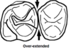
Over extended
How do you calculate O’Learys Index plaque score?

Number of surfaces with plaque present eg. M, L, B, D divided by no of surfaces examined eg. 32 teeth times 100
no. surfaces w plaque present / no. of surfaaces examined x 100 = %
What is this?

Materia Alba: it is associated with biofilm, loosely adherent mass of bacteria. Visible without use of disclosing agent.
Name the (5/6) treatment phases for treatment planning
- Preliminary Phase
- Phase 1 (non surgical phase).
- Evaluation
- Phase IV (maintenance phase)
- Phase II surgical phase / phase III restorative phase

Explain this furcation involvement

Class I. Concavity can be felt with probe. Probe tip cannot enter the furcation area.


