Pattern Recognition Flashcards

(309 cards)

1
Q

What are the 4 components of a medical image?

A

Equipment Patient Signal Receptors Signal Processing

2
Q

Describe equipment as a component of a medical image.

A

It is dealt with in relation to the patient It relates to: - positioning of equipment - equipment used - MRI specific or surface coils

3
Q

Describe signal processing as a component of a medical image

A

It tended to via a computer using specific algorithms Can be chemical in terms of film processing

4
Q

Describe signal receptors as a component of a medical image

A

Change between modalities - Transducers - Image recording plate - Film / Screen combination - Radioreceiver - PMTs

5
Q

Describe the patient as a component of a medical image

A

Need to understand the anatomy and physiology Positioning of the patient

6
Q

What needs to be understood to interpret an image?

A
  • Interaction of the source with various tissue types - Affect of beam geometry - Affect of patient position on structures - Anatomical structures (localisation, physiology and manifestation of disease)
7
Q

What is the effect of beam divergence? How is it reduced?

A

Image magnification Gives a geometric unsharpness Reduce by placing the recording medium as close to the object

8
Q

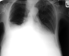
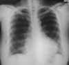
Why is a chest X-ray taken PA?

A

Allows for assessment of heart size It reduces unsharpness and magnification It reduces the effect of breast tissue If AP then the scapula can be projected into the lung field

9
Q

How is a chest X-ray taken?

A

PA (posterio-anteriorly) Remove the scapula out of the image view

10
Q

How does shade and colour alter perception of images?

A

Shade helps to identify structures The mind fills the gap to create the perception of depth 10% of colour interpretation is governed by context not wavelength

11
Q

What are the problems associated with radiography?

A

3D representation in a 2D image Summation of shadows Standardisation in positioning is vital to identify positioning and location

12
Q

What is the benefit of standardisation of positioning?

A

Allows you to identify the position and the location You become familiar with the orientation - familiar frame of reference

13
Q

How are hands X-rays usually acquired?

A

Dorsi-palmar Need to label right and left

14
Q

What is a reason for altering from a standard projection?

A

Makes it more comfortable for the patient If the patient is less likely to move

15
Q

What are the 4 different types of contrast?

A

Subject Recording medium/system - Image Objective Subjective

16
Q

What is the difference between subject and image contrast?

A

Subject contrast is the differences between X-ray intensities emerging from the patient Image contrast is the differences recorded in the radiographic image

17
Q

What is the difference between subjective and objective contrast?

A

Subjective - dependent on the eye of the observer, varies from person to person Objective - actual differences in densities or black and white

18
Q

How can subject contrast be changed?

A

Altering differences in attenuation using contrast agent - Barium, iodide, gadolinium

19
Q

What are examples of contrast agent?

A

Barium Iodide Gadolinium

20
Q

What are examples of contrast agent?

A

Barium Iodide Gadolinium

21
Q

Why is it important to get the patient, X-ray detector and beam source parallel?

A

Need to get a truly representative image Otherwise can get: foreshortening or elongation

22
Q

What is foreshortening in an X-ray image?

A

The image appears squashed Happens when the patient leans forward

23
Q

What is elongation in an X-ray image?

A

If the detector is angled you get a stretched/elongated image Can be used advantageously in the scaphoid

24
Q

Where can beam geometry be applied usefully?

A
  • PA chest radiography - Sacroiliac joints demonstration - Aid to identifying patient positioning
25
What are the identification points when assessing a radiographic image?
- Check patient ID - Time & date of examination - Check correct anatomical markers and appropriate legends - Facility name
26
What are the identification points when assessing a radiographic image?
- Check patient ID - Time & date of examination - Check correct anatomical markers and appropriate legends - Facility name
27
What anatomical features do you need to check when assessing a radiographic image?
- Check all relevant anatomy is included in the projection - Are all anatomical features accurately displayed? - Sharp definition of all cortical outlines and/or soft tissue - Protocols (differ between facilities) - Trace bony outline (disruption = pathology) - Compare both sides and projections
28
What are the points to check in terms of image quality in a radiographic image?
- Is there adequate contrast and density displayed? - Is there adequate penetration? - Is there any signs of unsharpness? - Is there any evidence of collimation? - Are there any artefacts present?
29
What is the sign that indicates adequate penetration in a chest X-ray?
Should be able to see the 4 thoracic vertebrae through the heart. If this is not visible, could be pathology or inadequate exposure.
30
What are the categories for artefacts?
Anatomical e.g. hair External e.g. clothing, pins, hearing aids Internal e.g. swallowed items Equipment/imaging related e.g. dust
31
What are the problems with X-ray interpretation that can lead to misdiagnosis?
Overlapping structures Projecting what you expect to see onto the image (e.g. if the patient is in pain, a soft tissue line can be viewed as a fracture)
32
What are the requirements for interpretation of an image?
- Understand what is in the image - Understand what is in the patient - Understand what was conducted - Be aware of the limitations of the procedure
33
What are the requirements for interpretation of an image?
- Understand what is in the image - Understand what is in the patient - Understand what was conducted - Be aware of the limitations of the procedure
34
What is pattern recognition?
It is an information reduction process. The assignment of visual logical patterns to classes based on features of these patterns and their relationship
35
What is clinical judgement?
It is the interpretation of X, being a real example of Y or support the conclusion reached
36
What is clinical judgement?
It is the interpretation of X, being a real example of Y or support the conclusion reached
37
What is the function of an X-ray examination?
- Accurate localisation of fracture and determination of number of fragments - Indicate the degree and direction of displacement - Provide evidence of pre-existing disease - May demonstrate a foreign body - May show indication of nature of injury (This can be linked to any imaging modality)
38
What are the limitations of an X-ray examination?
1. Contrast resolution 2. Spatial resolution 3. Sensitivity of the system - Need 30-50% bone loss to detect osteoporosis
39
What modalities can be used to detect osteoporosis?
- X-ray (poor) due to low sensitivity - DEXA - Radionuclide (very sensitive but not specific)
40
What are the points on a checklist for reviewing diagnostic image appearances?
- Size and shape of structure - Position of structures - Thickness of structures - Mineralisation - Trabeculation of cancellous bone - Vascular patterns - Contour of structure (smooth/sharp) - Look for symmetry - Joint space, size, shape, normal? - Alignment/articulation - Soft tissue assessment - Changes with development - Correct numbers of bones - Relationship with other structures
41
What are the points on a checklist for reviewing diagnostic image appearances?
- Size and shape of structure - Position of structures - Thickness of structures - Mineralisation - Trabeculation of cancellous bone - Vascular patterns - Contour of structure (smooth/sharp) - Look for symmetry - Joint space, size, shape, normal? - Alignment/articulation - Soft tissue assessment - Changes with development - Correct numbers of bones - Relationship with other structures
42
What are the starting points to interpreting an image?
- Examine general appearance of the bone - Trace the contour of each bone for irregularities - Examine joint space - Examine soft tissue (can provide indirect evidence of a fracture)
43
Why is it important to examine soft tissue in an X-ray?
Can provide indirect evidence of a fracture.
44
Why is it important to examine soft tissue in an X-ray?
Can provide indirect evidence of a fracture.
45
What are the 2 ossification centres and what are they responsible for?
Primary - controls width of bone Secondary - controls the length of bone
46
What is an additive disease?
Abnormal condition leading to an increase in fluid or bone. Fluid has the effect of distending tissue, which increases the thickness.
47
What are the results of having an additive disease?
- Bone has a higher than average atomic number (compared to normal bone) - Excessive bone growth or the replacement of cartilaginous tissue with bone - Increase n the amount of calcium present in the bone - Bone becomes denser in structure - Bone is more radio-opaque - Can be focal or general
48
What are the most common additive disease?
- Acromegaly - Paget's disease - Osteoarthritis - Osteochondroma - Osteopetrosis
49
What is a destructive disease?
Abnormal condition leading to an increase in air or fat OR a decrease in normal body fluid or bone
50
What are the results of having a destructive disease?
- Demineralisation of bone or an invasive destruction of bone tissue - Decrease in the amount of calcium in the bone - Bone becomes less dense in structure - Bone more radiolucent - Can be general or focal
51
What are the common destructive diseases?
- Osteoporosis - Rheumatoid arthritis - Osteomalacia - Exostosis - Osteomyelitis - Gout - Hyperparathyroidism - Ewings tumour
52
What are the 3 types of bone abnormalities?
1. Opacity - increased radiographic density e..g overlapping bone fragments 2. Radiolucency - decreased radiographic density e.g. fracture line 3. Distortion/displacement of normal structures e.g. - Bump/step/gap in cortex - buckle or bowing - collapse (vertebrae) - subluxation/dislocation - soft tissue swelling
53
What are the 3 types of bone abnormalities?
1. Opacity - increased radiographic density e..g overlapping bone fragments 2. Radiolucency - decreased radiographic density e.g. fracture line 3. Distortion/displacement of normal structures e.g. - Bump/step/gap in cortex - buckle or bowing - collapse (vertebrae) - subluxation/dislocation - soft tissue swelling
54
What is a Jones/avulsion fracture?
Tendon pulls off part of the bone it is attached to
55
What is a Jones/avulsion fracture?
Tendon pulls off part of the bone it is attached to
56
What are the points to note on a pelvis X-ray?
1. Sacro-iliac joints should be equal in width 2. Sacral foramina should not be disrupted 3. Superior surface of the symphysis pubis should be aligned (should be approx 5mm) 4. Acetabular regions should be compared for variation
57
What does a widening in the sacro-iliac joint represent?
Fracture of the main ring
58
What is the usual distance between the superior surface of the symphysis pubis?
5mm
59
What are the 2 rings of the pelvis?
1. Main pelvic ring 2. 2 Smaller rings formed from pubic and ischial bones
60
What are the frequent pelvic fractures?
Acetabular Sacral Coccygeal
61
What should be checked when looking at an X-ray of the cervical spine?
- Lines should be smooth and unbroken - Check vertebral alignment: - Along the anterior margins of vertebral bodies - Along the posterior margins of vertebral bodies - Along anterior bases of spinous processes (may be slight step at C2) - Vertebral bodies below C2 have a uniform, oblong shape - Intervertebral discs should be of equal height - The relationship between the anterior aspect of the odontoid peg and posterior aspect of the anterior arch of C1 should be no more than 3mm in adults and 5mm in children
62
What should be checked when looking at an X-ray of the cervical spine?
- Lines should be smooth and unbroken - Check vertebral alignment: - Along the anterior margins of vertebral bodies - Along the posterior margins of vertebral bodies - Along anterior bases of spinous processes (may be slight step at C2) - Vertebral bodies below C2 have a uniform, oblong shape - Intervertebral discs should be of equal height - The relationship between the anterior aspect of the odontoid peg and posterior aspect of the anterior arch of C1 should be no more than 3mm in adults and 5mm in children - Spinous process should be in a straight line - Distances between spinous processes should be equal - Atlanto-axial distance approximately equal distance - Lateral margins of C1 should align with lateral margins of C2
63
Describe the normal relationship between C1 and the odontoid peg.
The anterior aspect of the odontoid peg and posterior aspect of the anterior arch of C1 should be no more than 3mm in adults and 5mm in children Peg = 1/3 Space = 1/3 Cord = 1/3 Lateral margins of C1 should align with lateral margins of C2
64
Describe the soft tissue in the cervical spine
C1-C4 7mm approximately 30% of the vertebral body C5-C7 22mm approximately 100% of the vertebral body
65
Describe the soft tissue in the cervical spine
C1-C4 7mm approximately 30% of the vertebral body C5-C7 22mm approximately 100% of the vertebral body
66
What are the 5 lines to consider when assessing facial bones?
- Superior orbital margin - Inferior orbital margin - Inferior zygomatic - Superior mandibular - Inferior mandibular
67
What are the rules for trauma imaging?
- 2 views or possibly more required - Joints above and below the fracture must be visualised in case they may be involved - Ensure to continue the search even if a fracture is noted - When no fracture is noted, note any changes to the joint - Look for indirect evidence of fractures such as displacement of fat pads - Examine for a foreign body
68
If a decision cannot be made about a diagnosis based on an image, what should be done?
- Postpone the decision - Get more information - Get further views - Ask a colleague - Use a reference
69
How can you tell if a chest x-ray is foreshortened?
The clavicles appear higher than normal It alters the shape of the mediastinum It changes the shapes of the ribs
70
What condition is shown here?
Osteochondroma
71
What condition is shown here? What are the notable features?
Paget's disease Thickened trabecular and cortical bone The bone is weaker and as a result of it being a weight bearing bone, it bends. It doesn’t affect the fibular
72
What condition is shown here? What are the notable features?
Osteoarthritis - Reduced Joint space due to erosion of cartilage - Joint area more radio-opaque due to bodies protective mechanism (laying down extra bone to protect the joint) - Can have (but not present) bony outgrowths/osteophytes
73
What condition is shown here? What features can be noted?
Rheumatoid arthritis Z deformity in the thumb Swan neck deformity in the little finger Carpal bones, loss of joint spaces / decrease bone density
74
What pathology can be seen here?
Radial head fracture
75
What pathology can be seen here? What are the notable features?
Fractured Neck of Femur The left leg is shorter than the left due to the raised greater trochanter The leg is externally rotated as you can see more of the lesser trochanter
76
What pathology can be seen here? What are the notable features?
Osteoporosis - vertebral collapse The bone is thinner resulting in less attenuation of the Xray beam Only the trabecular bone is degraded so the framing occurs as the cortical bone remains - this gives low contrast
77
What sign is apparent in this image?
Sail sign It is a soft tissue sign. Due to the increased pressure within the joint, the fat pad is elevated. It is indicative of a radial head fracture
78
What can be seen in this image?
Bipartite patella - this is a normal variation
79
What can be seen in the image?
Multiple pelvic fractures - the likelihood of multiple fractures is increased due to the circular shape
80
What pathology can be seen here?
Facial fracture It can be identified as one of the sinuses has filled with blood, indicating a fracture.
81
What are the 7 stages to Chest X-ray interpretation?
1. Acceptability of the radiograph 2. Diaphragm, heart and mediastinum 3. Lung edges 4. Lung fields and posterior ribs 5. Anterior ribs & shoulder girdles 6. Neck and soft tissue 7. Assess for any tubes/wires/catheters
82
What should be checked to make sure a chest radiograph is suitable for use?
- Patient ID - Date of examination - Markers - Patient position (standard projection, is the patient erect) - Medial clavicles shoud be equidistant from the spinous processes - Scapula should be free from the lung field - Sufficient phase of respiration (count ribs) - Adequate penetration
83
How do you assess for adequate penetration in a chest X-ray?
Should be able to see down to T4 spinous process Beam should have enough power to display all features Need to be able to see behind the heart - if not could be a hidden pathology
84
What is the normal number of ribs seen in a X-ray when erect?
8-11
85
What is the normal number of ribs seen in a X-ray when seated?
7-9
86
In a chest X-ray what must be assessed in terms of technical quality of the image?
Projection Orientation Rotation Penetration Degree of inspiration
87
CXR: What needs to be assessed in stage 2?
Diaphragm, heart and mediastinum Trace around and assess: - heart size and shape - mediastinum - hilar vessels - fissures - shape of aortic knuckle - Free gas
88
CXR: Where should you look most closely for free gas?
Right hemidiaphragm Under the pericardium
89
CXR: How would aortic stenosis present?
Increased size in the left ventricle - the heart has to work harder against the resistance of the stenosis Can cause increase in the size of the ascending aorta and the aortic arch - depends on where the stenosis is
90
CXR: How would mitral disease present?
Increase in the size of the left atrium Deviated pulmonary trunk In later stages, the left and right atria can enlarge in addition to a larger left ventricle. Pulmonary trunk enlargement.
91
CXR: How do you check the mediastinum?
Check the shape - is it normal? Check the edge outline - it should be clear Some fuzziness is acceptable: - at the angles between the heart and diaphragm - apices - right hilum
92
CXR: What does fuzziness in the edge of the mediastinum indicate?
It could be normal (in expected places) Can indicate collapse or consolidation
93
CXR: In a normal image how do the hilar appear?
Left should be higher than the right Difference between them should be less than 2.5cm Should be concave in appearance Should have similar densities and shapes
94
CXR: In a normal image how does the trachea appear?
Should be central Slight deviation to the right at the aortic knuckle A shift is indicative of mediastinal problems Spinous processes should be in the centre of the trachea The white edge on the right should be no larger than 2-3mm on an erect film Right main bronchus is wider and steeper than the left
95
CXR: How would a right upper lobe collapse appear?
Change in aeration of the right upper lobe - becomes more radio-opaque (whiter) Trachea pulled slightly to the right Displacement of horizontal fissure Hilum displaced No change in heart border Minor fissure deflected upwards
96
CXR: How would a major right upper lobe collapse appear?
Right upper lobe is a flat wedge of opacity Trachea deviated to the right Aortic arch is tilted to the right Upper lobe opacity is against the superior mediastinum Right hilum is drawn upwards Compensatory overaeration in lower lobes (more radio-lucent)
97
CXR: What is the normal appearance of the diaphragm?
Right is higher than the left Difference should be less than 3mm Outline should be smooth The highest point of the right diaphragm should be in the centre of the right lung field Highest point on the left is slightly more lateral
98
CXR: What is the normal appearance of the costophrenic angles?
Well defined Acute angles
99
CXR: What are the early signs of a left lower lobe collapse?
Less heart shadow to the right of the spine Vague decrease in lucency of the lower left lobe Preservation of the left hemidiaphragm (slightly elevated medially) Displacement of the hilum
100
CXR: What are the signs of a major left lower lobe collapse?
Little or no heart shadow at the right of the spine Medial half of the border fo the left diaphragm is missing Left lower lobe is a wedge of opacity Left hilum is depressed Medial hemidiaphragm is obscured Upper lobes overaerated
101
CXR: What are the mediastinal lines and stripes that are visible?
Anterior pleural junction line Posterior pleural junction line Right paratracheal stripe Left paratracheal stripe Aortopulmonary window Para-aortic stripe Azygoesophageal stripe Paravertebral/paraspinal stripe
102
CXR: Describe the anterior pleural junction line
It is a result of parietal and visceral pleura meeting anteromedially Seen on 40% of frontal chest X-rays
103
CXR: Describe the posterior pleural junction line
Formed by the opposition of pleural surfaces of posteromedial surfaced of upper lobe of lungs Posterior to the oesophagus Anterior to T3-T5 Seen on 32% of PA chest X-rays
104
CXR: Describe the right paratracheal stripe
Normal on a frontal chest X-ray Represents right tracheal wall, adjacent pleural surfaces and any mediastinal fat Measures less than 4mm (widens in disease) Appears radio-opaque Lungs and pleura wrap around trachea Seen in 97%
105
CXR: Describe the left paratracheal stripe
Formed by the interface of medial pleura surface of the left upper lobe and the left lateral border of trachea. Less common to see due to aorta/subclavian/common carotid Seen in 20-30% of PA chest X-rays
106
CXR: Describe the aortopulmonary window
Lies between the aorta and pulmonary vessels Look to see if the window is obscured Can be obscured by lymph vessels
107
CXR: Describe the para-aortic stripe
Line that follows the aorta down
108
CXR: Describe the azygoesophageal stripe
Indicates the border of the pleura and fllows the oesophagus and azygous vein
109
CXR: Describe the paravertebral line
Refelection of the lungs around the vertebrae Only see if there is a pathology - haematoma, osteophytes RIGHT --\> interface of right lung and posterior mediastinal soft tissue (25% of frontal chest Xray) LEFT --\> left lung and left posterior mediastinal tissue, appears darker due to the heart shadow (35% of frontal chest X-rays)
110
CXR: What should be assessed in step 3?
Lung edges Look for evidence of effusion or pneumothorax Look for evidence of thickening of tagging of the pleura Don't forget to check behind the heart
111
CXR: What features demonstrate effusion?
Blunting of the costo and cario-phrenic angles
112
CXR: Descibe the hilar vascular markings
Hilar vascular markings are smaller in the top half of the X-ray field - this is due to gravity When the lung field is divided into thirds vertically: The 1/3 closest to the midline - vascular markings are prominent The middle 1/3 - vascular markings are visible but are less prominent The outer 1/3 - vascular markings are fine and difficult to visualise
113
CXR: What should be assessed in stage 4?
- Compare the right and left lung fields for similar densities - Compare zones on both sides - Compare vascular markings to surrounding features - Any changes in radio-opacity? - Check for lung tissue behind the heart - Count posterior ribs (DON'T MISS 1ST RIB) - Note the hilar shadows
114
CXR: Normally how many posterior ribs should be visible?
9-10 Can vary on each side Don't omit the first rib
115
CXR: What are the lobes and fissures that can be seen?
Oblique or major fissure Minor fissure Azygous lobe fissure Azygous lobe (NV) Superior accessory fissure (NV) Inferior accessory fissure (NV) (NV) - normal variant
116
Which lung is visible in this image? Why?
Right The right diaphragm is higher and is continuous from anterior to posterior and extends all the way from the sternum The right major fissure has union with the minor
117
CXR: Which lung is visible in this image?
The left diaphragm is lower and it extends to the heart shadow. Major fissures merge with the ipsilateral diaphragm.
118
Describe the left minor fissure of the lung
It only occurs in 8% of people but can only be seen in 1.6% of chest x-rays It separates the lingula from the rest of the left upper lobe Often resembles the right minor fissure
119
CXR: What is the companion shadow of the 2nd rib?
It is a dark shadow in the apical region approximately 2mm in width at the interior border of the 2nd rib
120
CXR: What should be assessed in stage 5?
Check the bony skeleton of the anterior ribs and shoulder girdle - Look for changes in density, compare side to side - Look for fractures and changes in shape - Any erosions
121
CXR: What should be assessed in stage 6?
Neck and soft tissues - Start at the neck and note any bony cervical abnormalities - Follow line around soft tissue (through axillary region and over the breast) - Look for evidence of surgery - Look for air in soft tissue - Consider skin folds in larger patients - Consider posterior and anterior axillary folds - Breast tissue - Can see the sternocleidomastoid on thinner patients - Can sometimes see nipples due to different densities
122
CXR: What should be assessed in stage 7?
Check for wires, catheters and foreign bodies
123
What should be assessed along these 3 lines?
1. Look for name, date and anatomical markers, consider the apical sections 2. Check for rotation. See if the lungs are of similar densities. Are they of equal size? Is the trachea central? Is it adequately exposed? 3. Assess the lung bases. Adequate inspiration (9-10 ribs)
124
CXR: What are the risky areas that need to be checked twice? Why?
Pulmonary apices Hila Retrocardiac areas Costophrenic angles Lesions hide here!
125
What is a tension pneumothorax and how does it present on a CXR?
Opening acts as a one way value Results in an increase in intrathoracic pressure with each breath Mediastinal shift away from the affected side Ipsilateral depression of the hemidiaphragm Mediastinal compression compromises venous return Black shadow
126
What is pleural effusion and how does it present on a CXR?
Depressed diaphragm Can contain air or fluid Massive collection can displace the mediastinum Look for upward curve against the lateral chest wall Get a fluid level appearance
127
CXR: What do you need to look for in lung collapse?
Movement of the horizontal fissure Deviation of the trachea Raised diaphragm (dependent on which lobe) Variation in radiodensity Overaeration of affected side to compensate
128
How does consolidation appear on a chest x ray?
Appears white Obliterates mediastinal line Lung becomes airless Similar in density to soft tissue Loss of diaphragm
129
CXR: How would consolidation of the left lower lobe appear?
Lose the diaphragmatic border Keep the left heart border
130
CXR: How would consolidation of the left upper lobe appear?
Lost the left heart border Keep diaphragmatic border
131
What pathology is visible?
Teratoma metastases
132
What pathology is visible?
Left pneumothorax
133
What pathology is visible?
Pleural effusion
134
What pathology is visible?
Calcified trachea and bronchi
135
What pathology is visible?
Fractured ribs and hydropneumothorax
136
What pathology is visible?
Lung abscess
137
What pathology is visible?
Acute TB with cavities query
138
What pathology is visible?
Sarcoidosis Progressive granulomatous recticulosis of unknown etiology. Invloves almost any organ. Characterised by non-caseating (caseation – tissue changed into a dry amorphous mass resembling cheese) epitheliod cell tubercules.
139
What pathology is visible?
Polycystic lung disease with fluid levels
140
What pathology is visible?
Right upper lobe collapse
141
What pathology is visible?
Lesion in the right upper lobe
142
What pathology is visible?
Consolidation of the right base Filling of air passages with exudate
143
What pathology is visible?
Infection in the right lower lobe
144
What pathology is visible?
Pleural effusion in right lung Fluid level - indicates effusion rather than consolidation
145
What pathology is visible?
Pleural effusion right lung Lateral view
146
What pathology is visible?
Left sided mass
147
What pathology is visible?
Metastases
148
What pathology is visible?
Emphysema with possible effusion
149
What pathology is visible?
pneumoconiosis
150
What pathology is visible?
Pulmonary calcification Query old TB
151
What pathology is visible?
Cardiomegaly
152
What pathology is visible?
Consolidation of lung bases
153
What pathology is visible?
Old TB - calcified deposits
154
What pathology is visible?
Cardiomegaly Increase in left ventricle size, possible increase in right atrium size, and left atrium. Consider mitral disease
155
What pathology is visible?
Free air under right diaphragm
156
What pathology is visible?
Right pleural effusion
157
What pathology is visible?
Opacification of the right lower lobe
158
What pathology is visible?
Congestive heart failure
159
What pathology is visible?
Diffuse opacities Fibrosing alveolitis
160
What are the 3 main scan planes used in ultrasound?
Longitudinal/saggital Transverse Coronal
161
Describe the orientation seen on a longitudinal US scan?
Top = skin Right = Feet Bottom = Back Left = Head
162
Describe the orientation seen on a transverse US scan?
Top = Skin Right = Patient's left Bottom = Back Left = Patient's right
163
How does fluid appear on an US scan?
Appears black Clear fluid should contain no echoes Some post-cystic enhancement behind the fluid If it has any internal echoes - suggestive of a thicker fluid
164
How does a solid mass appear on an US scan?
Usually well defined Full of echoes Mostly echogenic Can observe posterior shadowing
165
How does air/gas appear on an US scan?
Air/gas reflects US Can obscure the object you are trying to view Appears very bright Ususally see linear echoes
166
How do arteries appear in US?
Arteries should be pulsatile and have echogenic walls
167
How do veins appear in US?
Veins should be non-pulsatile Thin walls Should collapse on respiration/val-salva and compression
168
US: What are the components of patient safety to be considered?
No harmful effects Ensure the examinations are appropriate and necessary Always keep the US power as low as reasonably practicable Don't leave the transducer on the patient unless acquiring an image Keep scanning time to a minimum
169
US: Why is it necessary to use coupling gel?
Essential to obtain the image Eliminates the air interface to allow US transmission into the body Need to use sufficient for the transducer to glide smoothly Too much makes it harder to get
170
US: What are the effects of having the gain set too high?
Image is too bright
171
US: What are the effects of having the gain set too low?
Image is too dark
172
US: What is the procedure for setting the depth of the ROI?
Start scanning with the depth set to allow a full view of all major organs Adjust the depth according to the depth of the ROI Need to make the ROI as large as possible without losing information off the bottom of the screen If the depth is not correctly set up, landmarks will be difficult to assess
173
US: How is the focus area indicated and changed?
Indicated by a small arrow at the side of the image Can be moved up or down using the focus control
174
US: What is the effect of increasing the amount of focal zones?
Increases resolution Lowers the frame rate
175
US: When is it better to use only one focal zone?
Better for moving objects e.g. aorta
176
US: When is it better to use multiple focal zones?
Good for non-mobile superficial objects e.g. testes
177
US: What is the benefit of narrowing sector width?
Improves the image Need to not exclude any of the ROI Good for looking at the gall bladder and transverse aorta
178
US: What are the 4 movements possible with the probe?
Sliding Rotating Angling Dipping/rocking
179
US: Describe the probe movement of rotation
Rotation of the probe around a fixed point. Switching between LS and TS while keeping an organ in view
180
US: Describe the probe movement of angling
Alteration of the angle of the probe in relation to the skin
181
US: Describe the probe movement of dipping/rocking
Describes gently pusing one end of the probe into the abdoment
182
US: What are the steps for setting up the equipment?
- Power on - Enter patient details - Annotate the images - Select the probe - Select the preset - Orientate the scan - Select transmit frequency - Set overall gain - Set time gain control - Set focus - Set depth/magnification/FOV
183
US: How do you decide how to select the transmit frequency?
Always use the highest frequency that would provide adequate penetration This increases spatial resolution
184
US: What is the overall gain?
Controls the amount of amplification fiven to all returning echoes regardless of depth Should be set so soft tissue is mid-grey and fluid is black
185
US: What is time gain control?
It corresponds to specific depths within the patient and is used to compensate for increased attenuation with depth. Need to get similiar structures to appear at the same brightness at all depths
186
US: What is the effect of changing the focus to the depth of the ROI?
It increases lateral resolution
187
US: What happens to the frame rate as the field of view is decreased?
Increased frame rate
188
What position is this image taken in? Why?
The barium is raised at the top of the stomach. The patient is either lateral or supine.
189
What position is this image taken in? Why?
Patient is prone. The air is at the top of the stomach with the barium collected at the bottom.
190
What position is this patient in?
Supine
191
What position is this patient is in?
Prone
192
What position is this patient in?
Patient is prone The barium is the transverse colon with air in the ascending and descending colon
193
What position is this patient in?
Supine The air is in the transverse colon. The barium has collected in the ascending and descending colon
194
US: What is the examination technique for upper abdominal US?
- Need to fast for 6 hours before to assess biliary tree - Variable patient positions required - Start supine but can do erect, lateral, right and left anterior oblique - Minimum of 2 scan planes - Curvilinear transabdominal between 3-7MHz
195
US: On a longitudinal scan, what is in each direction?
Top: skin Right: feet Left: head Bottom: back
196
US: Describe how veins appear?
No bright walls Walls are indistinct All collapse on inhalation
197
US: How does the portal system appear?
Bright walls
198
US: What is the normal liver appearance?
Homogeneous mid grey echo texture Interrupted by vessels and ligaments Echogenic thin capsule around the liver Similar or slightly increased echogenicity when compared to the cortex of the right kidney Ligaments appear as echogenic linear structures
199
US: What is the portal triad and how does it appear?
Portal vein Hepatic artery Bile duct Double barrelled/ parallel doube channel only seen when dilated
200
US: Why does the diaphragm appear bright?
It is curved It focuses the US and therefore it appears bright
201
US: What are the 3 veins visible on a transverse section entering the IVC?
Right, left and middle hepatic vein
202
US: What direction should the blood travel in the portan vein and what colour would appear in Doppler?
It should travel towards the liver It should appear red
203
US: What are the consequences of portal hypertension?
Increased diameter of vessels Collaterals develop Reversed flow in the portal system Can get splenic varices or collaterals
204
US: In a longitudinal view of the kidney, where is the upper pole?
It is located at the bottom
205
US: How do you distinguish between the aorta and IVC?
Aorta walls are brighter than the IVC Normal views of the IVC do not have branches
206
US: What are the liver pathologies that can be seen using US?
Haemangioma Cirrhosis Hepatocellular carcinoma (HCC) Metastases
207
US: How does a haemangioma appear?
Extremely echogenic well circumscribed lesions Appear very bright
208
US: How does cirrhosis appear?
May appear normal Fat and fibrosis is hyperechoic There are textural changes - coarse and nodular Has a lobulated outline Asymmetrical hypertrophy/atrophy Haemodynamic changes e.g. portal hypertension and splenomegaly Ascites and HCC Image gets increasingly brighter over time and will see less of the liver Caudate lobe often spared so can look larger
209
US: How does a liver metastasis appear and where does it most likely come from?
May be solitary or multiple, appearance depends on primary Likely to be from the bowel or breast
210
US: How does the common bile duct appear?
Intrahepatic portion is demonstrated anteriorly and to the right of the portal vein Extraheptaic is harder to view and is often overshadowed by bowel Normal calibre = 6mm More ectactic in elderly = 8-9mm due to degeneration of the elastic fibre wall
211
What colour does the portal vein appear on Doppler?
Red
212
US: How would you see a stone in the common bile duct?
Often hard to see Use long oblique view to see the neck of the gall bladder Can sometimes just see the anterior surface as it is a strong reflector
213
US: What is the normal appearance of the gall bladder?
Variable positions, size and shape Distended anechoic pear shaped sac echogenic thin walls Can sometimes be absent or previous cholecystectomy
214
US: What are the potential gall bladder pathologies that can be visualised?
Choleithiasis (gall stones) Cholecystitis (inflammation) Polyps Adenomyomatosis Carcinoma
215
US: How do gallstones appear?
Calculi appears echogenic with posterior acoustic shadowing Postcystic enhancement due to decreased attenuation as it passes through fluid Usually mobile - move patient to clarify
216
US: What is the normal gall bladder wall thickness? How is measured?
Less than 3mm when fasting Measure the anterior wall in the transverse or longitudinal section Use the anterior wall as it is difficult to delineate the posterior wall from the stomach
217
US: What are the signs of acute cholecystitis?
Thickening of the gall bladder wall Gallbladder tenderness Gall bladder enlargement Pericholecystic fluid Gas in GB Acalculus in 5-10% of patients
218
US: What are the signs of chronic cholecystitis?
Can't distinguish between acute and chronic with US Recurrent RUQ pain Almost always in associations with gallstones Thickened gall bladder with narrow lumen
219
US: How do gall bladder polyps appear?
Small intraluminal echogenic structure Fixed to the gall bladder wall does not cast an acoustic shadow Common Can be: inflammatory, cholesterol or adenomyomas Cholesterol most common wth no malignant potential
220
US: How does adenomyomatosis appear?
gall bladder mucosa becomes hyperplastic and invaginates forming hypoechoic areaas called Rokitansky-Aschoff sinuses These sinuses have tiny cholesterol deposits Causes comet tail artefact Lumen commonly narrowed from wall thickening
221
US: What are the appearances of gall bladder carcinoma?
Variable ultrasound appearances - Solid mass occupying lumen - Irregular polypoid mass with lumen - Irregular thickening gall bladder wall, can be focal or diffuse
222
US: What is the normal pancreas appearance?
Homogeneous texture Echogenicity is age related Younger = bulky and hypoechoic Adult = hyperechoic Older = hyperechoic and tending to atrophy Margin should be smooth
223
US: What is the scanning technique for visualising the pancreas?
Transverse plane in the epigastrium Different angulations required for different views of the pancreas Left lobe of the liver can act as an acoustic window Align the transducer along the long axis of the pancreas to identify the anatomy A fluid filled stomach can be used to see the tail of the pancreas Need to see: uncinate process, head, body and tail
224
US: What are the pathologies of the pancreas that can be seen?
Pancreatitis +/- pseudocyst Calculi Pancreatic carcinoma
225
US: What is the appearance of pancreatitis?
Acute with bowel gas May appear normal at the onset or quickly resolve Assess for pseudocyst formation Necrotising pancreatitis may have focal complex lesions Enlarged oedematous gland +/- focal/lobular lesions Free fluid in the lesser sac or peritoneal spread of fluid Reduced echogeniciy
226
US: What is the pancreatic pseduocycst appearance?
Echo free mass New has thin walls, old has thicker walls Complex internal contents Irregular borders May have multiple sites
227
US: What is the normal appearance of the kidneys?
Size 10-12cm Smooth contour with bright line around Cortex slightly brighter than medually pyramids Collecting system appears bright and echogenic Capsule is thin and highly reflective Relectivity slightly less echogenic than the liver Pyramids are echo poor areas in the cortex
228
US: What are the renal pathologies that are visible?
Renal cysts Renal calculi Hydronephrosis Renal carcinoma
229
US: What are the renal cysts?
Echo poor circular area wiht posterior acoustic enhancement
230
US: What is the appearance of renal calculi?
Echogenic focus with posterior acoustic shadowing
231
US: What is the appearance of hydronephrosis?
Initially appears echogenic then echopoor
232
US: What is the appearance of renal dilation?
Echopoor area seen expanding the collecting system
233
US: What is the appearance of renal failure?
Maybe normal, increased or decreased in size Increased echogenicity
234
US: Why are renal stones harder to distinguish?
the brightness of the collecting system can make it difficult
235
US: What is the spleen scanning technique?
Examined from the left lateral aspect Coronal and transverse sectons are obtained with the patient supine/ left anterior oblique using an intercostal approach Gental respiration so as not to obscure the image with lung tissue
236
US: What is the normal spleen appearance?
Homogenous echo texture Smooth and mid-grey in echogenicity Smooth margins and pointed inferior border
237
US: What are spenunculi and how do they appear?
Accessory spleen Normal variant Near splenic hilum Usually 1-1.5cm in diameter Can enlarge and function as a normal spleen
238
US: What are the splenic pathologies that can be seen?
Splenomegaly \>13cm Cysts - echopoor with PCE Haemangiomas - well defined and echogenic Calcifications Abscesses Trauma Malignancy
239
What may be the cause of splenomegaly?
Non-specific sign May be due to: Trauma Portal venous congestion Systemic infection Neoplastic conditions Haematological disorders
240
US: What is the appearance of a spleen abscess?
Varies From echo free to mixed with solid and cystic components May contain septae and / or debris
241
What are the consequences of splenic trauma and what are their appearances?
Subcapsular haematoma Extra-capsular haematoma Splenic rupture - irregular area of reduced reflectivity Acute haematomas - well or poorly defined cresent shaped areas of increased reflectivity
242
US: What are the appearances of the spleen in leukaemia?
Acute - may be slightly enlarged with reduced echogenicity Chronic - grossly enlarged with reduced reflectivity
243
US: What is the appearance of lymphoma in the spleen?
Usually diffuse Enlarged Homogenous with decreased reflectivity Appearances vary
244
US: How is a patient prepared for a gynae exam?
Full bladder 1-1.5 pints of water 1-1.5 hours before the exam This moves bowel gas to enable the ovaries to be seen (uses the bladder as a window\_
245
US: What transducer is used in a gynae US?
Curvilinear transabdominal 3-7MHz OR Curvilinear transvaginal 5-8MHz
246
US: What is the normal vagina appearance?
Thin walled muscular H structure Can be used as a landmark during scanning 3 thin reflective echoes inferior to the urinary bladder
247
US: What are the 3 layers of the uterus and how do they appear on US?
Parametrium = outer serous layer. Highly reflective linear echo Myometrium - low level homogenous echoes of muscular tissue Endometrium = innter layer. Cavity lining. Changes throughout the menstrual cycle and in pregnancy
248
What are the 3 uterus positions?
Anteverted - most common. Cervix and vagina form a 90 degree angle Anteflexed = the angle between cervix and vagina is less than 90 degrees. Visualisation is increased with bladder filling Retroverted/retroflexed - uterine fundus is placed caudally and is a normal variant. Can result poor visualisation of endometrium TA (use TV)
249
What are the difference uterine variants?
Uterus develops from the fusion of the inferior aspec of the 2 Mullerian ducts at 4 weeks. Variants occur if this is disrupted Didelphic - complete failure of fusion. 2 uteri each with separate cervical opening +/- double vagina Bicornuate - partial fusion, double uterine cavities separated by a septum Unicornuate - only one duct due to the abscence of paramesonephric duct
250
US: What is the normal appearance of the ovaries?
Low reflectivity of outline Thin fibrous layer of tunica albuginea Cortex contains follicles Inner layer: medulla of connective tissue, contains vessels, strong reflectivity Must label R and L Often they are on different planes so can't be seen together
251
US: Why use a TV probe?
Allows further evaluation of uterus and ovaries Higher frequency transducer which increases resolution Decreases the distance to the area of interest means reduction in penetration is not a problem More detail
252
US: What are the uterus pathologies that are visible?
Myometrium - fibroids Endometrium = endometrial carcinoma and polyps
253
US: What is the appearance of fibroids?
Well defined mass of altered echogenicity May contain calcification Necrosis would causes a decrease in echogenicity
254
US: What is the appearance of endometrial carcinoma?
TV scan Increasingly common in postmenopausal women Thickened or irregular endometrium Loss of myo/endometrium differentiation
255
US: What is the normal thickness of the endometrium?
Less than 4mm
256
US: What is the appearance of polyps?
Echogenic area seen within the endometrial cavity May be surrounded by fluid (makes it easier to see) May see a feeder vessel with colour doppler
257
US: What are the ovarian pathologies?
Masses e.g. carcinoma Cyst e.g. simple/haemorrhagic/dermoid Functional - polycystic ovarian syndrome
258
US: How does a simple ovarian cyst appear?
Well defined Echo free Unilocular Thin smooth walls
259
US: What components of a cyst need to be considered?
Uni/multi-locular? Solid? Cystic? Papillary proliferations? Septae?
260
US: How does a haemorrhagic ovarian cyst appear?
Appearance varies with time Diffuse homogenous low level echoes, septated, clot retraction
261
US: What are the features of a benign mass?
Unilocular cyst Presence of a solid component less than 7mm Presence of acoustic shadowing Smooth multilocular tumour \<100mm No blood flow
262
US: what are the features of a malignant ovarian tumour?
Irregular Solid Ascites At least 4 papillary structures Irregular multilocular solid tumour\>100mm very strong blood flow
263
US: What is the appearance of endometriotic cyst?
Filled with homogenous low level echoes No loculations or solid elements Same internal echogenicity throughout Maybe depositsof endometrium in the pelvis
264
US: What are the features of polycystic ovaries?
Increased ovarian volume \> 10ml Follicle number \>12 Follicle diamete 2-9mm
265
What are the features of a dermoid ovarian cyst?
Cystic teratoma Tumour composed of a number of tissue e.g. skin, hair follicles and sweat glands Complex mass of different echogenicity and appearance
266
US: What are the reasons for thyroid ultrasound referral?
Palpable mass abnormal thyroid function test Biopsy of fine needle aspiration
267
US: What is the technique for imaging the thyroid?
Patient supine with neck extended TS and LS of entire gland from carotid to trachea If it extends retrosternally, scan during swallowing to lift above the thoracic inlet Should include vessels and nodes in the neck
268
US: What probe should you use to image the thyroid?
Linear probe 7-17MHz If patient is large use a lower frequency curvilinear
269
US: What is the normal appearance of the thyroid?
Homogeneous echotexture Greater reflectivity than adjoining musculature Thin reflective capsule Vascular structures may be visible In LS - lobes appear oval with a slender elongated upper pole and rounded inferior pole Can have normal colloid cysts
270
What is the role of US in the thyroid?
Identify signs associated with malignancy Visualisation of infiltration/spread to lympth nodes Small lesion detection Locating nodules for FNA and biopsy Enables histological evaluation
271
US: What are the thyroid pathologies that are visible?
Masses - cyst/carcinoma Thyroiditis Multi-nodular goitre
272
US: What are the appearances of a benign nodule of the thyroid?
Isoechoic or hypoechoic Well defined borders Echo poor halo May have cystic components - can be colloid or haemorrhage May be heterogeneous Comet tail artefact may be present Perinodular blood flow pattern Smooth walls, well defined
273
US: What is the appearance of thyroiditis?
Enlarged gland Heterogeneous Mixed echogenicity Increased blood flow
274
US: What is the appearance of thyroid malignancy?
Predominantly hypoechoic 70% solid 30% cystic component Microcalcifications Intranodular blood flow pattern
275
US: What are the indications for referral for aorta US?
pulsatile mass acute abdomen +/- back pain, collapse or trauma Screening Monitoring of AAA Post-op complications
276
US: What is the scanning technique for the aorta?
Easiest to find in TS 30-40mm above the umbilicus Assess whole length from diaphragm to bifurcation Look for origin or renal arteries Ensure TS image is as round as possible
277
US: How do you locate the renal arteries?
SMA may be used as a landmark Adapt technique to improve visualisation Use the right lobe of the liver or the right kidney as an acoustic window Right anterior oblique view Sometimes called a rose-thorn view
278
US: What is the scanning technique for a LS of aorta?
Turn transducer 90 degrees Assess from diaphragm to bifurcation Identify SMA and coeliac axis
279
US: What is the normal appearance of the aorta?
In TS - posterior to IVC, superior to renal arterie. Circular in shape Coeliac axis and branches appear like gull's wings Anechoic vessel with highly reflective border Follows the spine and tapers inferiorly On LS coeliac axis arises from aterio-superior aspect with SMA seen below that following the course of the aorta
280
US: What measurements of the aorta should be taken?
Measure just inferior to the renal arteries unless worried about AAA then measure at widest point TS - must be a 90 degrees (no salami slice) Measure antero-posteior inner edge to the inner edge in LS and TS Normal \<3cm in diameter
281
US: What is the appearance of AAA?
In LS aorta \> 3cm Loss of smooth tapering shape Usually seen central, anehoic true lumen surrounded by more echogenic thrombus Thrombus may calcify In TS large, roundish mass with anechoic true lumen Thrombus may be irregular / thickened If \>5.5cm consider for surgery
282
How do the different tissues appear on abdominal x ray?
``` Gas = black Fat = dark grey ``` Soft tissue = light grey Bone = white Metal artefacts = white
283
AXR: What can the location of organs depend on?
Anatomical location Affect on anatomical postion of body habitus Affect on anatomical position due to patient position and motility of organs
284
AXR: What fat stripes are visible?
Properitoneal fat stripe - outlines ascending and descending colon Can also see the fat stripes of abdominal wall separating muscles
285
What are the 9 areas of the abdomen?
R and L hypochondriac Epigastric R and L lumbar Umbilical R and L iliac Hypogastric
286
AXR: What are the 4 different types of body habitus? How common are they?
Hypersthenic 5% Sthenic 50% Hyposthenic 35% Asthenic 10%
287
Describe the hypersthenic body habitus?
Heart is nearly transverse Lungs are short Apices at or near the clavicles Diaphragm is high Stomach is high, transverse and central Colon is high around the periphery
288
Describe the sthenic body habitus
Heart moderately transverse lungs are moderate length diaphragm is moderately high stomach is high upper left colon spread evenly
289
Describe the asthenic body habitus
heart nearly vertical at the midline lungs are long apices are above the clavicles diaphragm is low stomach is low and medially (in pelvis on standing) colon is low and folds on itself
290
What are the differences between supine and erect abdominal radiographs?
Supine are easier to review, abdo contents more evenly spread and is of more uniform thickness Erect - abdo wall sags and is no longer of uniform thickness
291
AXR: Describe the appearance of barium and air in the stomach in erect, prone, supine and lateral images
Erect and prone - air rises to the top, barium sinks to the bottom In supine, the air is squashed down at the bottom In lateral the air is concentrated to the centre of the stomach with barium at the top and bottom
292
AXR: What is the distribution of air and barium in the large intestine when supine and prone
SUPINE - air in the transverse colon and sigmoid as well as base of stomach PRONE - air in the ascending and descending colon
293
AXR: WHat should be considered to determine the patient position and direction of x-ray beam?
Position of contrast agent Bony appearances Action of gravity Anatomical differences between right and left
294
AXR: What additional factors determine position of abdominal contents?
Phase of respiration Loss of muscle tone presence of pathology Age Quantity of contents in hollow viscera
295
AXR: What should an initial inspection check?
Correct patient and date of examination Correct markers Correct area Optimum contrast Optimum density Resolution Artefacts Collimation Are repeats required? Pathology
296
AXR: What are the 5 areas of interest to check?
Abdominal gas pattern Biliary tree and right urinary tract Left urinary tract and bladder Bones Soft tissues
297
AXR: What are the 3 types of abnormality?
Opacity - area of decreased image density Radiolucency - area of increased image density Distortion and displacement
298
AXR: What is the standard projection for GI X-ray?
AP with patient supine
299
AXR: What are the upper limits of the normal diameter of: Small bowel Colon Caecum
Small bowel = 3cm Colon = 6cm Caecum = 9cm
300
AXR: What are the defining features of the small bowel?
NO haustra Valvulae conniventes Many loops Small radius of curvature Loops are CENTRAL 3-5cm dimeter No solid faeces
301
AXR: What are valvulae conniventes?
Mucosal folds that cross the full width of the bowel Only found in the small intestine
302
AXR: What are the defining features of the large bowel?
Haustra (but not in sigmoid) NO valvulae conniventes Few loops Large radius of curvature Loops are peripheral 5cm diameter loops Solid faeces Have Taenia coli
303
AXR: What are the pathologies that can be found in the biliary tree and urinary tracts?
Increased opacity in = gall stones, renal calculi and ureteric calculus Increased radiolucency = gas in the biliary tree Distortion = size and the shape of the kidney
304
AXR: Where are the possible areas that calcification can occur?
Adrenal Renal Gall bladder Pancreas Aortic aneurysm Fibroid Bladder Prostatic
305
US: What are the relations of the ovary that can be seen?
Ovary, dominant follicle, follicles, bladder. ## Footnote anteriorly: broad ligament, mesovarium, ovarian vessels, obliterated umbilical vein posteriorly: ureter, internal iliac vessels, pelvic wall superiorly: external iliac vessels inferiorly: levator ani medially: ovarian ligament laterally: obturator vessels and nerves
306
US: What is the appearance of the ovaries?
homogenous echotexture with a central echogenic medulla volume on ultrasound can be calculated with following formula 5: 0.523 x length (cm) x width (cm) x depth (cm)
307
What are the relations of the vagina that can be seen?
anteriorly - cervix, bladder, urethra posteriorly - pouch of Douglas, Denonvillier's fascia, perineal body laterally - levator ani, pelvic fascia, ureters
308
What are the relations of the uterus?
anteriorly - bladder; uterovesical pouch posteriorly - rectum; pouch of Douglas laterally - broad ligament; uterine vessels uterine tubes open into its upper part inferiorly - uterine cavity communicates with that of the vagina
309