Physiology of the mouth, pharynx and oesophagus Flashcards
(23 cards)
State the functions of saliva
Lubricates and wets food for swallowing
Helps with taste
Begins digestion of starch and lipids using salivary amylases and lipases
Protects oral environment
Keeps mucosa moist
Contents destroy bacteria
Maintains alkaline environment → neutralizes acid produced by bacterial → prevents damage to teeth
State the composition of saliva
pH 6.2-8.0
Hypotonic solution
Contains:
Water
High [K+], [HCO3-] and [Ca2+] (relative to plasma)
Low [Na+] and [Cl-] (relative to plasma)
Mucous
Digestive enzymes – salivary α-amylase, lingual lipase
Antibacterial agents – thiocynate ions, proteolytic enzymes (e.g. lysozyme), antibodies
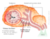
What are the 3 pairs of salivaty gland in thier head and the type of saliva they secrete?
3 pairs of salivary glands (exocrine glands)
Parotid – serous saliva, watery and rich in enzymes
Sublingual – mucous saliva, no enzymes
Submandibular – mixed serous and mucous
Von Ebner’s glands of tongue - produce lingual lipase

Describe the general structure of a salivary gland
“Bunch of grapes” appearance
Acini (single acinus - salivary gland) lined by acinar cells – initial secretion
Ducts lined by duct cells – modify secretion
Myoepithelial cells – contract to eject saliva
Describe the process of saliva secretion on a cellular level.
From acinar cells
Isotonic ultrafiltrate from plasma diffuses through acinar cells
Mixes with enzymes (serous cells) or mucins (mucous cells)
Secretion drains into ducts
In duct:
Net absorption of Na+ and Cl-
Net secretion of K+ and HCO3-
NaCl absorption > KHCO3 secretion → net absorption of solute
Ductal cells are H2O impermeable and so H2O cannot follow the solute → hypotonic solution

Describe the difference between resting and stimulated saliva
At rest saliva flow rate is lower → more time for ductal modification
At maximal salivation flow rate is higher → less time for ductal modification
HCO3- is the exception to the rule – it is selectively simulated when saliva production is stimulated so [HCO3-] increases with increasing flow rate
Resting saliva – low volume, highly modified, very hypotonic, neutral pH or slightly acidic, few enzymes
Stimulated saliva – high volume, less modification, less hypotonic, more alkaline, lots of enzymes

what impact does ADH and aldosterone have on hormone secretion ?
During dehydration or low vascular volume Na+ and water reabsorption increases -> saliva volume decreases
Describe the neural control of saliva secretion
Parasympathetic stimulation
Increases saliva secretion
In response to stimulation of taste and mechanoreceptors in mouth, sight and smell of food, nausea, and conditioned reflexes
Decreased by sleep, fear, and dehydration
Sympathetic stimulation
Initially stimulates the release of preformed mucous saliva but after that saliva flow decreases
Describe some symptoms of Xerostomia (dry mouth)?
Burning or scalding sensation in mouth
Dry and painful throat
Dry and rough tongue
Dry and cracked lips
Problems swallowing and speaking
Altered taste
Halitosis (bad breath)
Dental caries and periodontal disease
Oral infections, e.g. candidiasis
Difficulty with keeping dentures in place
What are the 5 classifications of taste?
Where are tastebuds found (location and within what structures)?
What is the anatomical structure of papillae?
What do taste buds contain?
sweet, sour, bitter, salty, umami
tongue, palate, larynx, and pharynx
On the tongue taste buds are found in papillae
Papillae anatomical structure: fungiform, foliate, circumvallate
Taste buds contain: taste receptor cells, supporting cells, basal cells
What are taste receptor cells?
How do they carry afferent info to the brain?
What do we need to taste?
( LO - Describe the process of taste)
specialised epithelial chemoreceptor cells which transduce a chemical stimuli into electrical signals, innervated by afferent to transmit info to CNS
Cranial nerves carry afferent information on taste
Signals carried to the medulla, and then to other regions of the brain including the sensory cortex
Saliva is needed as a solvent
Appreciation of flavour also involves olfaction
Describe the structures and processes involved in mastication
(LO-Describe the structures and processes involved in mastication)
Mastication = chewing
Physical digestion – breaking up food to increase the surface area for enzyme action
Teeth – cut (incisors) and crush (molars) food
Muscles of mastication
Masseter muscle
Temporalis muscle
Medial and lateral pterygoid muscles
(Suprahyoid muscles of neck also involved)
Movement of mandible, tongue, lips and cheeks help mix food with saliva and create bolus for swallowing
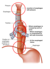
Describe the components of the pharynx
Extension of the alimentary canal
Nasopharynx – skull base to soft palate
Oropharynx – soft palate to epiglottis, posterior to the oral cavity
Laryngopharynx – epiglottis to cricoid cartilage, posterior to larynx

Describe the muscles of the pharynx
Muscular tube
Inner longitudinal muscles
External circular muscles – the pharyngeal constrictors
Contraction:
Shortens and widens pharynx when swallowing
Elevates larynx when swallowing
Forces bolus of food into oesophagus by peristalsis
Forms the upper oesophageal sphincter
Pharynx is a muscular tube that interconnects the nasal cavity, oral cavity, larynx, and oesophagus
Name the muscle layers of the oesophagus.
What structure does it pass through to become the stomach?
How does it transport food?
Internal circular and external longitudinal muscle layers
Superior third – striated muscle
Middle third – striated muscle and smooth muscle
Inferior third – smooth muscle
Passes through oesophageal hiatus in diaphragm at T10
Muscular tube that transports food by peristalsis
Name the 4 points of compression of the oesophagus
4 points of compression or narrowing: JABO
- Junction between pharynx and oesophagus
- In superior mediastinum where it is crossed by the arch of the aorta
- In the posterior mediastinum where the oesophagus is posterior to the left main bronchus
- At the oesophageal hiatus in the diaphragm
What is the lower oesophageal sphincter (LOS)?
Physiological sphincter at the gastro-oesophageal junction
Prevents reflux of gastric contents into the oesophagus
Higher resting basal tone
Components:
Right crus of diaphragm (contracts and acts like a pinchcock)
Acute angle at which oesophagus enters stomach
Mucosal folds at gastro-oesophageal junction (act like a “cork in the bottle”)
Positive intra-abdominal pressure - compresses and closes intra-abdominal oesophagus
What are the consequences of lower oesophageal sphincter dysfunction?
Pathological changes?
signs and symptoms?
Gastro-oesophageal reflux disease (GORD)
Reflux of acidic contents through LOS
Occurs when normal anti-reflux mechanisms are impaired
Characterised by:
- Heartburn - burning lower chest pain radiating upwards, related to meals, worse on bending and lying down, relieved by antacids
- Water brash (saliva overproduction)
- Acid brash (regurgitation of acid or bile)
- Regurgitation
If damage to mucosa occurs reflux oesophagitis can occur.
What can cause GORD?
Impairment of normal anti-reflux mechanisms, e.g. :
- Increased frequency of transient lower oesophageal sphincter relaxations (TLESRs)
- Increased intra-abdominal pressure, e.g. in pregnancy
- Low LOS pressure
- Hiatus hernia - prevents normal functioning of LOS – including disrupting diaphragmatic action which normally contracts and acts like a pinchcock
What is Barrett’s oesophagus?
- Metaplasia of squamous epithelium of oesophagus to columnar mucosa
- Proximal displacement of squamocolumnar junction ( junction between the squamous epithelium and the columnar epithelium)
- Complication of GORD or hiatus hernia
- Associated with increased risk of oesophageal adenocarcinoma (a malignant tumour formed from glandular structures in epithelial tissue)
Describe the phases of swallowing.
Oral phase
- •Mastication produces bolus which must then be swallowed
- •Oral (voluntary) phase – duration up to 1 second
- •Tongue moves bolus back towards oropharynx
- •Sensory receptors in palate and anterior pharynx send information via cranial nerves to swallowing centre in medulla
- •Swallowing reflex initiated
Pharyngeal phase
Duration: 1 second
Involuntary
Soft palate elevates – blocks of nasopharynx
Respiratory tract protected:
- Respiration inhibited
- Glottis closes
- Larynx elevates
- Epiglottis tilts to cover opening of larynx
- Upper oesophageal sphincter opens
Peristaltic wave of contraction propels food through sphincter into oesophagus
Oesophageal phase
Primary peristaltic wave spreads along oesophagus, propelling bolus towards stomach.
The lower oesophageal (cardiac) sphincter and proximal stomach relax and bolus enters the stomach.
Secondary waves clear residual material.
Sphincter constricts to prevent reflux
What is Achalasia?
- Loss of coordination of peristalsis of lower oesophagus and spasm of LOS
- Functional obstruction
- Dilation and hypertrophy of oesophagus
- Characterised by:
- Intermittent dysphagia (swallowing problems) for both solids and liquids
- Regurgitation of food
- Retrosternal chest pain
Lecture key points
- The composition of saliva reflects it’s function
- Saliva is secreted by acini and undergoes ductal modification in salivary glands
- The composition of resting saliva is different from stimulated saliva
- Taste receptor cells are chemoreceptors found in taste buds
- The organisation of muscles in the pharynx and oesophagus reflects their role in swallowing
- The lower oesophageal sphincter consists of a number of components and is important in preventing reflux of gastric contents into the oesophagus
- There are a 3 phases to swallowing, both voluntary and involuntary
- The swallowing reflex is an important brainstem function
