Pigmentations & Tissue Deposits Flashcards

(74 cards)

1
Q

What are the 4 main categories of pigments?

A

1) Hematogenous pigments
2) Melanin
3) Lipofuscin
4) Exogenous pigments

2
Q

What are 4 types of Hematogenous pigments?

A

1) Hemoglobin
2) Hemosiderin
3) Bilirubin
4) Porphyrins

3
Q

What are the 3 main categories of Tissue depositis?

A

1) Calcification
2) Amyloid
3) Uric Acid

4
Q

What are 3 type of Calcification?

A

1) Dystrophic
2) Metastatic
3) Calcinosis cutis

5
Q

What is the cause of the jaundice color that comes from RBC catabolism?

A

Bilirubin

6
Q

What animal species do not have Biliverdin reductase so they accumulate Biliverdin instead of Bilirubin?

A

Birds

7
Q

What is the end product of the breakdown of the Heme group?

A

Bilirubin

8
Q

In Hemoglobin Catabolism, what are the 2 pathways that Conjugated bilirubin will go once in the bile duct?

A

1) Most will be secreted into the GI tract
2) Some will go back into the blood

9
Q

Term used when too much bilirubin in the blood

A

Hyperbilirubinemia

10
Q

Term used when Bilirubinemia >2mg/dl

A

Jaundice

11
Q

Definition of Jaundice/Icterus

A

Increased bilirubin in tissues

12
Q

What are 2 gross descriptions of Jaundice/Icterus

A

1) Yellow-green discoloration of tissue or fluid
2) Most prominent in mucous membranes, adventicial surfaces

13
Q

What is the ONLY organ tissue that you DO see pigment in jaundiced tissues?

A

Cholestatic liver

14
Q

What is a microscopic description of Jaundice/Icterus?

A

Yellow-brown intracellular (hepatocytes, kupffer cells) or extracellular pigment (bile canalliculi)

15
Q

What are 3 types of hyperbilirubinemia and main characteristic of each one?

A

1) Pre-hepatic -> too much breakdown of RBCs
2) Hepatic -> Hepatocyte dysfunction
3) Post-Hepatic -> buildup of conjugated bilirubin in blood

16
Q

In Prehepatic hyperbilirubinemia, what happens and what is the cause?

A

Bilirubin production exceeds hepatocellular uptake

Cause: Hemolysis (intravascular or extravascular)

17
Q

In Hepatic hyperbilirubinemia, what are the 3 types of hepatocellular dysfunction and the cause of it all?

A
  1. Decreased bilirubin uptake
  2. Decreased conjugation
  3. Decreased secretion in bile

Causes: Hepatic insufficiency, hepatitis, hepatocellular degeneration

18
Q

In Posthepatic hyperbilirubinemia, what happens and what causes it?

A

Reflux of conjugated bilirubin into blood

Causes: Biliary obstruction (cholestasis) or rupture

19
Q

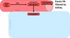
Which mechanism of jaundice is to blame with this sheep?

A

Prehepatic hyperbilirubinemia

20
Q

What are 2 gross descriptions of Hemoglobinuria?

A

1) Red-brown coloration of kidney and urine
2) Pink serum

21
Q

What is a microscopic description of Hemoglobinuria?

A

Red-orange material in renal tubules

22
Q

What are 2 ways of hemoglobin catabolism?

A

1) Extravascular hemolysis
2) Intravascular hemolysis

23
Q

What happens in Extravascular Hemolysis?

A
24
Q

What happens in Intravascular Hemolysis?

A
25
Extravascular and Intravascular hemolysis BOTH result in jaundice True or False?
True
26
What are the 5 etiologies of Intravascular hemolysis?
1) Oxidative damage (most common) 2) Erythrocyte metabolic deficiency 3) Infectious 4) Immune-mediated 5) Direct membrane damage
27
Copper Toxicosis falls under which etiology of Intravascular hemolysis?
Oxidative damage
28
Neonatal Isoerythrolysis falls under which etiology of Intravascular hemolysis?
Immune-mediated
29
What is the end result of Acute Copper Toxicosis in small ruminants?
Hemoglobinuria
30
Which is most severe (Prehepatic, Hepatic, or Posthepatic)?
Posthepatic
31
Which has the slowest onset (Prehepatic, Hepatic, or Posthepatic)?
Hepatic
32
Prehepatic, Hepatic, or Posthepatic? (IMHA)
Prehepatic
33
Prehepatic, Hepatic, or Posthepatic? (infectious canine hepatitis (adenovirus))
Hepatic
34
Prehepatic, Hepatic, or Posthepatic? (Hepatic lipidosis)
Hepatic
35
Prehepatic, Hepatic, or Posthepatic? (Biliary calculus)
Posthepatic
36
What are 4 things that can be done clinically to determine the cause of jaundice?
1) CBC 2) Urinalysis 3) Chemistry Panel 4) Diagnostic Imaging
37
What is the end result of Neonatal Isoerythrolysis?
Immune mediated intravascular hemolysis
38
How is Hemosiderin stored as?
Iron stored intracellularly as ferritin
39
What is a gross decription of Hemosiderin?
Must have alot to impart gross brown color
40
What are 2 microscopic descriptions of Hemosiderin?
1) Dark-yellow-brown, coarse granular cytoplasmic pigment 2) Stains blue-black with prussion blue/Perls
41
What are 2 types of Hemosiderin accumulation and what does it contain?
1. Local: - Chronic congestion (heart failure cells) - Hemorrhage 2. Widespread: - Hemolysis - Inherited disorders of Fe storage
42
Erythropoietic porphyria has an inherited deficiency of what enzyme and what does this result in?
uroporphyrinogen III cosynthetase porphryins accumulate in dentin and bone
43
What are 2 gross decriptions of Erythropoietic porphyria?
1) Pink-red discolored bones and teeth 2) Fluoresces with UV light
44
What is a gross description and microscopic description of Melanin?
Grossly -\> Black/brown tissue Microscopically -\> Fine brown/black cytoplasmic granules
45
What enzyme is needed for Melanin synthesis?
Tyrosinase
46
What are 3 things that activates Tyrosinase activity?
1) UV light 2) Hormones (MSH) 3) Inflammation
47
What is Lipofuscin derived from and commonly found in?
Derived from breakdown of lipids Found in aged cells and injured cells (Wear and tear pigment)
48
What is a gross description and microscopic description of Lipofuscin?
Grossly: - Usually nothing - Large accumulation = brownish color tissue Microscopically: -Golden-brown, fine granular cytoplasmic pigment
49
Accumulation of vitamin A causing yellow-orange discoloration leads to what type of exogenous pigment?
Carotenoid pigmentation
50
Diagnosis when Carbon deposits accumulate in cells
Anthracosis
51
What is a gross description and microscopic descrition of Calcification?
Grossly: -Hard, white gritty granules/plaques Microscopically: - Basophilic amorphous granules - Stain black with von Kossa stain
52
Local deposit of Ca2+ in areas of injury happens in which type of Calcification?
Dystrophic
53
Widespread deposit of Ca2+ in otherwise normal tissues happens in which type of calcification?
Metastatic
54
What causes Dystrophic calcification?
Ca2+ ions interact with fatty acids producing insoluble calcium soaps (Saponification)
55
What causes Metastatic calcification?
Hypercalcemia
56
What are 5 favorite sites for Metastatic Calcification?
1) Vascular intima/adventicia 2) Gastric mucosa 3) Renal tubular epithelium 4) Pulmonary interstitium, pleura 5) Basement membranes
57
What is the best morphological diagnosis for our dog case?
Metastatic calcification
58
What are 2 causes of Hypercalcemia?
1) Excess PTH 2) Excess Vitamin D
59
Dystrophic or Metastatic? (acute pancreatitis)
Dystrophic
60
Dystrophic or Metastatic? (cow liver)
Dystrophic
61
Dystrophic or Metastatic? (granuloma)
Dystrophic
62
Dystrophic or Metastatic? (cow heart)
Metastatic
63
Dystrophic or Metastatic? (calcinosis circumscripta)
Dystrophic
64
Dystrophic or Metastatic? (dog skin)
Trick Question: Its neither This is Calcinosis Cutis and seen with dogs with Hyperadrenocorticism
65
What is Amyloid made up of?
Fibrils made of stacked β- pleated sheets
66
What are 2 gross decriptions and 2 microscopic descriptions of Amlyoid?
Grossly: - Enlarged, firm, waxy organs - Stains blue violet with iodine and sulfuric acid Microscopically: - Amorphous homogenous eosinophilic extracellular material (hyaline) - Stains pink and has green birefringence with polarized light when stained with Congo Red
67
What are 4 types of Amyloid?
1) Amyloid A (most common) 2) Amyloid Light Chain (AL) 3) Endocrine Amyloid 4) AB Amyloid
68
What type of amyloid is deposited in ‘reactive systemic amyloidosis’?
Amyloid A
69
What 2 breeds is Reactive Systemic Amyloidosis hereditary in?
1) Sharpeis 2) Abyssinians
70
Term used for accumulation of uric acid in tissues
Gout
71
What animal species does not have uricase (uric acid is end product)?
Birds and reptiles
72
Decreased renal function or dehydration results in gout in what animals species?
Birds and reptiles
73
Diet, genetic disorders or chemotherapy results in gout in what animal species?
Mammals
74
Term used for granulomatous inflammation surrounding deposit
Tophi