Pleural Disease Flashcards
(32 cards)
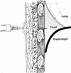
Pleural Anatomy
- 2 layers made of mesothelial cells
- Visceral pleura ⇒ lines lungs
- Parietal pleura ⇒ lings chest wall
- Normal pleural fluid production ~ 16.8 nl/day for 70 kg adult
- Fluid flows from visceral to parietal pleura
- Lymphatic drainage ~ 470 cc/day
- 28x more than production
- No fluid in pleural space normally
Pleural Effusion
Definition
Accumulation of fluid in the pleural space.

Pleural Effusions
Pathophysiology
-
↑ fluid accumulation
Entry of fluids into pleural space:- ↑ systemic venous pressure
- ↑ pulmonary venous pressure
- ↑ permeability of pleural vessels
- ↓ pleural pressure
- ↓ microvascular oncotic pressure
-
↓ fluid removal
Blockage of lymphatics:- Central lymphatic obstruction
- Obstruction of lymphatic channels at pleural surface by tumor
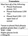
Transudates
Characteristics
Light’s Criteria
Exudates
Characteristics
Light’s Criteria

Transudates
Etiologies

Exudates
Etiologies

Pleural Fluid
Analysis
- Cell count and differential
- Chemistry
- Proteins, LDH, albumin, amylase, pH, glucose
- Obtain concurrent serum values
- Gram strain and culture
- Cytology
- Other tests as indicated
- Lipids, fungal culture, triglycerides, Ig
Pleural Effusion
History
- Asymptomatic
- Dyspnea ⇒ d/t compression of underlying lung
- Pleuritic CP ⇒ see w/ some exudative effusions
Pleural Effusion
Physical Exam
- ↓ tactile fremitus
- Dullness to percussion
- ↓ or absent breath sounds
- Tracheal shift to contralateral side w/ very large effusion
- Tubular breath sounds, egophany (E to A changes)
CHF Related
Pleural Effusions
- Most common cause of transudates
- D/t ↑ pulmonary venous pressures from LV dysfunciton
- Usually bilateral, R > L
- Thoracentesis often not needed
- Unless atypical or fail to resolve w/ medical treatment
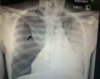
Image 1
Left-sided Massive Pleural Effusion
With contralateral shift of mediastinum and trachea.
Most common cause of non-traumatic massive pleural is cancer.
Parapneumonic Effusions
Definition
Exudative effusions in setting of bacterial PNA or lung abscess.
- Often very high WBCs and LDH levels
- Effusion on same side as PNA
Uncomplicated Paraneumonic Effusion
Characteristics
- Negative gram stain and culture
- pH > 7.30
- Glucose > 40
- Resolves w/ simple abx treatment of PNA
Complicated Parapneumonic Effusion
- pH < 7.20
- Glucose < 40
- Requires chest tube or surgical drainage for resolution
Empyema
“Pus” in the pleural space.
- Will usually have a positive gram stain or culture
- Treatment same as complicated parapneumonic effusion
RA
Pleural Effusions
- Pleural glucose < 30
- P/S ratio < 0.5
- pH < 7.3
- High LDL level > 700
TB
Pleural Effusion
- Exudative effusiosn d/t hypersensitivity rxn
- Usually neg. AFB smear
- Lymphocytic predominance
- Culture neg. for AFB
- Adenosine deaminanse in fluid consistent w/ TB
Malignant Pleural Effusion
- 2nd most common cause of exudative effusion
- Malignant cells seen in 60% of 1st thoracentesis
- Yield inc. by 20% on second tap
- Common etiologies
- Lung Ca
- Breast Ca
- Lymphoma
- Ovarian Ca
Chylothorax
Disruption or obstruction of thoracic duct ⇒ leakage of chyle fluid.
- High TAG
- Milky appearance
- Lymphocytic predominance
- Pleural triglyceride > 110 mg/dL ⇒ 85% of pts
- Etiologies ⇒ trauma, surgery, malignancy (lymphoma)
Cholesterol Effusion
“Pseudochylothorax”
- High cholesterol ⇒ milky appearance
- Chronic inflammatory process such as TB or RA
- Pleural cholesterol > 200 mg/dL ⇒ 75% of pts
- Cholesterol crystals in fluid
Nephrotic Syndrome
Pleural Effusions
Caused by PE and renal vein thrombosis
Pleural Effusion
Management
- Treat underlying cause of effusion
-
Thoracentesis
- Can remove up to 1,500 ml to relieve dyspnea
- Removing more inc. risk of re-expansion pulmonary edema
-
Chest tube
- Drain complicated parapneumonic effusion/empyema
- Palliate SOB d/t recurrent large pleural effusion e.g malignancy
- Perform chemical pleurodesis to obliterate pleural space & prevent reaccumulation of fluid ⇒ rarely done
-
Surgery ⇒ thoracoscopy or thoracotomy
- If interventions via chest tube fails
Pneumothorax
Definition
Air in the pleural space.