Practical: Horse Flashcards
(106 cards)
Foramen mandibulae: Nerve block

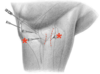
Describe the nerve-block of the bicipital bursa
- 6cm distal from the cranial part of major tubercle
- 7cm cranial from the deltoid tuberosity
- Proximomedial direction
- Aim at the intertubercular groove
Difficult to reach, accessed in a standing horse

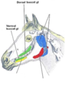
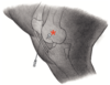
Foramen supraorbitale
Found in a dimple: Root of the zygomatic process of the frontal bone
Describe the primary nerve-block of the shoulder
- Between the cranial and caudal pars of the major tubercle
- Horizontal needle
- Caudomedial direction

The horse should be in a standing position
Trochanter major
- Divided into low cranial and high caudal portions
- Palpated under the biceps femoris


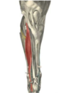
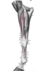
M. extensor digitorum lateralis
(FL)
Lymph node of the head
- Mandibular: Intermandibular space, forms a ‘V’ shape near the facial notch
- Lateral retropharyngeal: Found in clumps around the pharyngeal wall, caudal to the guttural pouch


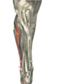
M. extensor digitorum lateralis
(HL)
Caudoventral border of the lung
- 16th IC space - Tuber coxae
- 14th IC space - Tuber ischiadicum
- 10th IC space - Shoulder joint
Diaphragm is located between ribs 8-17
Tuberositas deltoidea
- Craniolateral aspect of proximal humerus

Age ‘determination’
2.5 years → 10 years
- I1 erupts : 2.5 years
- I2 erupts : 3.5 years
- I3 erupts : 4.5 years
- Dental star on I1 : 5 years
- Dental star on I2 : 6 years
- Dental star on I3 : 7 years
- White spot apears in I1 dental star : 8 years
- White spot apears in I2 dental star : 9 years
- Cup disappears from I2 maxilla : 10 years
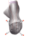
Describe the arthrocentesis of the centrodistal joint
- Small depression halfway between:
- Tuberculum of the talus
- Os tali centrale
- Long medial tarsal collateral ligament
- Needle directed perpendicular to the limb
- 1-2 cm deep

Describe needle access to the carpal joint (dorsal approach)
- Lateral/medial from the common digital extensor
- Palpable depressions between the bone rows
The limb should be flexed
Describe needle access of the distal digital flexor tendon sheet (Dorsolateral approach)
- Slightly dorsal from the lateral collateral ligament
- Eminences of Ph-1-2 are palpable
- Needle directed lateromedially under the digital extensor tendon
The limb can be weight-bearing or extended whilst it is held

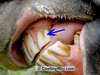
Nasal opening of the nasolacrimal duct
- Found on the floor of the nasal vestibule
- Red/pink dot at the base of the septum
Give the borders of the conchal sinus
- Dorsal border: Fuses with the conchofrontal sinus
- Middle border: Communicates with the ethmoidal meatus
- Ventral border: Connects with rostral maxillary sinus
Ostium ileocaecale
May be detected with a stethoscope a few cms ventral to the paralumbar fossa

List the patellar ligaments
- Lateral
- Intermediate
- Medial
- Lateral femoropatellar

Age ‘determination’
6 days → 18-24 months
- Id1 erupts : 6 Days
- Id2 erupts : 6 Weeks
- Id3 erupts : 6 Months
- Id1 cup disappears : 10 months
- Id2 cup disappears : 12 months
- Id3 cup disappears : 18-24 months
(Id1 = First Deciduous incisor)
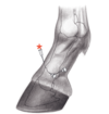
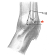
Describe needle access of the coffin joint (dorsal approach)
- Just over the tip of the extensor process
- Needle direction either:
- Parallel to the ground
- Perpendicular to the skin
The limb should be weight-bearing, in the midline

Maxillary sinus
- Caudal border: Opens into: Conchofrontal + sphenopalatine sinus
- Rostral border: Opens into the ventral conchal sinus

Accessory ligament
- Attaches DDF to the metacarpus
- Approx. 4cm proximal to the head of the splint bone

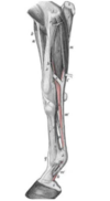
M. extensor carpi radialis
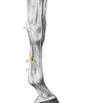
Describe the needle access of the fetlock joint (distal palmar/plantar approach)
- Between:
- Lateral proximal sesamoid bone
- Base of Ph-I
- Dorsoproximal needle direction
Dorsal from the lateral digital artery
The limb should be weight-bearing