Practicals Flashcards

1
Q

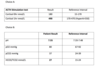
Feline platelets appear to aggregate (clump) more readily than other species. If platelet clumps have formed during blood sampling, they can be best appreciated while examining the feathered edge. Why is this finding important to recognize in all species, not just cats?

A

Have to consider DIC, sequestration, or lack of production by the bone marrow

* Anything about 100 is adequate for clotting anyway

2
Q

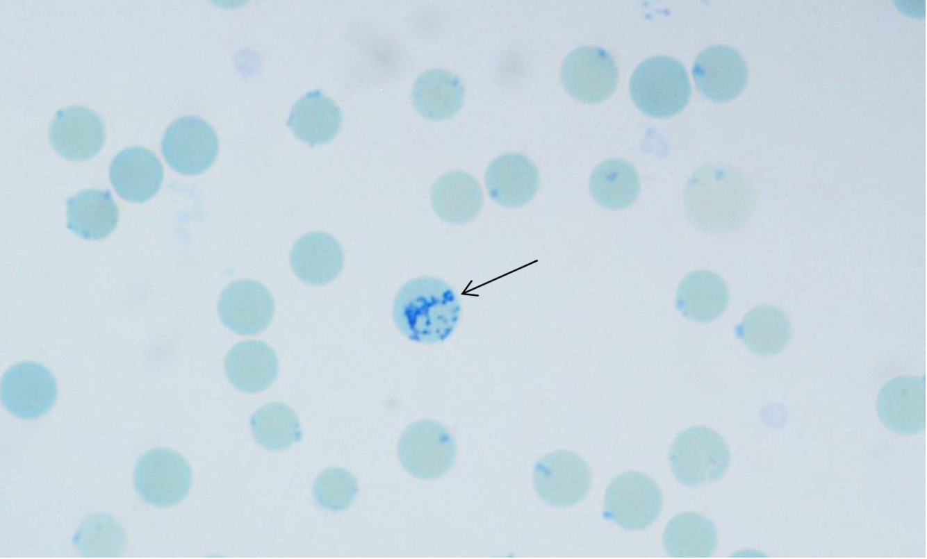
Note the morphology of feline erythrocytes- how do they compare to canine erythrocytes? e.g. size shape colour central pallor inclusions

A

canine- relatively large, uniform, biconcave disc

feline- not as uniform, smaller, no central pallor, Hb of cats is more susceptible to oxidant injury, low numbers of Heinz bodies can be seen in normal cats

3
Q

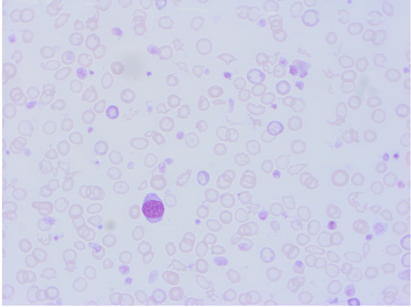
What disease process does the presence of these atypical cells most likely indicate?

A

Neoplasia

4
Q

How is the presence of agglutination likely to affect the RCC? Would this affect any of the calculated indices?

A

False low reading, false readings on MCV MCH MCHC

* If the RCC is wrong, the other readings would be incorrect as well

5
Q
A

Polychromatophil are our reticulocytes

And no arrow- Heinz Bodies (points to oxidative damage)

6
Q
A

Howell Jolly Bodies

Sign of regenerative anaemia, if we have a lot of them then we think about the marrow– dyserythropoeisis

* random ones no problem

7
Q
A

Basophilic stippling

* Lead poisoning

* In ruminants, basophilic stippling is common and has the same significance as polychromasia

8
Q

Dark stool in a dog DDX

A

Ulcers in the gastrointestinal system

Tumors of the esophagus or stomach

Infections

Foreign body in the gastrointestinal system

Disorders involving inflammation of the intestinal system

Kidney failure

Drug toxicity (e.g., anticoagulant drugs)

Diet containing raw food

Pneumonia

Trauma

Disorders involving abnormal clotting of blood

9
Q

Causes of thrombocytosis

A

Haemhorrage

Excitement- splenic contraction

Neoplasia

  • especially Hepatic carcinoma, malignant histiocytosis, lymphoma, mast cell tumor

Inflammation - e.g. Lymphocytic-plasmacytic enteritis

Hyperadrenocorticism

Immune-mediated hemolytic anemia

Chronic allergies

Iatrogenic - glucocorticoids, vincristine

10
Q

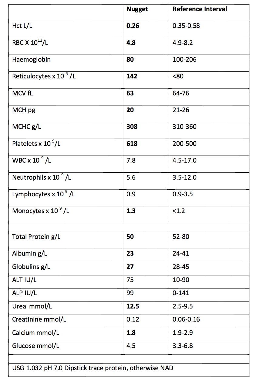
Most likely mechanism for hypocalcaemia given the results?

A

Hypoalbuminaemia

11
Q

What is pica?

A

Eating strange things- could mean vitamin deficiency

12
Q

* Platelet clumping- spurious thrombocytopaenia, common in cats as an artefact (EDTA)

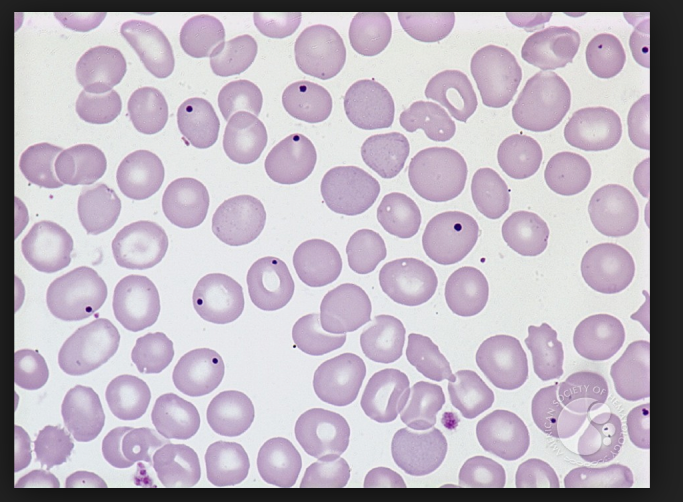
* atypical cells in the feathered edge- round nucleus, cytoplasm packed with dark granules– MAST CELLS (other ddx is basophil– in cats lavender granules)

* Howell Jolly body- normal for occasional sitings in cats
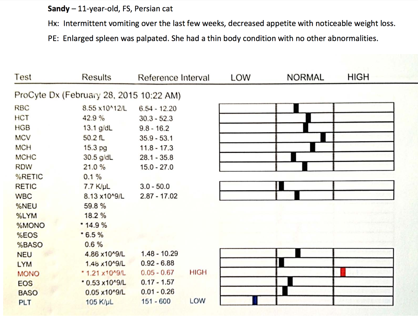
A

MCT in the spleen

* Histamine release–> gastic ulceration

13
Q

6 yo MN Standard Poodle

Hx: Acute onset of severe exercise intolerance

PE: Temp: 39.7 C RR 56. Pale and icteric mucous membranes. Enlarged spleen and liver.

* Very high WCC because of the NRBC– but still massive leukocytosis not uncommon in haemolytic anaemia

* Ghost cells, spherocytes

* Marked neutrophilia with regenerative left shift

* Severe inflammation but not so severe that the marrow has become depleted

* Marked monocytosis because they are eating red cells– increasing in number to migrate into the different organs
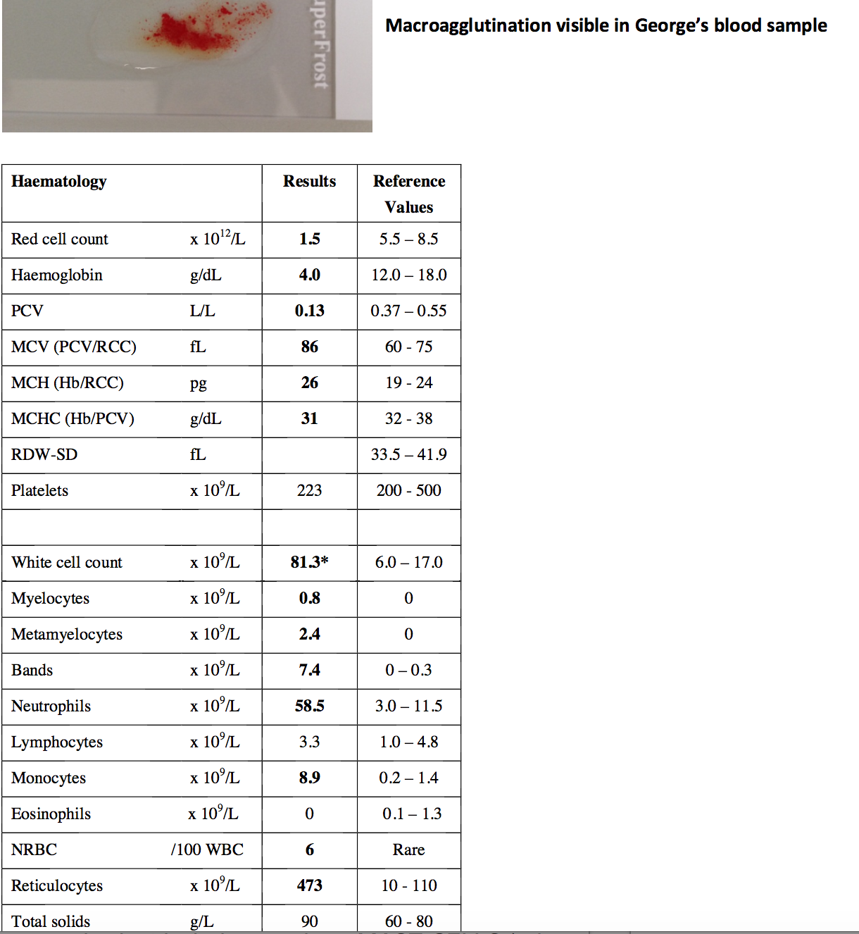
A

IMHA

* Enlarged liver and spleen due to attempt to produce haematopoietic cells in the spleen and liver just like they did when they were young (also microthrombi causing liver damage– swelling because of liver necrosis)

* Haemolytic regenerative anaemia– causes massive inflammation

* Microthrombi often occurs–> causes areas of necrosis in lots of organs particularly liver and kidney– often also a profound leukocytosis (inflammation) AND haemolysis due to the excess free haemoglobin

14
Q

2 yo neutered female Pug

Acute weakness, lethargy, and inappetance

PE: pale MM, passed reddish urine in consult room

* astericks mean something isn’t right according to the analyzer

* Dipstick from urine- positive for blood (not myoglobin)

* Thrombocytosis– due to the inflammation (though you can see thrombocytopaenia), chronic haemorrhage, splenic contraction (in response to hypoxia, anaemia)

A

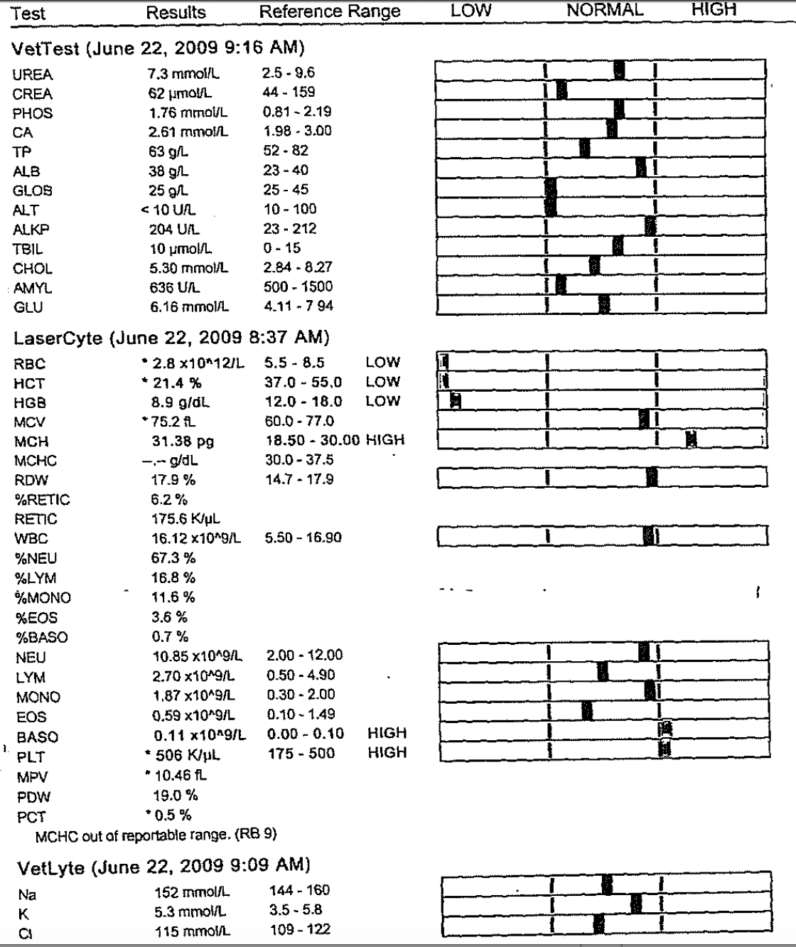
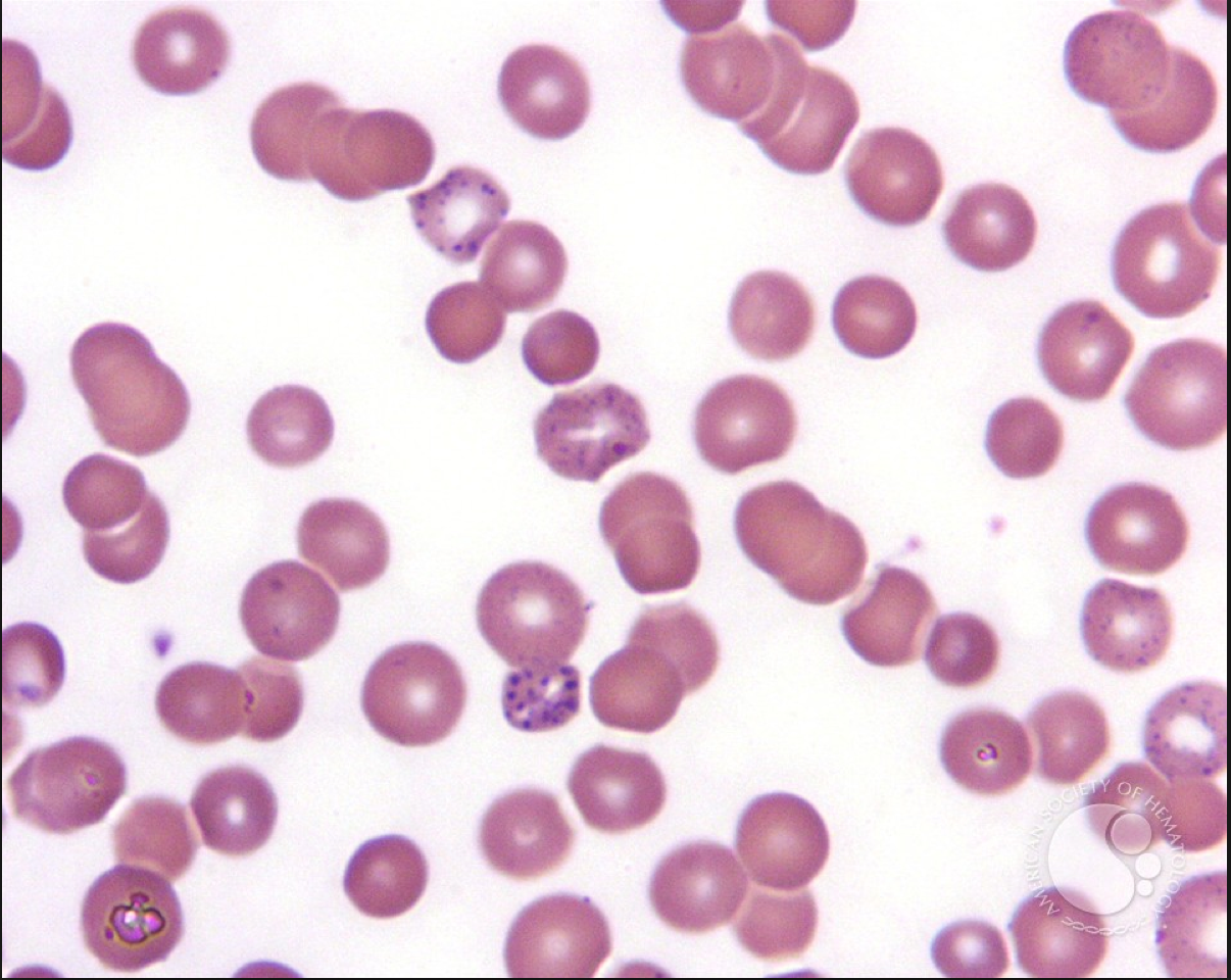
Due to eccentrocytes and Heinz Bodies with everything else–> oxidative damage–> THEREFORE ONION TOXICITY

* Regenerative anaemia normocytic– is there haemorrhage or haemolysis?

15
Q

When do you see a falsely high MCHC?

A

Free haemoglobin (haemolysis), lipaemia interferes with measurement of haemoglobin, Heinz bodies intefere with measurement of Hb as well

MCHC can’t truly be increased

16
Q
A

Eccentrocytes

Heinz Bodies (sticking out noses)- damaged Hb sticking out

17
Q

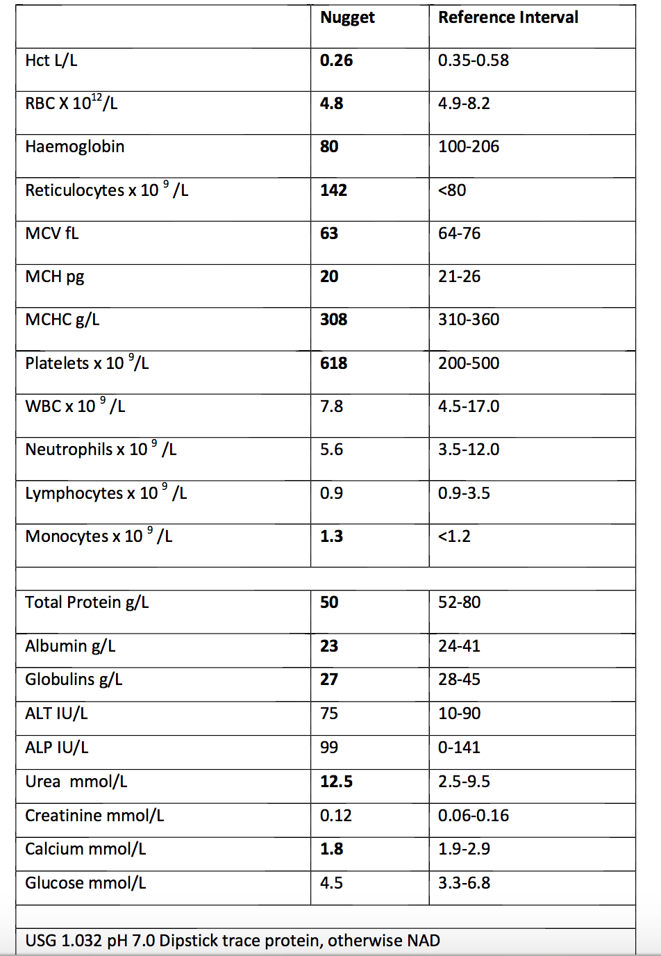
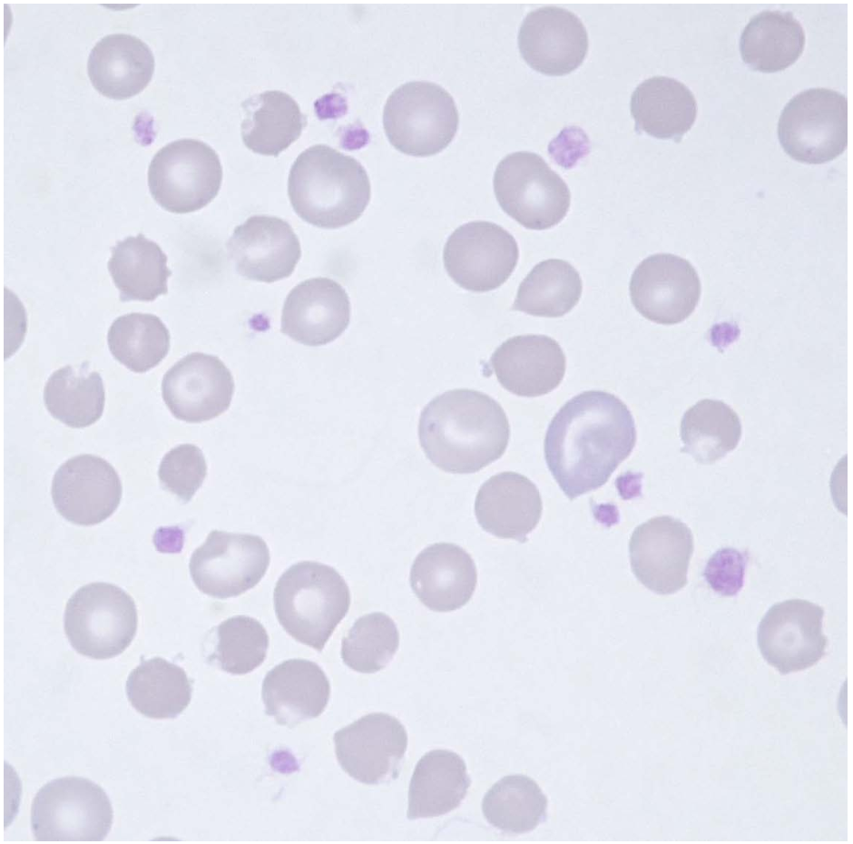
8 yo neutered male Staffy

Chronic lethargy, pica, dark faeces

pale mucous membranes, otherwise NAD

Microcytic, hypochromic = iron deficient (or breed specific with low MCVs e.g. Chowchow)

A

Bleeding from gastrointesintal tract causing iron deficiency– if it was internal it would recycle

Bleeding tumour, chronic intussusception that is not fully blocking

18
Q
A

Activated lymphocyte

Schistocytes

Hypochromic

Acanthocytes

Echinocytes

(IRON DEFICIENCY)

19
Q

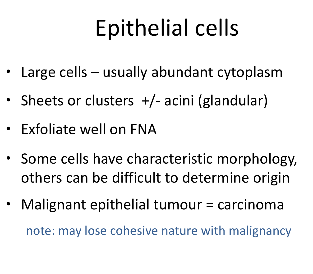
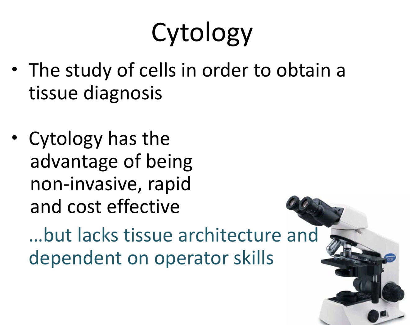
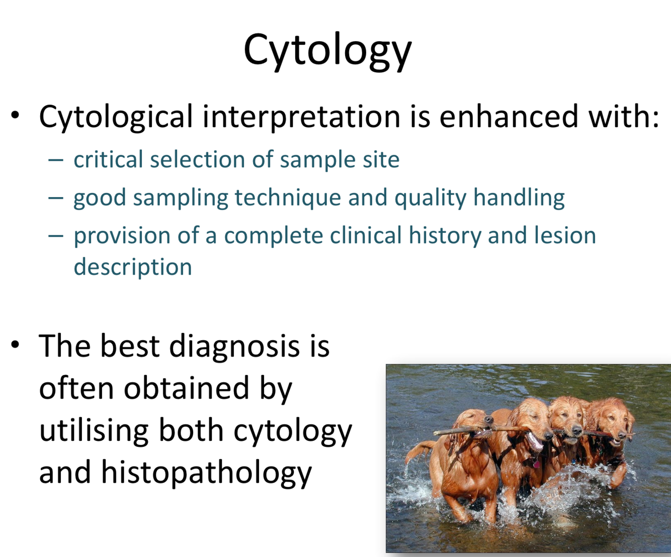
What is cytology? Advantages and disadvantages?

A
20
Q

How do you enhance cytological interpretation? Best diagnostic information by??

A
21
Q

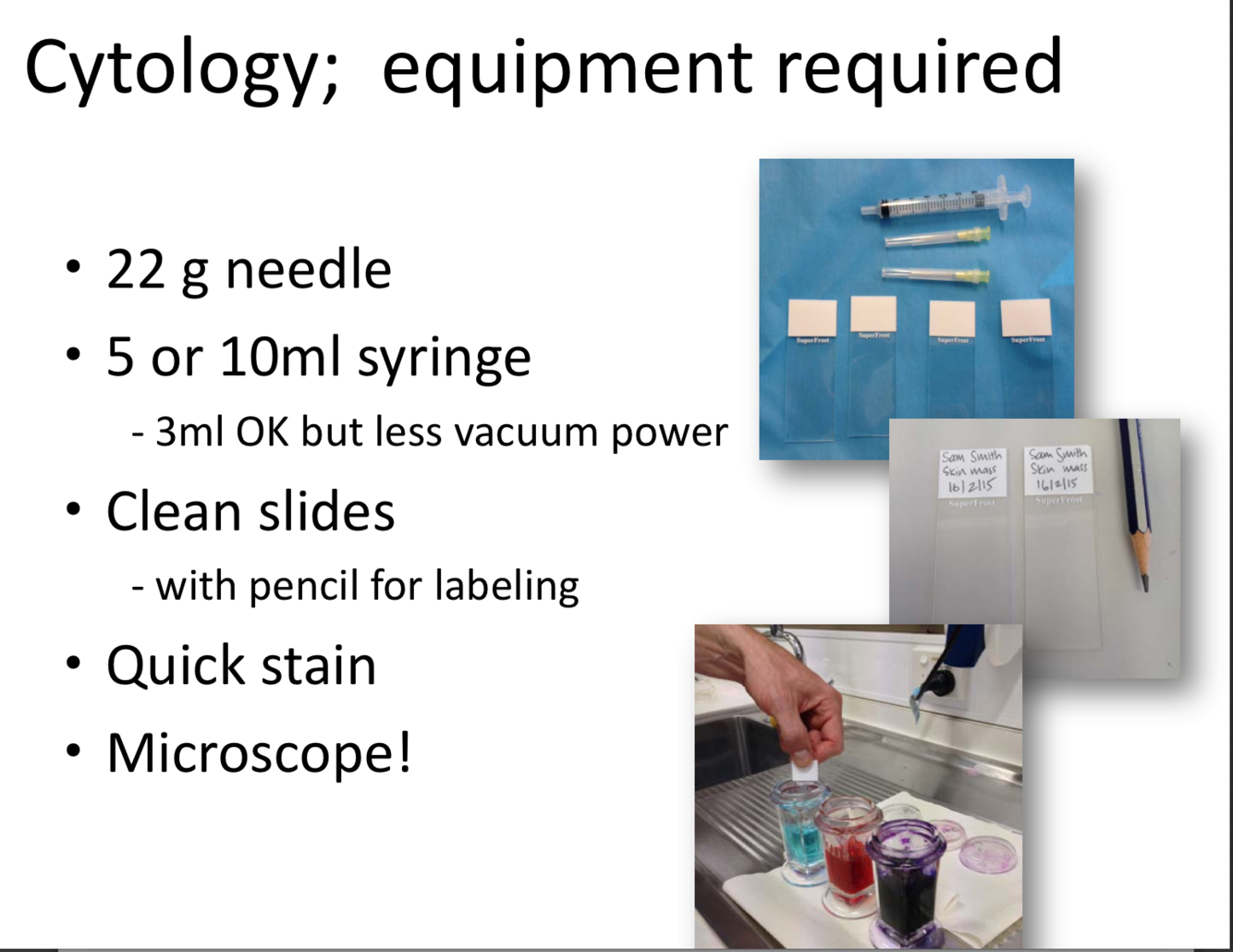
Equipment required for cytology

A
22
Q

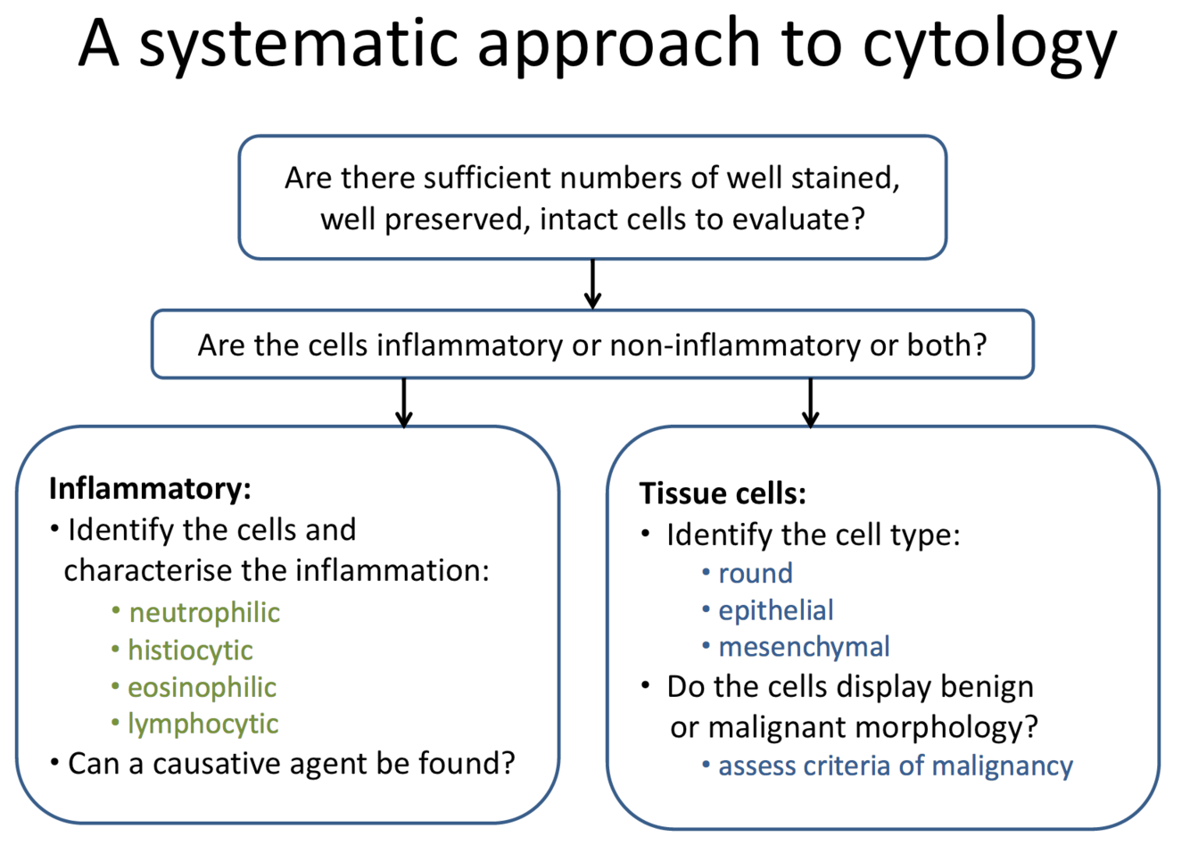
Systemic approach to cytology

A
23
Q

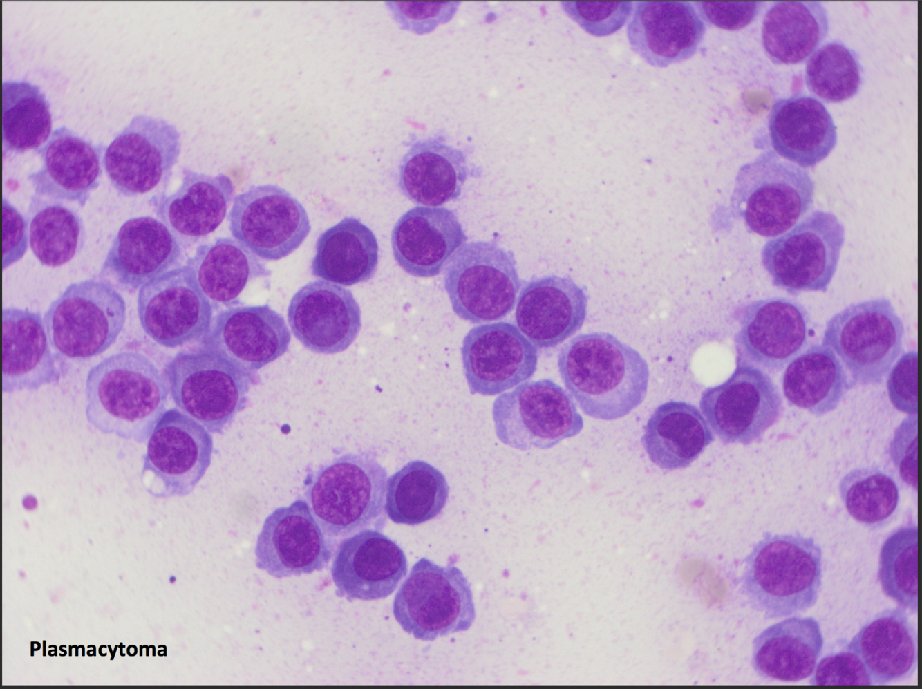
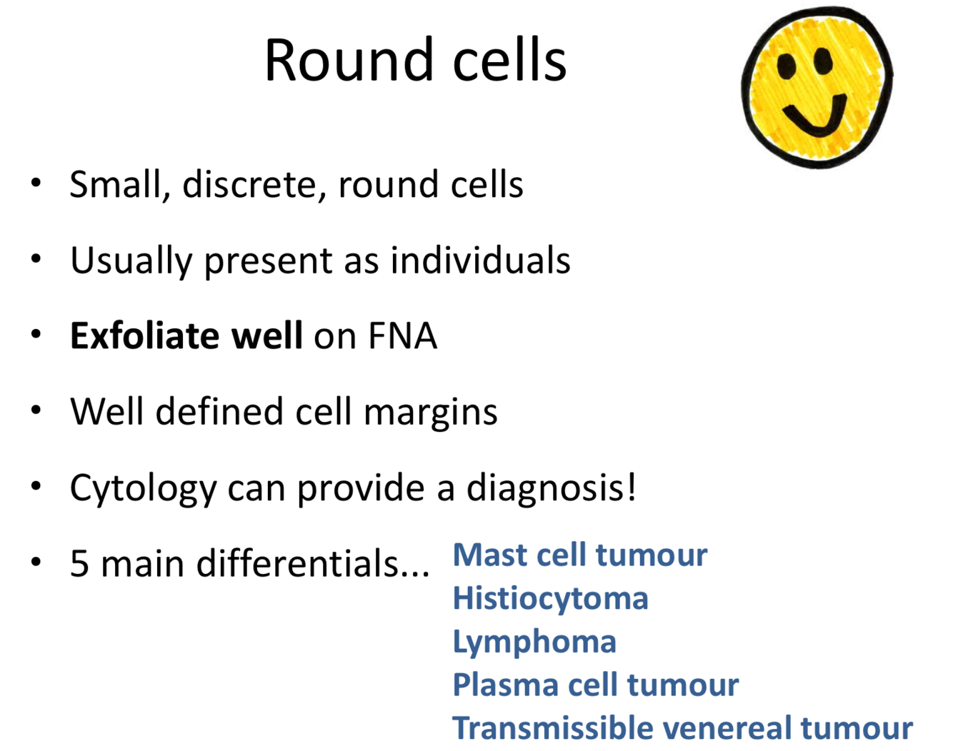
5 main differentials with round cells seen in cytology

A
24
Q

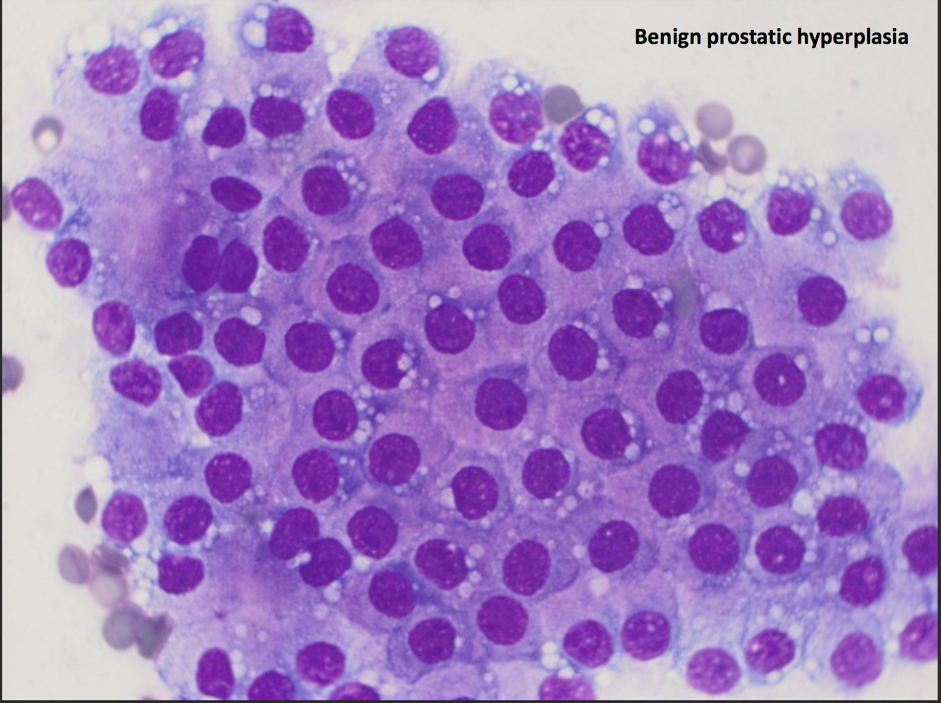
What is a carcinoma- what kind of cell? How do they go with cytology?

A
25
what type of cell does a sarcoma derive from? how do they go with cytology?
26
27
28
Fred 12 yo MN Cock Spaniel Abdominal distension noticed a week ago, otherwise normal \* Lesion: generalized lymphadenomegaly, hepatosplenomegaly, fluid in abdomen. Atypical lymphocytes seen in blood film. FNA of LNs and spleen
\* Microscopic findings: the smears highly cellular, monomorphic large lymphocytes (92%) with fewer smaller lymphocytes (8%)... occasional mitotic figures... background abundant cytoplasmic fragments (lymphoglandular bodies). Rare neutrophils and well differentiated mast cells noted. \* Cytology: large cell lymphoma
29
Buster, 4 yo MN American Bulldog Lesion: a large ulcerated skin mass ventral perianal region
\* Microscopic findings: plump pleomorphic mesenchymal (spindle) cells, blood, leukocytes... mesenchymal cells indistinct whispy borders... large round oval nuclei with stippled to coarse chromatin and 1-3 often prominent but small nucleoli. Frequent mitotic figures. \*\* Sarcoma... histopath: Myxosarcoma
30
31
How does buffy coat analyzer work?
Okay white cell count Struggles with any sort of abnormalities Does not adequately differentiate the different layers Repeatedly centrifuge to get the buffy coat to spread out evenly
32
How does impedance technology work?
when running WCC, lyse red cells newer ones use laminar flow- histograms counting the number of cells, proportionate to the deflection of electrical current-- when the machine has separated platelets from red cells appropriately they are two separate curves-- when they overlap a lot both counts are dubious e.g. when red cells are small \*\* looking at the impedance graph helps us decide if we can believe what the machine is telling us
33
Normal or abnormal?
Normal \*\* none of them are good at basophils
34
Normal or abnormal?
Abnormal impedance graph (WBC histogram)
35
What is flow cytometry?
36
Feline eosinophils highly distributed Cows have big lymphocytes and monocytes
37
What does it mean when there is a shark fin?
It means bands! Which means look at the smear and look for toxic change and bands! toxic change + bands (mostly due to toxic change as it is the change in the cytoplasm) in neutrophils makes them fluoresce more and merge with the lymphocytes
38
What does the abnormal dot plot mean?
The line drawn is arbitrary, not nice separate clouds \*\* in this case it means Leukaemia-- difficult to tell if they are myeloid or lymphoid on the smear and the machine has the same drama
39
What does this mean?
Regenerative anaemia
40
What might you think?
Strong regenerative response but you couldn't see polychromasia on the blood smear
41
Thrombocytopenia
42
Platelet clumping
43
the feathered edge
44
monolayer
45
WBC and platelet estimate from smear?
46
Boxer 6 yo FN Hx: lethargic and anorexic past few days, vomited dark fluid a few times over past week, loves chewing sticks, plastic, etc. Occasionally raids the garbage PE: tense distended abdomen, 39.8 T, pale mucous membranes, HR 172, tacky MM 1. are there enough platelets for haemostasis? 2. what are the most likely causes of hypochloridemia and hypernatraemia? 3. mild azotaemia? 4. calculate globulins? hypoalbuminaemia? 5. DDX?
1. no should be 150 x 10^9 (it's 107 x 10^9) 2. vomiting would lose HCl and therefore cause dehydration that would increase Na (hone in on upper GI or pancreatitis) 3. Mild azotaemia from dehydration 4. TP- albumin= Globulins = 33 g/L 5. Internal bleeding, peritonitis + sepsis (magnitude of white cells-- possibly after eating a FB, resulting in a perforation), chronic myeloid leukaemia, necrotising pancreatitis (20% of dogs with pancreatitis have a normal amylase and lipase-- pancreatic lipase snap test)
47
Cocker spaniel 9 yo MN \* lethargic, vomited a few times, mild weight loss despite good appetite \* Mild enlargement prescapular LN, T 39.1, pale MM, HR 168 1. Anaemia is regenerative? Potential causes for anaemia? 2. Potential causes of low urea in this case? 3. Potential causes of hyperglycaemia and which is the most likely in this case? 4. Primary DDX? 5. If there was a Lymphocyte count \> 10?
1. Chronic disease, neoplasia 2. Urea cycle occurs in the liver and kidneys 3. Hyperglycaemia due to liver failure (does not clear 4. DDX: 5. Lymphocyte count \> 10 (1-4.8)-- do not ignore-- highly unusual only exception young animal... start wondering about lympho proliferative disease
48
9 yo Crossbreed MN \* fasted pre anaesthetic screen before routine dental 1. What cells are included in the granulocyte count? 2. Differentials for increased urea? 3. What is a right shift? 4. when do you see activated lymphocytes?
1. Neutrophils, eosinophils, basophils and mast cells 2. dehydration-- USG concentrated \> 1.030 we are happy, pre renal azotaemia 3. \> 5 segments mature neutrophilia (right shift)--\> cortisol levels elevated 4. activated lymphs can be seen with inflammation, low numbers can be normal, after vaccination
49
11yo FN lethargic anorexic for 48 hours 38.9T, slightly pale MM, HR 102, possible hepatomegaly \*\* # means unsure \*\* dot plots say it is wrong
\* increased nucleated reds-- disrupted bone marrow \* you can see dead cell during apoptosis where the nucleus is undergoing karryorexis (sp?) not normal to see unless it has been sitting over night \* large atypical cells-- requires a pathologist to diagnose \*\*Lymphoma organ origin (spleen, liver, lymph node origin); leukaemia means bone marrow origin
50
If your platelet count is 44 x 10^9/L (200-500), DD? How many are enough platelets?
Immune mediated (can be due to lymphoma), Spleen platelet sequestration (can be due to lymphoma), Consumption with DIC, reduced marrow production, sequestration, acute inflammation \*\* Over 100 platelets is enough
51
Difference between lymphoma and leukaemia? How do you determine which it is?
Lymphoma organ origin (spleen, liver, lymph node origin); leukaemia means bone marrow origin \*\* determine based on looking at the patient
52
Hypoglycaemia DDX
Neoplasia, Addisons, Sepsis
53
Two types of neoplasia that make globulins
B cell lymphoma or leukaemia, plasma cells- Multiple myeloma
54
small to intermediate cell lymphoma or leukaemia
55
Neutrophils above 50, think what?
Cancer or inflammation??
56
\* Dipylidium caninum "flea tapeworm" \* How did the cat become infected? - cat ate a flea \* What sort of treatment do you recommend? - Praziquantel \* What precautions can be taken to prevent re-infection? - long-term flea control - warn the owner that it is a zoonosis
57
In March 2011, a local vet called to investigate an increase in abortions, mortality and morbidity in beef cattle (Angus and Hereford) on a property in NSW. A number of animals died over 1-2 months. A number of animals had aborted or calved prematurely. At the initial visit, one animal was examined and had pyrexia, tachycardia, tachypnoea, and icteric mucous membranes. Anaemia is most affected animals. Moderate hyperbilirubinaemia, hypergammaglobulinaemia, leucocytosis, neutrophilia with left shift and lymphocytosis. Stained blood smears revealed that 3% of RBCs contained internal spots or larger structures. Illness occurred in cows within 1 to 2 months of calving and most animals became inappetant. The cows had icteric mucous membranes. There was no known access to any toxic plants that could explain the icteric mucous membranes (e.g. lantana, fireweed or bracken fern). 1. What was the most likely cause? 2. What measures would you implement to treat and control this problem? Outline a strategy. Is there a need to notify authorities? 3. How would you monitor the problem and the progression of disease? 4. How would you assess the economic impact of this disease? 5. Could there be possible long-term consequences or issues to consider? 6. Is this problem specific to beef cattle?
1. What was the most likely cause? Ixodid ticks (Haemophysalis) transmitting Theileria orientalis 2. What measures would you implement to treat and control this problem? Outline a strategy. Is there a need to notify authorities? - Tick control-- dipping (have to convince them it is a problem and should do this when the cattle are preggo for example) - supportive care - Naphthaquinone compounds but not available in Australia (IM injection, 1-2 doses) 3. How would you monitor the problem and the progression of disease? - Monitor CBCs, monitor tick population, watch for new cases, make sure tick treatment is given if you're buying cattle from a non-endemic area to endemic area 4. How would you assess the economic impact of this disease? - Keep records of local affected cattle 5. Could there be possible long-term consequences or issues to consider? - endemic stability: long term is positive for local cattle that develop resistance.... naive cattle brought in is where the longterm epidemics could occur 6. Is this problem specific to beef cattle? - for this specific species, Theileria orientalis, only cattle (beef and dairy) but sheep, goats, deer, and horses theileriosis spp. do infect them but not in Australia
58
Found on a 13 yo cat
DDX: Felicola subrostratus, Demodex Underlying disease, old, unable to groom itself... uncommon
59
Rabbit severe dandruff DDX? After fur scraping? TX?
DDX: Fur mites-- Cheyletiella parasitovorax (most common), Leporacus gibbus, Fleas: Ctenocephalides felis or C. canis, Saroptes cuniculi Fur scraping: Cheyletiella parasitovorax (claws are very prominent) TX: revolution (Advantage is ineffective), severe cases shampoos and fur clipping \*\* does survive off the host so clean the enclosure
60
Beef weaner calves and cattle scouring Dairy cows developed scouring Two of the dairy cows died Post morem ileal lesions DDX? TX?
DDX: Strongyloides, Giardia-- extracellular, Cryptosporidium-- epicellular but intracellular and extraplasmic \* Cryptosporidium-- GI or intenstinal, direct faecal oral route, thick walled oocyte passed in faeces \* TX: Trisoprim is an systemic antibiotic, not an anti protozoal... so can try hyper immune bovine colostrum, supportive care.. control the outbreak biosecurity and quaratine, ID animals that have it and separate them, treat them with fluids, sterilize the area with steam, flame gun, and dry ammonia \*\* C. parvum is a zoonosis!!
61
Pet chickens and similar parasite on the cat, concerned daughter has itchy bites on her limbs DDX? Tramission? TX? Should she also treat her rabbits?
DDX: Dermanyssus or Ornithonyssus \* Different life cycles and different management implications Ornithonyssus \* Transmission by contact-- handling the bird TX: treat env and animals- exclude exposure to the vectors-- remove rubbish or shelters for the mites, spray with 1% malathion, vacuum, wash cats bed \* Chickens-- topical ectoparasiticides \*\* Rabbits? Yes treat by bathing, selamectin, ivermectin
62
Dermanyssus galinae
63
Red nematodes in the faeces of a horse
DDX: Strongylus, Cyathostomes, Triodontophorus, Draschia, Habronema \* Cyathostome
64
One year old Alpaca post mortem severe pallor of MM, pale skeletal musculature and kidneys, mild submandibular oedema, severe subcutaneous oedema ventral trunk \* 3rd compartment of the stomach-- nematodes \* Farmer lost 8 adult alpacas over the last 5 months-- affected were in good condition but died quickly 1-2 days \* anthelmintics and antibiotics
\* Haemonchus contortus \* Anthelmintic resistance \* faecal egg reduction test -- \* Closantel drug of choice-- do not want to over use....
65
Histomonas meleagridis \* If you control the Heterakis (pinworms) you control the Histomonas \* Separate chickens and turkeys \* Disease transmission by egg of worm or earthworm- especially imp. with intensive rearing \* Broad spectrum anthelmintics
66
Anorexic sheep, bottle jaw, decreased production in cattle, liver pathology If Fasciola... treatment with short witholding period?
Haemonchus contortus, Ostertagia, Hepatotoxins (Sporidesmin on pasture), Fasciola hepatica TX: Oxyclozanide is a salicylanilide anthelmintic
67
68
69
Treatment?
70
Is this parasite responsible for the owner’s dermatitis?
71
Are the owner’s other pets at risk of infection?
72
DDX?
DDX ´Anoplocephala magna ´Anoplocephala mamillana ´A. perfoliata: length:4-8 cm,small-rounded scolex,lappet behind each of four suckers,mostly found in terminal ileum,Caecum,large intestine ´A. Magna:length:20-80 cm,large scolex,wide suckers open anteriorly,no lappet,mostly found in jejunum,occurs rarely ´A.mamillana: length:4cm,large scolex,suckers slit-like,found in small intestine and rarely stomach
73
Treatment?
74
Prevention?
75
DDX ## Footnote žHaemochous contortus ¡Signs in sheep: anaemia, bottle jaw”, anorexia ¡Seasonality matches (infections in spring) ¡Could cause poor production in cattle žOther nematodes: Ostertagia ¡Can causes anorexia in sheep ¡Sheep did not have signs of scours. So this dx is unlikely žHepatotoxins (e.g. sporodesmin on pasture) ¡Could cause liver pathology ¡Other signs and seasonality don’t match žFasciolar hepatica ¡Signs matched(anaemia, “bottle jaw”, decreased production in cattle) ¡Liver pathology matches (scarring and congestion) ¡Seasonality matched (from spring to early winter) ¡Can infect cattle AND sheep Diagnosis: Fascioliasis
76
DDX žHaemochous contortus ¡Signs in sheep: anaemia, bottle jaw”, anorexia ¡Seasonality matches (infections in spring) ¡Could cause poor production in cattle žOther nematodes: Ostertagia ¡Can causes anorexia in sheep ¡Sheep did not have signs of scours. So this dx is unlikely žHepatotoxins (e.g. sporodesmin on pasture) ¡Could cause liver pathology ¡Other signs and seasonality don’t match žFasciolar hepatica ¡Signs matched(anaemia, “bottle jaw”, decreased production in cattle) ¡Liver pathology matches (scarring and congestion) ¡Seasonality matched (from spring to early winter) ¡Can infect cattle AND sheep Diagnosis: Fascioliasis
77
Fasciola hepatica life cycle
78
Fascioliasis signs and symptoms in sheep? In Cattle?
79
Diagnosis of Fasciola hepatica
80
Treatment of Fasciola hepatica
81
Prevention of Fasciola hepatica
žSTRATEGIC tremotocidal drench ¡1. Treat in late winter with Closental®. ¢Reduce the pasture contamination before snails become infected with miracidia ¡2. treat again in mid-summer with Triclabendazole® ¢ Kill immature flukes acquired over summer ¡3. again in 3 months(April/May) with closentel® ¢ Kill the immature flukes that weren't kill in summer(back-up) ¡None registered for lactating cattle. žRemove snails from pasture ¡Fence off snail infested areas ¡Drain marshy areas ¡Snail bait ¢Not if you have farm dogs! žImmature flukes travel through liver. This can cause scaring and activation of spores. ¡Cirrhosis of liver cannot be treated ¡5-in-1/6-in-1 vaccinations can reduce damage to liver due to Clostridium spores(Prevents “blacks disease”- sudden death in sheep
82
83
Life cycle of a flea tapeworm (Dipylidium caninum )-- how did a cat become infected?
84
Differentials to proglottid in cat faeces
Dipylidium caninum Taenia
85
Treatment for Dipylidium caninum
Praziquantel Treat for fleas too
86
DDX for
87
Treatment for Theileriosis orientalis
88
Monitoring for Theiriolosis orientalis
\*Record keeping! \*Number of animals effected \*Number of abortions reported \*How many fatalities? \*Monitoring the situation across the state
89
Longterm consequences of Theileria orientalis
90
2 yo Boxer Progressive coughing, reduced exercise intolerane and decreased appetite over the last few days. Harsh lung sounds and RG showing bronchial to interstitial lung pattern. CBC-- eosinophilia and basophilia. Sample is BAL
91
3 yo MN Lab 2 week history shifting lameness and pyrexia. Multiple swollen joints (elbow, hocks, carpus). FNA fluid from five sites, with similar cytological findings at each site.
92
9 yo Lab 2 month history of lethargy, inappetance and weight loss. Regurgitating food. Pleural effusion and mediastinal mass. Ctyology sample: Cytospin smear of milky appearing pleural fluid
93
12 yo MN Lab Week of inappetance, occasional vomiting, posturing abnormally when urinating. Prior to the consult, he passed red turbid urine.
94
13 yo MN Shih-Tzu Enlarged submandibular lymph node
95
Enlarged lymph nodes
96
12 yo MN Bull Terrier Presented with large lip mass (approx 5 cm diameter) which appeared ulcerated on the ventral aspect (mucosal surface within the mouth)
97
1 yo F Birman Week long history of ascites and icterus Peritoneal fluid: Increased nucleated CC Some red cells in peritoneal fluid Increased TP, albumins and globulins Rivalta test- positive -- correlates to increased Acute phase proteins
98
4 yo MN Golden Retriever Three day history of anorexia and vomiting. U/S large volumes of abdominal fluid and free gas in the abdomen with a suspected small intestinal FB.
99
What are toxic neutrophils? What are degenerate neutrophils?
Toxic neutrophils are simply kicked out early from haemapoeisis-- eventually become clear and normal as they age in the blood--simply haven't gotten rid of DNA yet-- May have Dohle bodies. Degenerate neutrophils were affected by cytokines +/- bacteria and are unhealthy, expanded (taken on water)-- think of NECROSIS, NEOPLASIA, or BACTERIA
100
4 yo female Bengal cat Lethargy, poor appetite one week w/ intermittent vomiting PE: Subdued but responsive HR 142 RR 28 MM yellow CRT \<1s T 38.8C Tense abdomen Regenerative anaemia or non regenerative?
101
4 yo female Bengal cat Lethargy, poor appetite one week w/ intermittent vomiting PE: Subdued but responsive HR 142 RR 28 MM yellow CRT \<1s T 38.8C Tense abdomen DDX for hepatopathy and icterus? Most likely cause of hyperglycaemia? How would you differentiate between potential causes of hyperglycaemia?
102
4 yo female Bengal cat Lethargy, poor appetite one week w/ intermittent vomiting PE: Subdued but responsive HR 142 RR 28 MM yellow CRT \<1s T 38.8C Tense abdomen \* Potential causes of hypercholesteraemia? \* Intrepret hypercalcaemia and hyperproteinaemia \* What further tests would you like to perform to further evaluate Ambah? Rank these in order of priority. \* What is your interpretation of the U/S results?
103
4 yo female Bengal cat Lethargy, poor appetite one week w/ intermittent vomiting PE: Subdued but responsive HR 142 RR 28 MM yellow CRT \<1s T 38.8C Tense abdomen \*What is the snap test? \*Prolonged poor appetite what other tests?
104
4 yo female Bengal cat Lethargy, poor appetite one week w/ intermittent vomiting PE: Subdued but responsive HR 142 RR 28 MM yellow CRT \<1s T 38.8C Tense abdomen \* 2 days later developed an effusion \* What are you ddx for effusion of dark yellow? \* How would you classify this effusion? Nucleated cell count x 10^9/L 5.5 Protein 27 g/L RCC 0.02 x 10^12/L Bilirubin 266 umol/L
105
2 yo Holstein cow History: calved 3 weeks ago, one week later diagnosed with uterine infection and treated with antibiotics. After antibiotics were finished became inappetant and milk production dropped. Now anorexic and dull. PE: Quiet but responsive, poor body condition (1.5/5), dehydrated, hypothermic, hyperaemic, MM \* DDX for erythrocytosis \* Leukogram description \* Calculate globulins-- what process could account for hyperglobulinaemia and hyperfibrinogenaemia and which is most likely? \* Calculate anion gap from the biochemistry results? 13-20 mmol/L expected range. Reasons for increased anion gap? \* Do we have evidence of selective chloride loss in this patient? Calculate corrected Cl or Na-Cl difference... common causes for Cl loss in cattle?
\* 5. Also renal disease and Ptyalism
106
2 yo Holstein cow History: calved 3 weeks ago, one week later diagnosed with uterine infection and treated with antibiotics. After antibiotics were finished became inappetant and milk production dropped. Now anorexic and dull. PE: Quiet but responsive, poor body condition (1.5/5), dehydrated, hypothermic, hyperaemic, MM \* Animals with severe hyponatraemia and/or hypochloridaemia can have decreased renal concentrating ability-- what is the mechanism for this? \* What does the elevated Beta-hydroxybutyrate indicate?
107
2 yo Holstein cow History: calved 3 weeks ago, one week later diagnosed with uterine infection and treated with antibiotics. After antibiotics were finished became inappetant and milk production dropped. Now anorexic and dull. PE: Quiet but responsive, poor body condition (1.5/5), dehydrated, hypothermic, hyperaemic, MM \* ID type of urine cast in the photo-- what cell is next to the urine cast? \* What is the significance of these sediment findings for this cow? \* In a cow what USG is considered concentrated (e.g. appropriately concentrated in response to dehydration)? \*Is the severe azotaemia in this cow likely pre renal, renal or post renal? \* What are the potential causes for hepatopathy? \* GIven the serum elevation in BOHB why might the urine ketones result be negative?
108
2 yo Holstein cow History: calved 3 weeks ago, one week later diagnosed with uterine infection and treated with antibiotics. After antibiotics were finished became inappetant and milk production dropped. Now anorexic and dull. PE: Quiet but responsive, poor body condition (1.5/5), dehydrated, hypothermic, hyperaemic, MM \* Describe the cow's acid base abnormalities \* Potential causes of hyperlactaemia \* Interpret the rumen fluid analysis findings. What are DDX for abnormalities?
hypercapnia- CO2 retention
109
1.5 yo Budgie Blue Boy Found dead in aviary containing six other birds Post mortem: reasonable body condition, staining of the feathers around the vent - thickened crop mucosa with marked ulceration, very small areas of necrosis on oral cavity and oesophageal mucosa - very enlarged right lobe of liver, splenomegaly - dark contents in intestinal tract \*\* Main DDX for crop lesions? \*\* Diagnosis?
\*\* Wet smear of the crop mucosa revealed a very large number of Trichomonas spp organisms Dx: Chronic ingluvitis (inflamm of the crop) due to Trichomonas gallinae
110
Trichomonas spp
111
3 yo male X dog History: Vomiting past 2-3 days, increasingly lethargic and has not eaten for 24 hours PE: Subdued but responsive. HR 112 RR28. MM orange-yellow and tacky, CRT \< 1 s. T 39.4C. Tense mildly distended abdomen \* What are your DDX for a fluid with this gross appearance? \* Does the fluid bilirubin level support a bile peritonitis? \* Examine the direct smear of the fluid. What cell types can you identify and can you see a cause for effusion? \* What further tests would you consider on the fluid?
112
2 yo Holstein cow History: calved 3 weeks ago, one week later diagnosed with uterine infection and treated with antibiotics. After antibiotics were finished became inappetant and milk production dropped. Now anorexic and dull. PE: Quiet but responsive, poor body condition (1.5/5), dehydrated, hypothermic, hyperaemic, MM
113
5 yo FN Shih Tzu History: Owner noticed her jaw was swollen PE: Mass around the left side of the mandible, soft with ill-defined margins Viscous blood tinged fluid was aspirated from the mass \* What are the predominant cell types present? Note light basophilic material in the background? What is it? Is the mass likely inflammatory or neoplastic? \* Main DDX and how would you further eval?
114
Blood gas analysis ph? Primary acid base disturbance? ID compensatory acid- base response? \* Base excess?
115
Biochemistry panel: provides info on metabolic disturbances... what do you look at?
116
Metabolic Acidosis
117
Metabolic alkalosis
118
Respiratory acidosis
119
Respiratory alkalosis
120
2 yo Staffy, Collapse History: Last normal this morning. After work O found Max collapsed in the backyard. Known scavenger, some green material in faeces a few days ago.
pH- Acidotic (7.22, normal 7.4) SBE- supporting the pH because it is -8 which means lack of HCO3 \* Metabolic (SBE has to do with metabolic system) PaCO2 is low because you would be hyperventilating because you are trying to blow off excess CO2
121
PCV numbers for transfusion
Most would transfuse \< 15% Most wouldn't above 25% But if it occurs acutely or if you're sending the patient to surgery this might change your mind
122
10 month old DSH Vomiting and lethargy Vomiting yesterday, few days of anorexia Outdoor cat so may have been vomiting longer, two small children, does eat children's toys MBA: MM pale pink and dry CRT \> 2s HR 220 short and narrow pulse skin turgor decreased Dehydration 10% RR 45, RE normal Clear lung fields Mentation dull but responsive, unwilling to ambulate, stand weekly, ventro flexion head and neck, tense in cranial abdomen on palpation
\* metabolic alkalosis \* Cl low-- fits with alkalosis Hypochloraemic metabolic alkalosis If it was just an alkalosis, would expect SBE to be higher 4-5 Lactate is very high-- so maybe two processes to balance each other out to give a normal SBE Hypoperfusion-- lactate is an indicator-- causing metabolic acidosis-- but the alkalosis is a bit stronger so pH is going up \* PvCO2- if SBE is normal body may not know it needs to compensate and pH is higher than normal.. resp system kicks in, but not a big difference. \*Hypokalaemia--- ventral flexion of head and neck-- next stop respiratory muscles stop working (no more than 0.5 mL/hr)
123
Examples of obstructive shock?
GDV and Pericardial effusion Reduced venous return (caudal vena cava) with GDV
124
Respiratory alkalosis and pain
Hyperventilation- may blow off more CO2
125
6 yo German Shepherd Unproductive retching unsettled behaviour and panting Fine two hours ago, ate his food really quickly
pH 7.1 SBE -12 PvCO2 25-- alkalotic (venous CO2) metabolic acidosis Compensatory respiratory
126
Special considerations in a massive dog that would need fluids fast? (shock)
Give hypertonic first-- because pulling fluid out of the interstitium and EC space (300 mL)... then you would give isotonic... if you only gave isotonic you'd have to give it fast 2.5 L for example
127
\* distributive shock- sepsis + hypovolaemia from prolonged dehydration \* Tx: Amoxicillin \* Acute resuscitation rate: 80 mL/kg/hr (GDV could go 120 mL/hr)
128
Fluids rates in shock?
Mild dog- 20 mL/kg/hr; cat- less 3/4 Moderate- 40 Severe- 80
129
Assessing a patient to stabilize (fluid focus)
1. Is it shock? 2. What type? 3. Mild- moderate-severe--\> % blood volume 4. Fluids? 5. Risk factors/ safety limits (pulmonary oedema, TBI, hypoalbuminaemia, etc.) 6. Resuscitation targets-- shock volume rates derived from blood volume (lactate, mentation, BP, MM colour, extremity temp, pulse quality... safety limits monitoring RR RE pulse oximetry) 7. Anything else?
130
Trouble shooting persistent hypotension
\* If you've given entire blood volume and they still haven't responded \* DDX: continued loss (haemorrhage), blood vessel tone, heart contractility, endocrine diseases that can limit ability to respond normally-- Addison's
131
Causes for hyperventilation
Anaemia and hypoxaemia, pain, nausea, hyperthermia
132
Tachycardia in a cat v. dog
Lesser tachycardia in a cat is more important than in a dog e.g. 220
133
9 yo MN German Shepherd X Husky History: lethargic for past few months, reluctant to exercise. PD last few days. Abdomen looked bloated a few days ago but then returned to normal. PE: Pale MM, HR 160, CRT 2s, RR 32 T 38.5C, abdominal fluid wave 1. Describe the anaemia? DDX 2. Evaluate the blood smear. Erythrocyte abnormalities ID and what processes?
134
small arrow on top- Mesothelial cell (reactive) small arrow on bottom- same Other arrow on top- Macrophages
135
136
E= equal spaced bumps= echinocytes can be normal
137
10 yo MN Burmese History: PU/PD, weight loss, ravenous appetite for weeks but slightly decreased over last few days \* PE: slightly pale MM, HR 160, CRT 1.5 s, T 38.5, abdominal effusion \* Describe the leukogram and the likely cause of the leukogram changes \* Potential causes for abnormalities in cats? \* How would you interpret the biochemistry changes in this cat given the clinical history?
Overall these findings support diabetes mellitus with secondary hepatopathy and possible pancreatitis
138
Most likely cause of erythrocyte abnormalities?
\* Diabetes Mellitus
139
140
Calculate the anion gap. Reference interval for AG in cats is 15-25 mmol/L how would you interpret this result? What acid- base disorder does this suggest?
141
What further tests would you consider? Napolean suspected DM+ liver hepatopathy + pancreatitis
continued: \* Pancreatic lipase 2.1 ug/L (reference 0.1-3.5) \* If hyperglycaemia was more mild fructoasmine could be measured to assess longer term glycaemic control to differentiate from stress hyperglycaemia \* Total T4- polyphagia and weight loss could reflect hyperthyroidism though he is younger than typically seen TT4 result 13 nmol/L (reference 11-46)
142
2 yo MN DSH History: Lethargic and inappetant for past 2-3 days. Indoor and outdoor access. No other pets in household. PE: Pale MM, CRT 1.5s , HR 210 with grade I/VI early systolic murmur, RR 40 with mildly increased effort. T 39.3 C \* Describe the anaemia. What are you DDX? \* What erythrocyte morphologic abnormalities can you ID? \* What leukocyte morphologic abnormalities can you ID? \* The analyser indicated thrombocytopenia. Is this confirmed on evaluation of the smear? Are numbers adequate for haemostasis? \* Why might the analyser have given error results for the GGT, Albumin, and TP?
\* Why might the analyser have given error results for the GGT, Albumin, and TP? The sample haemolysis may be interfering with measurement or the machine may have short sampled due to a small fibrin clot or small sample. This could reflect in vivo intravascular haemolysis or an in-vitro artefact.
143
\* DDX for the severe hypocalcaemia and hyperkalaemia in this case? How would you confirm the cause? \* What is the likely cause of hyperphosphataemia/ \* How would you interpret the elevated lipase in this cat? \* What is the significance of the low ALT reading? \* What is the likely cause of the hyperbilirubinaemia and mild icterus? \* What are your primary differentials for this case?
144
2 yo MN DSH History: Lethargic and inappetant for past 2-3 days. Indoor and outdoor access. No other pets in household. PE: Pale MM, CRT 1.5s , HR 210 with grade I/VI early systolic murmur, RR 40 with mildly increased effort. T 39.3 C \* What further tests would you recommend to investigate this case further? \* How would you treat Zeus given the owners finances are limited?
145
Mycoplasma haemofelis causing haemolytic anaemia
146
6 week old puppy with parasites found in faeces.. possible transmission? When do you deworm puppy? Control less than 8 weeks? Over 10 weeks old?
Trans mammary 2, 4, 6, 8 weeks of age for treatment Drontal (Pyrantel + Febantol) or Febendazole or Milbemycin (expensive) \*\* Ancylostoma has some resistance if you only gave Pyrantel Now it is 10 weeks old so we have more things we can use... what do we use now? Moxidectin spot on, Revolution is not an all wormer (not effective against Trichuris)-- not registered for dogs as all wormer, Milbemycin, Febendazole \* Deworm pregnant bitch too to prevent transmammary infection \* Pick the poo up (can take at least 3 months for larvae to die) \* Hookworm PPP can be as early as 2 weeks
147
Urine Specific Gravity
148
USG Scale
149
Direct observation of urine
150
What do urine dipsticks assess?
151
Factors affecting dipstick results
152
Protein lab assessment in urine
153
Evaluating proteinuria
154
Glycosuria
155
Ketonuria
156
Bilirubinuria
157
Blood portion of the dipstick
158
pH of urine
159
Urobilinogen
160
Dipstick pads to ignore
161
Urine sediment evaluation
162
Urine sediment cells
163
Urothelial cells and erythrocytes
164
Squamous epithelial cells
165
Fat globules
166
167
168
169
170
Urine crystals
171
Magnesium ammonium phosphate (struvite)
172
173
Calcium carbonate
174
Bilirubin
175
Ammonium biurate
176
Cystine
177
Dog- struvite crystals and bacteria
178
Fungal hyphae- can reflect infection or sample contamination (particularly with voided samples)
179
180
Urinary tract neoplasia
Urothelial cell carcinoma- urine
181
182
Describe the lymphocyte shown. What process could account for these lymphocytes? Describe the anaemia? What could cause? Evaluate urine sediment below. In light of the urinalysis and biochemistry findings interpret Piglet's azotaemia?
183
Piglet is hyperglycaemia. Is he likely to have diabetes mellitus? What mechanisms could be causing the hypoalbuminaemia? Interpret the urine protein: creatinine ratio results below. When the fresh urine sample was evaluated the above structures noted. Identify these structures and discuss their significance in this case.
184
185
1. What are the potential mechanisms for erythrocytes? 2. Calculate the Na:K ratio. What processes could cause the noted electrolyte changes in this dog? 3. Perform a USG and dipstick on the urine. Sediment exam findings have been provided below. Interpret the azotaemia in light of the urinalysis findings. Is it likely prerenal, renal or postrenal? 4. What is the likely significance of the 1+ protein? How could you further evaluate?
186
5. When the fresh urine sample was evaluated the following structures were noted. ID these structures and discuss their potential significance in this case. 6. What are the differentials for Benji's hypoglycaemia? 7. What are the potential mechanisms behind the hypercalcaemia in this patient? 8. Can you think of two disease processes that could cause hyperkalaemia, hyponatraemia and hypercalcaemia? Are there any clues in the leukogram that would also support one of your diagnoses being more likely? 9. Interpret the ACTH stimulation tests below.
187
10. In light of these results what is the most likely diagnosis for Benji based on all the above information? 11. Consider an alternative scenario. What if Benji's ACTH stimulation test result had been as follows. what would your most likely diagnosis have then been?
188
189
Eimeria- caecum (rarely in kidney in liver) \* birds, ruminants, lagomorphs 1 oocyst: 4 sporocysts:2 sporozoites High mortality Cannot cross contaminate between turkeys and chickens-- highly host specific Confirm by post mortem looking for lesions as they occupy specific parts of the GI tract OR you can measure the tiny eggs and use morphometric data to determine what eggs you are looking at Control: reduce moisture, clean the environment, reduce faeces, rotate the free range poultry locations, avoid multi-aged flocks (older--\> younger).... biosecurity- quarantine and on-farm biosec. Vaccination-- live and live attenuated---given to the chicks in the hatchery Monensin: cocciodiostatic extracellular stages Toltrazuril: targets all stages But consider if they are using turkeys for meat there are withholding periods (Heterakis-- carries histomonas nematode-- careful that if you do rotate that you let the pasture dry (cloacal drinking)- requires moisture to transmit)
190
\* ascarids, nematodes, or cestodes \* Toxocara canis (roundworm)-- large, light \* Ancylostoma (hookworm)-- red and small, hooks on buccal cavity \* Why infected tx with Pyrantel? Doesn't kill larvae, so if larvae encysted larve develop into adult worms, deworm every 2 weeks to take care of encysted larvae, could have gotten the worms from contaminated environment-- transplacental or transmammary if bitch was not dewormed \* mix pyrantel with ivermectin or pyrantel and praziquantel \* Treat preg bitch from 40th day of gestation to 14th day of gestation-- febendazole OR high dose ivermectin \* Puppies treated 2, 4, 6, 8, 12 weeks of age then monthly after that-- spot on like Advocate (all wormer)-- do it for transplacental and transmammary (Ancylostoma can kill puppy in pre adult form-- 10 or 12 days old haemorhaggic diarrhoea)--\> nothing may show up in faecal float PPP is usually 14 days so if you are just before that-- treat anyway-- deworm with something quick like Drontal (pyrantel and febantel converted in the gut to febendazole which is more effective at the kill.... febendazole metabolizes to oxbendazole... if you're giving a dog febantel in drontal it goes to febendazole.... oxbendazole is getting to the worms or febendazole if you're giving febantel--- fendbendazole has a greater binding ability then oxbendazole)-- then you can look at the poo a few days later and will find worms after that \* recommend faecal exam 2-4 times for the first year.. then 1-2 x per year after \*\*Drontal every 2 weeks (least expensive and effective)
191
Aelurostrongylus abstrusus - nematode with hook for ID Faecal analysis- Baermann's technique Cat DH L1 ingested by snails or worms-- remain infective for 6 months in the environment-- consumed by cat PPP 6 weeks L3 goes into SI penetrates the wall--\> lungs via blood stream Transport host are lizards Treatment: Febendazole, Advocate, Emodepside, Eprinomectin, Milbemycin
192
DDX: IBD, protozoal, Worms (trichuris, ancylostoma), viral: parvo, corona, bacterial: salmonella, e. coli, food allergy \*\* Giardia in faecal smear \* Diagnostic testing: confirmation by looking at the poop-- faecal smear (specifically looking for giardia) first then faecal float, watery and fatty, faecal float in zinc sulphate, Giardia SNAP test (most sensitive for Giardia), or PCR \*\* Drontal in this case does not have 100% efficacy-- dronal for 3 days and metronidazole for a week \* Zoonosis \* Transmitted orally directly or contaminated food or water \* Villous atrophy, malabsorption, vomiting \* Supportive care, Metronidazole \* Prevention: improve hygiene, reduce overcrowding stress, cleaning water bowl, filtered water
193
\* Babesia vogeli, Haemotropic Mycoplasmas, Anaplasma platys-- may be difficult to find in the blood smear All transmitted by the brown dog tick-- since we don't have the brown dog tick many Victorian vets aren't worried but we do find cases •Is this parasite the only potential cause of this animal’s presenting signs? –Babesia vogeli –IMHA and immune mediated thrombocytopaenia may arise secondarily •What are your other differential diagnoses? –Babesia spp. –Anaplasma spp. –Haemotropic mycoplasmas –Hepatozoon –IMHA –Zinc Toxicity –Rickettsial diseases –Bartonellosis –Leptospirosis –Many more! •What further tests would you perform to confirm your diagnosis? –PCR –Stained blood smear •How would you treat this animal? –+/- Blood Transfusion based on full clinical picture –IV Fluids –Imidocarb dipropionate –Diminazene aceturate (B –Atovaquone and azithromycin (B. gibsoni and B. conradae). canis) •What would be your future recommendations to the owner? –Ensure tick prevention is part of prophylaxis (topical acaricides) –Examine dog’s skin and hair coat for ticks especially if in known endemic area * 1 in 4 dogs may have a canine tick-borne pathogen in northern Australia! * This can happen here in Victoria (travel or not!) * If you find an animal has IMHA and always just give corticosteroids without attempting to find the underlying cause… you are taking a huge risk! \*\* Always do a PCR for tick borne diseases before or soon after you've put your dog on Corticosteroids--- or try Doxycycline-- Babesia you need Imidocarb B. gibsoni -- dog finding and Haemophysalis ticks \* B. vogeli- larger than gibsoni \*\* subtly ask has your dog recently been attacked or ever in it's life
194
Rhipicephalus sanguineas-- smiley face (anal groove behind the anus)-- transmits Babesia spp. \* Not a paralysis tick, not going to cause anaemia directly or paralysis (Ixodes- frown face and banana shaped palps) \* test for it: blood smear-- bilobed structure of B. vogeli \* Monitor for signs of Babesiosis pyrexia, anaemia, lethargy, icteric \* Imidocarb +/- other tx if severe \* Prevention with advantage, bravecto, frontline
195
Tick prevention or treatment
Kill on contact v. kill on biting or sucking blood \* Takes seconds for the tick to transmit these organisms to the dog \* Nexgard or any systemic would be ineffective \* Effective against ticks and passing on tick borne diseases: Advantix, Preventix, Amitraz (collar), Frontline \* Systemic would be effective against paralysis tick
196
Ctenophalides felis because first two spines are similar length \* What happened? Most flea products last a month protection may have waned, unlikely cause of flea plague because the fleas were probably already in the house and the vibrations activated the pupae.... fleas can survive in the environment for 9 months \* What to do now? Clean out furniture and soft furnishing- wash and outside in the sunlight, flea bomb but won't kill pupae that are still there, treat their pup regularly-- may be some hiding in the carpet \* Prevention for recurrence-- fleas can hide in cool areas in the backyard-- under the house, prevent infected wildlife entering and reinfesting \* Why important? Flea anaemia (kittens), hypersensitivity, autoimmune disease (Pemphigus), welfare-- vectors for Dipylidium, Rickettsia, Bartonella, Haemotropic Mycoplasma \* Dipylidium caninum- flea tapeworm- zoonosis- asymptomatic but can cause anal pruritis, passage of proglottids \* Bartonella henseale- zoonotic, cat scratch disease, transmission through adult flea faeces, human can contract the disease by being scratched by the cats.... mild fever and swollen LNs in humans-- immunocompromised people at risk- kids and the elderly. If can't get rid of an infection ask for this test. \* Rickettsia felis- cat flea typhus, zoonosis, first cases in Australia 09, contracted after exposure from fleas from kittens, clinical signs to humans: fever, severe abdo pain, thrombocytopaenia, etc. very serious--cooler climates--vasculitis so can affect any organ-- most flu like symptoms and get over it therefore have antibodies if you ever become immunocompromised later down the track may have issues \* Haemotropic mycoplasma- zoonosis, transmitted by infected blood or vertical transmission, fights, immunocompromised people at risk... clinical signs in cats: asymptomatic but can cause haemolytic anaemia \* Acanthoceilonema reconditium- nematodes of dogs- global distribution- unique route of transmission: full development of microfilariae into infective form (c.f. Dirofilaria-- must differentiate-- through mossies)-- clinically asymptomatic but can cause HS rxn.
197
Echinococcus granulosis, or Taenia spp. (hydatigena, multiceps, ovis, pisiformis, serialis) \* E. granulosus- dog is DH, adult tapeworm is not pathogenic and thousands could be present in dog without clinical signs... livestock well tolerated in the liver or lung, risk of haematogenous transfer to CNS, kidney, etc. Humans-- major PH concern-- hydatids in lung or liver-- rspiratory problems-- risk of death.... Prevent eating offal by dogs, basic hygiene and hand washing after contact with dog, anthelmintic treatment for dogs, wash fruit and veggies before eating, bury carcasses to eliminate risk of scavenging. \* Taenia hydatigena-- little damage to intesines, occasional reports of obstructions T. ovis- young dogs \* Taenia ovis- sheep measles predilection site masseter and heart muscle. Downgrading carcass $$ loss Taenia hydatigena- liver predilection site \* Taeniids advice for control-- same advice with anthelmintics every four weeks
198
Heartworm
Heartworm PPP 6 months!!! \* Oral: ivermectin (heartguard) and milbemycin \* Topical moxidectin and selamectin \* Moxidectin (instead of Milbemycin)- kills larvae (L3) 60-90 days If under 7 months start on HW prevention--\> test in 6 months If after 7 months test now--\> start on HW prevention--\> retest in 6 months Sydney, first vaccination, not on HW prophylaxis yet-- no need to test because under 6 months, start on Moxidectin now \* 2 yo dog moved from QLD to Melb owner missed a couple months of HW tx - HW necessary? Yes now--\> test in 6 months; drug choice any ML depending on owner's preference... Moxidectin since longest reach back-- taken up in high quantity by fat (collie- milbemycin and moxidectin) \* 7 yo Lab move from WA to Brisbane, cough and lethargy, diagnosed with HW-- HW test necessary? Yes double check as previous vet said HW positive. Test for L1 in blood OR antigen test-- do BOTH!!! Pathogenesis-- not the presence of the worms that cause the problems (pulmonary artery going into the right atrium)-- response of the host's immune system to the presence of the antigens causing the pulmonary arteries to thicken which causes a decrease in compliance which can lead to pulmonary hypertension. Some worms get old and break off and emboli can exacerbate the condition. Tx right sided heart failure otherwise you'll get the full blown ascites, etc. So you're trying to reduce inflammation more than get rid of the worms-- first priorty to lessen the work of t he heart. Most cases of heartworm in different classes-- wait to treat if you suspect in Class I (say positive antigen test here in Melb with no clinical signs)-- wait for clinical signs to show, don't waste the client's money. Could be a false positive. PPV is very low in a low prevalence area. Can send to a different laboratory-- 3 x.. then do radiographs if all positive. Advice come back in 6 months recheck the animal and retest. If comes back with exercise intolerancec within that time. \*Recall that you have to differentiate microfilariae from the look alike. \* Ag tests are pretty sensitive now can detect even one female... but specificity is lacking, a lot of false positives. \* Imaging- enlargement of the heart, abnormal lung parameters, U/S Treatment protocol: Strict cage rest or play pen 6 to 8 weeks (don't want thrombi and increase BP), doxy (can harbor Wolbachia- bacteria similar to Rickettsia-- month on month off 10 mg/kg for 3-4 weeks), Pred (to reduce inflammation (staged inflammatory release of endotoxins-- gives the body a chance to respond to one thing at a time) because it is what causes clinical signs and very painful, pred because it is short acting, alternative day tx may go for 3-4 months), Melarsomine (adulticide) give 2 months after starting doxy -- 3 dose regime with t he Melarsomine \*\* Start them on MLs (larvacide) Ideally 3 months prior to adulticide but depends on risk-- kills developing larvae up to 30 days old. Kills mff caution- anaphylaxis-- keep under observation for 24 hours. \*\* window where neither MLs or Melarsomine won't kill American Heartworm Society good reference Rebecca Traub
199
Anoplocephala perfoliata, Anoplocephala magna, Oxyuris equi \* A. perfoliata- lappets peter pan collar- how pathogenic? historially thought to be non-pathogenic but hang around ileocaecal valve and can cause ulceration and predispose to blockage, intussusception, rare but can do \* Tx and prevention: Praziquantel 1 mg/kg, Pyrantel pamoate 13.2 mg/kg, Treat other horses in the paddock, treat autumn and spring- only intermittently shed-- only 50% of infected horses with evidence in faeces, the other 50% didn't have evidence... Pick up faeces. Goal is to reduce numbers. If you don't see evidence can't say whether they have it or don't.
200
Boxer pup, neuro signs 3 weeks progressive. Unwillingness to jump, clumsiness. Ended up dying.
DDX: Neospora caninum, Toxoplasma gondii, Paralysis tick Boxers are predisposed to Neospora caninum- hind libm paralysis and neuro signs. Mild response to clindamycin. Presence of tissue cysts in histopathological section. \* Toxo gondii-- tissues cysts similar-- can also cause disease in older dog immunocompromised but in a puppy neospora... kittens it would toxoplasma more likely. Always check for both antibodies in older dogs. \* Lab diagnosis: live-- detection of tachyzoites, biopsies of effected muscle... CSF cytology and PCR. Detection of antibodies IgM or IgG to neospora. Faecal float is not useful extra intestinal phase. Dead dog-- histo of brain, spinal cord, muscles \* Pathogenesis and prognosis: puppies vertical transmission from Mom (tachyzoites reproduced intracellularly causing tissue inflammation and necrosis)-- myositis, necrosis etc. \* Prognosis: early good prognosis, once arthrogryposis occurs poor prognosis. Tx: Pyrimethamine + Clindamycin + Sulphonamides- drugs to suppress tachyzoite replication \* dont' breed from the same bitch again-- likely future litters would be affected. Puppies of the same litter may be sub-clinically affected. Adult dogs can be affected if they become immunocompromised such apregnancy (how they pass it on) otherwise asymptomatic
201
Choanotaenia, Amoebotaenia, Raillietina, Davainea, Hymenolepis But they are large so likely Railientina, Davainea \* Davainea- haemorrhagic enteritis \* Rail...- Catarrhal enteritis The others diarrhoea \* Treatment: Don't use Praziquantel in free range chickens (would have to euthanise) but otherwise OK to use plus control IH \* Can also see eggs in faeces
202
203
Firm swelling near stomach and chronic weight loss Intra-abdominal posterior mass most likely neoplastic Coprological exam of the snake and full post mortem Lymphoid tumour found
HIstopath slide- cryptosporidium-- epicellular position (intracellular but extra cytoplasmic) Evidence of epithelial destruction Zinc or sucrose floats (see oocysts) ELISA or immuno chromatography (dipstick) Smear or PCR Tx: No cure, supportive, hyper immune bovine colostrum Potential zoonosis-- C. parvum... other Crypto immunocompromised including children.. waterborne and direct infection
204
1. You request the owner measure his water consumption over 3 days to confirm his polydipsia. They report he has drunk 2.4 L over the 3 days. Does this confirm he is polydipsic? 2. Perform a USG and dipstick on the urine. Sediment results are provided below. Is the urine hyposthenuric, isosthenuric, or hypersthenuric?
205
3. What are the potential causes for glucosuria? 4. Formulate a list of DDX that would account for his clinical signs given this urinalysis result. 5. List the tests that would help further differentiate the causes.
206
6. Describe the leukogram. What is the likely mechanism behind this leukogram? 7. What is the most likely reason that the total solids level is significantly higher than plasma protein level? 8. Given the marked hyperglycaemia and glucosuria, what disease process is likely in this case? 9. Would this disease process account for all his clinical signs? Is there a potential concurrent disease.
207
10. Is the azotaemia likely prerenal, renal, or post renal? 11. What do increases in serum levels of ALP and ALT enzymes indicate? 12. Formulate a list of DDX for Sakura. What tests would you like to do next?
208
13. You elect to do an ACTH stimulation test once the diabetes is stabilised. Interpret the following results for Sakura. The owner has been reading up on the internet and is worried he has Cushing's disease? 14. Sakura requires a high dose of insulin to control his diabetes mellitus and his polydipsia persists. The owners are getting frustrated as he is now occasionally urinating inside the house. You elect to do a low dose dex suppression test. Interpret the results (stays high at 4 and 8 hours and is high at 0 hours) 15. Is there any further testing you would recommend?
209
210
211
Metabolic alkalosis? Respiratory acidosis? Respiratory alkalosis?
212
213
214
PPP Clin Path Prac 9 - Metabolic and Endocrine Case 1 – Sam the dog 6-year -old MN Labrador Retriever History: His owner is worried he has arthritis as he can’t walk as far now . Appetite normal. PE: Obese. HR 70 bpm, panting, T 37.3C, pink mucous membranes, CRT \<1s. BWT 40kg. Dull dry coat 1. List the problems identified on the laboratory results-- for each problem you ID, list the most common disease conditions that may be associated with this abnormality.
215
Examine the image from the blood film below. What is the main erythrocyte morphology abnormality present (arrow) an d what pathophysiological mechanisms may result in this appearance ?
Marginal increase in total lipase-- most likely normal for the dog (5% normal distribution) - No azotaemia, unlikely pancreatitis as not painful abdomen or vomiting, unlikely diabetes mellitus as not hyperglycaemic, not hypocalcaemic, no GIT signs, no steroid leukogram
216
217
218
219
220
221
222
223
224
225
High pH supporting alkaemia and with the elevated HCO3 this reflects the presence of primary metabolic alkalosis. There is no evidence for respiratory compensation.