Radiography pictures - Abdominal Flashcards
(82 cards)
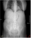
What muscle in red?

If can see psoas then unlikely to have fluid accumulation
What muscle in red?

If can see psoas then unlikely to have fluid accumulation
What in red?

Colon
What are arrows pointing to?

Colon above, small bowel below
Supine or standing?

Supine. Normal image.
Supine or standing?

Standing. Notice the air/fluid levels. Don’t see liver, pancreas, spleen.
What is in red?

Baby. If doing an abdominal xray in a female get a pregnancy test! Abd film is 6x radiation of chest xray.
What is arrow pointing to?

Colonic obstruction. Those are magnets that PT swallowed and obstructed colon.
What kind of gas pattern? Where found?

Wavy. Coiled spring appearance. Jejenum.
What kind of gas pattern? Where found? Indented serosa?

Rectangle. Found in ileum.
What kind of gas pattern? Where found? Indented serosa?

Semicircular. In colon. Serosa indented by haustra (small pouches that give the colon its segmented look).
What can be said about this gas pattern?

Normal.
What can be said about this gas pattern?

Possible LBO on left flank.

What do you see here?

Large bowel. Ask PT if they are farting.

Do you see something wrong here?

Large Bowel Obstruction. Note the air/fluid level.

What kind of pattern do you see?

Small Bowel Gas Pattern. Very tender abdomen, need surgical consult. No owl, no psoas, kinda see SI joint.
What kind of gas pattern? Can see owl? Psoas?

Small Bowel Gas Pattern. No psoas shadow, can see owl.
Is there an obstruction? See any levels?

Small Bowel Obstruction. Air fluid level “coiled spring look”.
See an obstruction? Liver, gall bladder, psoas, SI joint?

Small bowel obstruction. Liver yes, gall bladder no, psoas no, barely see SI.
What kind of sign? See any fluid levels? Obstruction?

String of Pearls Sign, pathoneumonic for SBO. Multiple air/fluid levels.
String of Pearls Sign pathoneumonic for?

Small Bowel Obstruction
What is the name of this sign? What does it mean? What seen and not seen?

Coffee Bean Sign means volvulus (a twisting of gut). Do see liver but not gall bladder, pancreas, spleen. maybe see psoas.
What name of this sign? What does it mean?

Coffee bean sign. Volvulus, twisting of gut.
Assessment?

Free Air in the Abdomen. Liverer not seen due to diaphragm perforation and air leak. No psoas, no SI joints, no owls