Radiography pictures - Musculoskeletal Flashcards

1
Q
A

Black triangle. Possible ACL tear.

2
Q
A

T2 MRI showing edem at femoral head. Often won’t show on xray “plain film”.

3
Q
A

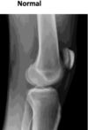
Lateral femoral condyle slamming into tibial groove causing a bone bruise. heals faster than fracture but doesnt show up on xray

4
Q
A

Fx of posterior element of back. Sensitive (shows a fracture) but not specific (not what kind or why). Doesn’t tell what it is but can tell it’s a break.

5
Q
A

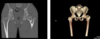
Bone Scan. 88 y/o male with metastatic prostatic adenocarcinoma.

6
Q
A

Ultrasound showing Achilles’ tendon rupture, Quad tendon rupture

7
Q
A

Ultrasound. Used to locate foreign body in soft tissue. Left is AP, right is Lateral.

8
Q
A

4th metacarpal looks narrower than others

9
Q
A

Xray on left (fine), MRI on right showing bone bruise

10
Q

Which under, which normal, which over penetrated?

A

Under, normal, over

11
Q
A
12
Q
A

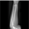
Lucent fracture lines. Most fractures appear as a radiolucent line.

13
Q
A

Not a lucent line. Not a fracture. Flare of tibia.

14
Q
A

Yes, fracture lucent line.

15
Q
A

Compression fractures may appear as a sclerotic line. Most common in distal radius and vetebral bodies.

16
Q
A

Important to know. Lucent line goes into joint.

17
Q
A

Displacement fx. NOT angulated! If both ends are touching then nondisplaced, if not touching then displaced.

18
Q

Displaced or angulated?

A

Displaced.

19
Q
A

Nondisplaced Fracture. still lines up.

20
Q
A

Nondisplaced Fracture. still lines up. can see a lucent line.

21
Q
A

Angulated. Described in terms of position of the distal fragment with respect to the proximal.

22
Q
A

Rotation Fx.

23
Q
A

Distraction. Some fractures separated by a gap with no overlap: described as distracted. Due to tug of different muscles.

24
Q
A

Anterior dislocation of 5th metacarpal base. No fx.

25
Q
A

Impaction. Shoulder shorter than normal.

26
Q
A

Impaction.

27
Q
A

Avulsion fx. when tendon pulls bone off.

28
Q
A

Avulsion fx. when tendon pulls bone off.

29
Q
A

Joint Effusions. Presume joint effusion if no hx of arthritis and acute PT then hemarthrosis. Blood may be the result of an intra- articular injury to either the soft tissues or adjacent bone.

30
Q
A

Blood (effusion)

31
Q
A

Open fx on left, closed fx on right

32
Q
A

Transverse FX

33
Q
A

Linear FX

34
Q
A

Nondisplaced

35
Q
A

Displaced, oblique.

36
Q
A

Spiral. Oblique looks like spiral but they aren’t the same.

37
Q
A

Greenstick

38
Q
A

Comminuted. More than 2 places.

39
Q
A

Transverse Fracture. Perpendicular to the long axis of the bone, due to direct or tension force. Most common forearm/leg. Most stable fx.

40
Q
A

Oblique Fracture, due to indirect force. Shearing force from compression and angulation forces. Unstable fracture, needs OR.

41
Q
A

Spiral Fracture. Created by twisting movement through the long bone axis. Hard to tell apart from oblique. Least common.

42
Q
A

Spiral Fracture. Broke in two areas. two points of fracture. displaced and won’t heal right by itself. needs help to heal.

43
Q
A

Spiral Fracture. Broke in two areas. two points of fracture. displaced and won’t heal right by itself. needs help to heal.

44
Q
A

Comminuted Fracture. Contain more than 2 fracture fragments.

45
Q
A

Comminuted Fracture. Contain more than 2 fracture fragments.

46
Q
A

Comminuted Fracture

47
Q
A

Butterfly Fragment. Due to bending/indirect force. Butterfly fragment along the concave, compression side

48
Q
A

Greenstick Fracture. Usually in kids. Incomplete fx of long bone. Usually forearm because of fall and go boom resulting in bending force. Incomplete, transverse fracture.

49
Q
A

Greenstick FX usually in kids. incomplete fx of long bone. usually forearm because of fall and go boom resulting in bending force. Incomplete, transverse fracture.

50
Q
A

Buckle Fracture (Torus fracture). Type of incomplete fractures. Typically occur at metaphyseal diaphsyeal junction after a FOOSH. Very stable fracture, heals well. Only in kids. very subtle. cortex fractures but are stable and don’t actually need casting but do so for protection

51
Q
A

Buckle Fracture (Torus fracture). Type of incomplete fractures. Only in kids. Very subtle. Cortex fractures but are stable and don’t actually need casting but do so for protection

52
Q
A

Salter-Harris System Type 1, 5-7%. Type 1 not too bad. As number gets closer to 5 it’s really bad. May not be able to see very easily or almost at all. In kids.

53
Q
A

Salter-Harris Type 2, 75% most common. Fracture passes across most of the growth plate and up through the metaphysis. Fx goes away from growth plate.

54
Q
A

Salter-Harris Type 3. Through physis and epiphysis 7-10% Fx away from growth plate toward joint. Poor prognosis.

55
Q
A

Salter-Harris Type 4, combo of 2 and 3. 10% of salter-harris. Contiguous through the metaphysis, physis, and epiphysis.

56
Q
A

Salter-Harris Type 4, Uncommon < 1%. Crush injury, worst prognosis.

57
Q
A

Salter-Harris Type V, Uncommon < 1%, crush injury of the physis. Worst prognosis.

58
Q
A

Salter-Harris Type V, Uncommon < 1%, Crush injury of the physis

59
Q
A

Fracture of clavicle midshaft. Very common. Load from side of shoulder and common midshaft fx. Can heal on own.

60
Q
A

Bayonet fx of clavicle

61
Q
A

Bayonet fx of Clavicle.

62
Q
A

Clavicle fracture with ORIF

63
Q
A

acromion and clavicle. joint is disrupted. usually doesnt need surgery.

64
Q
A

Half moon is where humeral head sits in glenoid and should be able to see it.

65
Q
A

Scapula looks like Y

66
Q
A

Greater Tuberosity Fracture. MRI on left, xray on right.

67
Q
A

Proximal Humerus Fracture. Humeral head is free floating. No good.

68
Q
A

Proximal Humerus Fracture. Unstable fracture neck of humerus, treated by open reduction and internal fixation.

69
Q
A

Proximal Humerus Fracture. Needs internal fixation.

70
Q
A

Comminuted Humerus Fracture

71
Q
A

Anterior shoulder dislocation. 90% of shoulders dislocate anteriorally. most common by far. no hemicircle moon thing. need to reduce.

72
Q
A

Anterior shoulder dislocation. 90% of shoulders dislocate anteriorally. most common by far. no hemicircle moon thing. need to reduce.

73
Q
A

Anterior shoulder dislocation. 90% of shoulder dislocations.

74
Q
A

Humerus Fracture

75
Q
A

Radial Head Fracture, (note the “Sail Sign”). Anterior fat pad sign in elbow, fills with blood.

76
Q
A

Radial Head Fracture

77
Q
A

Positive fat pad sign (Sail sign) on lateral radiograph of elbow. Indicates that fluid is in the elbow joint. In acute setting, the fluid is blood, most commonly from a fracture.

78
Q
A

Fracture of the lateral condyle of humerus. Elbow Fracture.

79
Q
A

Monteggia Fracture Dislocation. Fracture of the ulna shaft + Dislocation of the radial head.

80
Q
A

Monteggia Fracture Dislocation, displaced and overlapped fracture of the ulnar shaft is present. Radial head is dislocated anteriorly.

81
Q
A

Elbow Dislocation of child. most elbow dislocations are posterior.

82
Q
A

Elbow dislocation of adult. Most elbow dislocations are posterior.

83
Q
A

“Both Bones” forearm Fracture

84
Q
A

Midshaft Radius fx

85
Q
A

“Nightstick Fracture” Results from a direct force on forearm. Defensive wound with throwing arm up over forehead to protect self.

86
Q
A

Midshaft Radius Fracture

87
Q
A

Wrist. Buckle fracture.

88
Q
A

Wrist. Buckle fracture.

89
Q
A

Wrist. Buckle fracture.

90
Q
A

Intra-articular wrist fracture

91
Q
A

Intra-articular wrist fracture

92
Q
A

Colles Fracture. Very common fractures of the distal radius due to FOOSH.

93
Q
A

Smith Fracture, Fracture of the distal radius. Fall onto a flexed wrist. Direct blow to the back of the wrist.

94
Q
A

Smith Fracture, Fracture of the distal radius. Fall onto a flexed wrist. Direct blow to the back of the wrist.

95
Q
A

Smith Fracture, Fracture of the distal radius. Fall onto a flexed wrist. Direct blow to the back of the wrist.

96
Q
A

Smith Fracture, Fracture of the distal radius. Fall onto a flexed wrist. Direct blow to the back of the wrist.

97
Q
A

Galeazzi Fracture Dislocation. Consist of fracture of the distal part of the radius with dislocation of distal radioulnar joint and an intact ulna. Can also be distal radial fracture with a distal ulnar physeal fracture.

98
Q
A

Galeazzi Fracture Dislocation. Consist of fracture of the distal part of the radius with dislocation of distal radioulnar joint and an intact ulna. Galeazzi equivalent fracture is a distal radial fracture with a distal ulnar physeal fracture

99
Q
A

Metacarpal Fracture. Common; account for 10% of all fractures and 40% of all hand fractures. Fractures of 5th MC make up 25% of all metacarpal fractures (10% of hand fx)

100
Q
A

Metacarpal Fracture. Common; account for 10% of all fractures and 40% of all hand fractures. Fractures of 5th MC make up 25% of all metacarpal fractures (10% of hand fx)

101
Q
A

Metacarpal Fracture. Common; account for 10% of all fractures and 40% of all hand fractures. Fractures of 5th MC make up 25% of all metacarpal fractures (10% of hand fx)

102
Q
A

Subcapital Hip Fracture

103
Q
A

Subcapital Hip Fracture

104
Q
A

Hip Fracture

105
Q
A

Hip Fracture

106
Q
A

Hip Fracture

107
Q
A

Hip Fracture

108
Q
A

Degenerative Joint Disease - Hip

109
Q
A

Hip Arthritis

110
Q
A

Avascular Necrosis AVN with secondary Degenerative Joint Disease - Hip

111
Q
A

Avascular necrosis. Cysts in femoral head.

112
Q
A

Slipped Capital Femoral Epiphysis (SCFE). Happens in chubby kids. Might complain of hip or knee pain. Femoral head slips off and loses blood supply causing bone necrosis. Vague hip or knee pain with no hx of trauma.

113
Q
A

Slipped Capital Femoral Epiphysis (SCFE). Happens in chubby kids. Might complain of hip or knee pain. Femoral head slips off and loses blood supply causing bone necrosis. Vague hip or knee pain with no hx of trauma.

114
Q
A

Slipped Capital Femoral Epiphysis (SCFE). Happens in chubby kids. Might complain of hip or knee pain. Femoral head slips off and loses blood supply causing bone necrosis. Vague hip or knee pain with no hx of trauma.

115
Q
A

Slipped Capital Femoral Epiphysis (SCFE). Happens in chubby kids. Might complain of hip or knee pain. Femoral head slips off and loses blood supply causing bone necrosis. Vague hip or knee pain with no hx of trauma.

116
Q
A

Transverse distal femoral shaft fracture. Transverse distal femoral shaft fracture with one shaft-width lateral displacement and overriding of the fracture fragments. High velocity injury.

117
Q
A

Knee Degenerative Joint Disease.

118
Q
A

Knee Degenerative joint disease

119
Q
A

Knee. Bipartite patella.

120
Q
A

Knee. Bipartite patella.

121
Q
A

Knee. Bipartite patella

122
Q
A

Oblique Tibia Fracture. Indirect shearing force, combo of compression and angulation forces. Results in an oblique fracture of a long bone which tends to be less stable than others.

123
Q
A

Medial Malleolar Ankle Fx

124
Q
A

Ankle. Bimalleolar Ankle Fracture.

125
Q
A

Maisonneuve Fracture. Combination of spiral fracture of the proximal fibula with ankle injury of one or more: widening of the ankle joint due to distal tibiofibular syndesmosis, OR, deltoid ligament disruption, OR fracture of the medial malleolus.

126
Q
A

Maisonneuve Fracture, proximal fibula fracture

127
Q
A

Maisonneuve Fracture Fibula

128
Q
A

Maisonneuve Fracture Ankle

129
Q
A

Calcaneus Fracture. Most commonly fractured tarsal bone and accounts for about 2% of all fractures, ~60% of all tarsal fractures. Extra-articular: 25-30%, intra-articular: 70-75%

130
Q
A

Calcaneus Fracture. Most commonly fractured tarsal bone and accounts for about 2% of all fractures, ~60% of all tarsal fractures. Extra-articular: 25-30%, intra-articular: 70-75%

131
Q
A

MRI. Achilles’ Tendon Rupture.

132
Q
A