Radiology Midterm Flashcards
(59 cards)

Lamina Dura
A thin, hard layer of commpact alveolar bone that appears as a continuous white line on radiograph: Lines the socket and strongly signifies a vital pulp.

PDL space/ Periodontal Ligament Space
Radiolucent line between root and lamina dura
Widening due to:
1) Tooth Mobility
2) Disease

Alveolar Bone
Consists of inner and outer cancellous (spongy) bone
Alveolar Crest
Thin, White, radiopaque line

Incisive Foramen
Oval or round shaped radiolucency that transmits the nasopalatine nerve and vessels
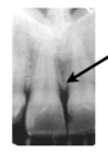
Median Maxillary Suture/ Median Palatal Suture
Thin, Radiolucent line

Nasal Cavity / Nasal fossa
Radiolucent cavity above central incisors
*Nasal Conchae
Shell shaped bones that appear as diffuse radiopaque masses

Nasal Septum
Vertical Bony wall that appears as a radiopaque line that divides the nasal fossa
Floor of Nasal Cavity
Radiopaque wall of cortical bone

Anterior Nasal Spine

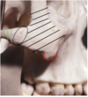
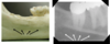
Soft Tissue Shadows
Nose (Yellow)
Lip (Green)

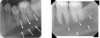
Maxillary Canine Area/ Y-line of Ennis
Black arrow= lateral wall of nasal fossa
Grey arrow= Maxillary sinus

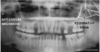
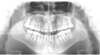
Maxillary Sinus/ Sinus Septa (Septum)

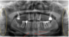
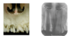
Maxillary Tuberosity
Radiopaque bluge distal to the third molar region
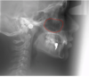
Star= pterygoid plates

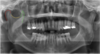
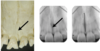
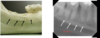
Black Arrows= Genial Tubercles
Bumps of bone appearing as a radiopacity for muscle attachment
Red Arrows= Lingual Foramen

Mental Ridges

Mental Foramen
An oval/round radiolucency where the inferior alveolar nerve divides into its terminal branches- incisive and mental nerves
Can run close to third molar roots, causing problems upon extraction
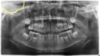
Mandibular Canal
Contains the inferior alveolar nerve and blood vessels that extend from the mandibular foramen ro the mental foramen

Mandibular Canal
Contains the inferior alveolar nerve and blood vessels that extend from the mandibular foramen ro the mental foramen
Can run close to third molar roots, causing problems upon extraction
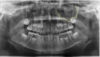
Internal oblique ridge / Mylohyoid ridge
For muscle attachment and appears as a dense radiopaque band underneat the apicies of the molars

External Oblique Ridge
A continuation of the anterior border of the ramus appearing as a distinct radiopaque band just underneath crown of the molars
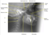
Submandibular Gland Fossa
Depression on the lingual side of the mandible below mylohyoid ridge.
Trabecular pattern of bone is sparse and appears as a radiolucency
Differs from pathology in that it occurs bilaterally
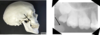
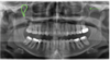
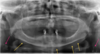
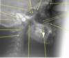
Coronoid process of the mandible

Yellow arrows=Zygomatic Process of Maxilla: “U” shaped radiopacity
Red Arrows= Zygomatic bone