Radiology - Pictures Flashcards
(151 cards)
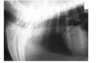
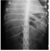
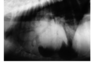
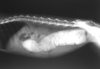
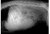
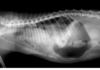
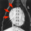
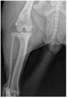
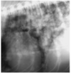
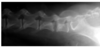
What can be seen?
tracheal collapse
tracheal hypoplasia
none of them
both of them

Tracheal collapse
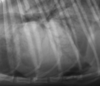
When was the contrast medium administered? (R301)
a. there was no contrast medium administered
b. half an hour ago
c. 2 hours ago
d. cannot be told based on the image

b. half an hour ago
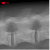
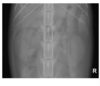
What kind of pulmonary pattern is visible in the picture? (R302)
a. nodular
b. interstitial
c. both a and b true
d. none of them are true
a. nodular
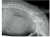
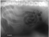
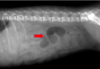
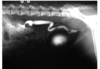
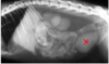
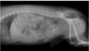
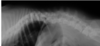
What abnormality is visible in the picture? (R303)
Intestinal obstruction
air swallowing
gastric torsion
gastric dilatation

gastric torsion
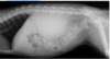
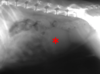
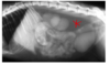
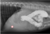
Which statement is true? (R304)
the stomach is empty
the size of the liver is small
there are probably struvite and calcium oxalate stones in the bladder
this is a radiograph of a male cat
there are probably struvite and calcium oxalate stones in the bladder
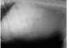
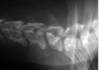
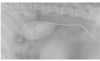
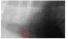
What could not cause the abnormality in the picture? (R305)
cervical penetrating skin wound
esophageal perforation
diaphragmatic rapture
tracheal injury
diaphragmatic rapture
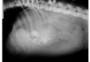
Which statement is false regarding the image? (R306)
this is a couple of month old young animal
vascular ring anomaly can be suspected
this abnormality can be diagnosed the best with solid food mixed with contrast
the complete blockage of the oesophagus is suspected

the complete blockage of the oesophagus is suspected
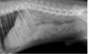
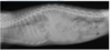
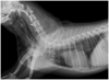
What abnormality is visible in the picture? (R307)
diaphragmatic hernia
pneumothorax
cardiomegaly
no abnormality is visible

no abnormality is visible
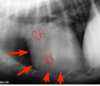
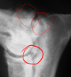
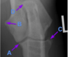
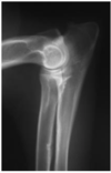
Which statement is true regarding the image?(R308)
this is the forearm of a young animal
the asterix marks a gastrocnemius sesamoid bone
the arrow marks an epiphysis
there is a healing fracture in the picture

there is a healing fracture in the picture
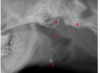
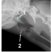
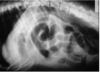
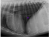
Which statement is false? (R309)
1- larynx
2- os basihyoideum
3- bulla tympanica
4- ala ossis atlantis

1- larynx
Which statement is false regarding the image? (R310)
this is a growing animal
the arrow shows towards the head of the animal
this is a lumbar vertebra
no abnormality is seen in the picture

the arrow shows towards the head of the animal
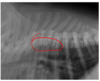
What abnormality is visible on the thoracic spine? (R311)
kyphosis
spondylosis deformans
discospondylitis
lordosis
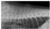
lordosis
Which statement is true regarding the image? (R312)
the thorax is rotated
the liver is small
the heart is elevated from the sternum
all 3 are true

all 3 are true
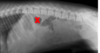
What abnormality is visible in the picture? (R313)
vertebral tumor
discospondylitis
discus hernia
protrusion
discospondylitis
What abnormality is visible in the picture? (R314)
scoliosis
hemivertebra
extrusion
all the 3

all the 3
What abnormality is visible in the picture? (R315)
lumbalisation
thoracoisation
there can be both
cannot be told based only that picture

cannot be told based only that picture
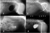
This radiograph is typical of which dog breed? (R316)
Dachshund
Yorkshire terrier i
Great Dane
bulldog
bulldog
Which statement is true? (R317)
this abnormality is common in boxers
this abnormality generally causes very severe clinical signs
this abnormality is caused by a septic process
this abnormality generally causes severe pain
this abnormality is common in boxers
Which statement is false? (R318)
1 - for. intervertebrale
2 – proc. spinosus
3 – proc. articularis caudalis
3 – proc. articularis cranialis

1 - for. intervertebrale
Which statement is true? (R319)
the animal „B” has heart disease for sure
the animal „A” has tracheal collapse for sure
the animal „B” may have tracheal collapse
there are severe pulmonary congestion in both animals

there are severe pulmonary congestion in both animals???
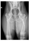
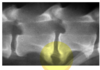
Which statement is true? (R320)
A-pylorus, B-fundus, C-spleen, D-liver
A-colon, B-fundus, C- liver, D- liver
A-pylorus, B-colon, C- spleen, D- liver
A-fundus, B-colon, C- spleen, D- liver

A-pylorus, B-fundus, C-spleen, D-liver
Which statement is true? (R321)
the thorax is slightly rotated
intestinal obstruction is confirmed
the contrast medium was barium sulphate for sure
the contrast was administered at least 12 hours ago

the contrast medium was barium sulphate for sure
Which statement is true? (R322)
1- epiglottis, 2- thyroid
1- epiglottis, 2- hyoid
1- soft palate, 2- thyroid
1- soft palate, 2 – hyoid

1- soft palate, 2 – hyoid
Which statement is true? (R323)
this is a female dog
this spleen is enlarged
the urinary bladder is full
this is an intravenous urography

the urinary bladder is full