Schwarzenberger - Skin Intro Flashcards
(58 cards)
1
Q
What are some important factors in the skin PE?
A
- Use all available clues -> look at the patient first, then look at the skin
- Complete skin exam: good lighting and a naked patient (incl: skin, hair, nails, mucous membranes)
- Identify the lesion type and note any modifying factors (color, size, number)
- Describe the distribution and pattern: generalized, localized to, linear, grouped
2
Q
What is this?

A
- Papule: palpable lesion elevated above skin surface
- <0.5 cm in diameter
- Do not have to be perfectly round
- Can also be umbilicated
3
Q
What is this?

A
- Nodule: firm (indurated) lesion thicker or deeper than 0.5cm
- Cyst would be even bigger
4
Q
What is this?

A
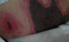
- Macule: flat, non-palpable lesion <0.5cm in diameter
- Patch if >0.5 cm
5
Q
What is the main role of the epidermis? Dermis?
A
- Epidermis: Barrier (protection)
- Dermis: Structural and nutritional support
6
Q
What is this?

A
- Plaque: palpable lesion elevated above skin surface >0.5cm (classic example is psoriasis)
7
Q
What are these?

A
- Vesicles: blister <0.5 cm
- Herpes: multiple, coalescing vesicles in a herpetiform distribution -> see attached image
- Bullae are bigger
- Pustule if it has white/yellow fluid in it

8
Q
Can Staph infect the skin?
A
- Not unless you have a scratch or surgically cut the skin
- Can infect the hair follicles
9
Q
What is this?

A
- Wheal = hive: temporary raised areas of the skin surrounded by a red base
10
Q
What is this?

A
- Spider angiomas: capillary malformations
11
Q
What is this?

A
- Lichenification: result of repetitive scratching
- Granular layer becomes thickened (hyperkeratotic)
12
Q
What are these?

A
- Excoriations -> much more superficial than ulcers, which extend down into the subcu tissue (see attached)

13
Q
What is this?

A
- Atrophy: cigarette/tissue paper wrinkling
- Epidermis can virtually be holding on by blood vessels, and can sometimes get ecchymoses quite easily
14
Q
What are the important functions of the skin?
A
- Barrier function
- Immune recognition and surveillance: by antigen presenting cells (Langerhans cells in epidermis; lymphocytes and dermal dendritic cells in dermis)
- Damage repair: keratinocytes proliferate in epidermis in response to injury or inflammation, fibroblasts in dermis
- Thermoregulation: vasculature in the dermis
- Protection from UV radiation: malanocytes
- Communication
- Failure at any level can result in damage/disease
15
Q
How does the skin function as a barrier? What can happen if it is defective (4)?
A
- Physical barrier, regulating water loss, protecting against mechanical, chemical and microbial insults from outside world
- Possible consequences of defective skin barrier:
1. Dehydration
2. Infection
3. Injury of skin, such as ulcers
4. Inflammation - Example: atopic dermatitis (eczema) due to mutation in fillagrin gene (see attached image)

16
Q
What condition is this? Associated mutation?

A
- Atopic dermatitis (eczema)
- Fillagrin gene mutations
17
Q
Why is skin a poor host for growth of orgs?
A
- Intact skin is poor host for growth of organisms:
1. Dry
2. Impermeable
3. Sheds off
4. No blood vessels in the epidermis - Both innate AND adaptive immunologic processes important in skin
18
Q
What can happen if skin immune regulation is compromised?
A
- Consequences of impaired skin immune function include:
1. Infection
2. Skin cancer
3. Inflam and/or autoimmune skin diseases
4. Allergic reactions - Skin can be involved/injured, either primarily or secondarily (“innocent bystander”) by immunologic functions
19
Q
What condition is this?

A
- Bullous pemphigoid: auto-antibodies to hemidesmosomes (adhesion molecule)
- IF: frozen skin overlayed with fluorescently labeled Ab’s

20
Q
What conditions is this?

A
- Pemphigus vulgaris: auto-Ab’s to desmosomes
- More erosions than blisters
- Chicken-wire IF

21
Q
How does this happen?

A
- Neuropathic ulcers: people with diabetes are more prone to pressure ulcers because they don’t “feel” that they need to move around
- Trigeminal trophic syndrome: trigeminal N loss of sensation, leading to scratching (can’t feel the pain they are inducing)
22
Q
How is the skin involved in photoprotection?
A
- Epidermis contains melanin, which helps protect against damage from ultraviolet radiation
1. UV light can also damage the immune system - Genetic and/or acquired conditions can reduce or eliminate pigment in skin
- Loss of photoprotection increases risk of burning and skin cancer
23
Q
What are the acute effects of UV radiation on skin?
A
- Inflammation (sunburn)
- Immunomodulation: can even cause chills, headaches
- Epidermal hyperplasia
- Vitamin D photosynthesis
- DNA damage: apoptosis or cell cycle arrest to repair
24
Q
What are the chronic effects of UV radiation?
A
- Photoaging: sun is really what ages the skin (more so than just age)
- Photocarcinogenesis:
1. Basal cell and squamous cell carcinomas
2. Melanoma
a. Lifetime incidence of melanoma has gone from 1 in 3500 to 1 in 50
b. AA more likely to get melanoma on hands and feet

25
What histo changes do we see w/chronic UV radiation?
- **Solar elastosis**: elastin fibers that have been damaged by UV radiation, and are broken and clumped
- Collagen is what keeps your skin young

26
What is xeroderma pigmentosum?
- Failure in the DNA repair process -\> start getting skin cancers before they are 10 years old

27
What is hypohidrotic ectodermal dysplasia?
- Genetic condition due to _mutations in EDAR_ (“Ectodyplasin A receptor”) gene: can't sweat, so they _overheat easily_ bc they can't regulate temp
1. Protein critical for proper interaction between developing ectoderm and mesoderm
- Results in abnormal hair follicles, sweat glands, and teeth
28
What pathologic process causes this?

- Small vessel vasculitis
- Diseases that injure blood vessels disrupt circulation, resulting in cutaneous necrosis and/or ulceration
1. Antiphospholipid Ab syndrome can cause widespread cutaneous necrosis (see attached image)
29
What types of symptoms can disorders of nerve sensation in the skin cause?
- Itch
- Dysesthetic pain/burning (postherpetic neuralgia: nerve pain caused by varicella zoster virus)
- Hyperesthesia: excessive sensitivity
- Loss of sensation can also result in insensitivity to injury (think: diabetic peripheral neuropathy)
30
Friendly reminder.

Good job!
31
What are the 3 layers of the skin, and their cells?

- _Epidermis_: Keratinocytes (\>90%)
1. Melanocytes, Langerhans, Merkel cells (light touch sensory cells; can cause lethal cancers)
- _Dermis_: Fibroblasts, collagen, elastic
1. Blood vessels, nerve endings: if you scrape knee and it bleeds, you're in the dermis
- _Subcutis_: Fat, blood vessels, fibrous septae (cause cellulite)
32
What is this?

- Normal skin
- Blood vessels go up into the dermal papillae: this is why you can get "needle-point bleeds" when you get a scrape
33
What are the main functions of the epidermis vs. the dermis and subcutis?
- Epidermis: primarily a barrier function, protection, and wound healing (must be able to "fix itself")
- Dermis/subcutis: structural and vascular support and innervation
34
How often does the epidermis regenerate? How does this happen?
- Epidermis is a self-renewing tissue that “sheds” itself on average _every 28+ days_
1. 14 days to reach stratum corneum
2. 14 days to desquamate
3. This happens a lot faster in psoriasis as a result of inflammation
- Keratinocytes grow from stem cells in the basal layer and are shed from the surface -\> terminally differentiate as they move upwards
- Apoptosis (“programmed cell death”) normally low in epidermis, but can increase in some situations
35
What are the four layers of the epidermis?

- Organized based on position and structural properties of keratinocytes:
1. Stratum corneum
2. Stratum granulosum: granular cell layer
3. Stratum spinosum: spiny layer
4. Stratum basale: source of stem cells (division starts here)
a. Adhere to dermis (BM zone) through hemidesmosomes
36
What are the 4 major types of cell junctions?
- _Desmosomes_: KC-KC adhesion
- _Hemidesmosomes_: epidermis-dermis adhesion
- _Adherens junctions_: links actin filaments KC-KC
- _Gap junctions_: connexin proteins, important in cell-cell communication
37
What layer of the epidermis is this? What happens here?

- _Spinous layer_: cells stop dividing and start terminal differentiation
- Develop lipids (lamellar granules) important in barrier function
- “Spiny” due to visible desmosomes with which one KC adheres to another

38
What layer of the epidermis is this? What happens here?

- **Stratum granulosum**: IC keratohyaline granules synthesized (including profilaggrin)
- Lipids in lamellar granules secreted into IC space to form _water barrier to keep water in skin_
39
What layer of the epidermis is this? What happens here?

- **Stratum corneum**: nuclei, organelles degenerate, and cells flatten
- Profilaggrin processed into filaggrin, which help keep water in cells
- Keratins (structural cytoskeletal proteins) combine with filaggrin into macrofibrils that create protective layer
40
What are keratins?
- Major fibrous structural proteins in hair and nails; \>40 different kinds
- Combine to form **intermediate filaments** (KNOW THIS)
- Pairs differ by location in body
- Mechanically stabilize cell against physical stress
- Have lg amounts of sulfur-containing amino acid **cysteine** (esp. hair and nails -\> this is why there is a sulfur-y smell when you burn your hair)
41
What kinds of filaments to keratins combine to form?
INTERMEDIATE
42
What 2 sites are these histo pics from?

- Note the *stratum corneum differences*
1. _Trunk_ on the left: basket-weave, loose
2. _Palm_ on the right: more protective, dense
43
Why is stratum corneum described as "brick and mortar?"
- _Bricks_: flattened keratinocytes filled with keratin and filaggrin
- _Mortar_: lipid mixture surrounding keratinocytes, providing water barrier

44
What type of cell is this? What does it do?

- **Melanocyte**: pigment-producing dendritic cells
- Derived from _neural crest_; migrate in embryonic development
1. Defect in neural crest migration can lead to melanoma in unsuspected places
- "Live” along basal cells, w/about one per 10 keratinocytes (see attached image)
- Produce _melanin_, a radiation-absorbing pigment, and transfer it to surrounding keratinocytes via dendritic processes
- _Primary defense against ultraviolet radiation_

45
Appreciate this melanocyte-keratinocyte unit

Good job!
46
What are these? What do they do?

- **Langerhans cells**: dendritic cells in mid-epidermis
- One of major immunologic players in skin
- Recognize abnormal Ag's in skin; take up, process, and present to lymphos in regional lymph nodes
1. _Migratory_: move to lymph tissue to present (processing and presentation)
- Important in _allergic reactions, tumor surveillance_
47
What are Merkel cells?
- Epidermal cells
- Associated with light touch sensation
- Can develop into malignant tumors (rare, but lethal)

48
How thick is the dermis? What is in it?
- Thickness varies significantly by site: _1-4 mm thick_, and functions primarily as a support layer
- _Contains_: blood vessels and lymphatics, nerves, sweat and oil glands, and hair follicles
- Note: sebaceous glands follow hair follicles, but eccrine do NOT
49
What do fibroblasts do in the dermis?
- **1o cell** in the dermis (mesenchymal origin)
- Responsible for _synthesis and degradation of CT_ proteins, incl collagen, elastin, glycosaminoglycans and other glycoproteins
- Injury to skin triggers fibroblast mitosis
- Responsible for _wound healing and scar formation_
50
What are these?

- **Mast cells**: specialized tissue cells rich in histamine and heparin granules
- Release granules when triggered by injury or binding of IgE antibodies during allergic reactions
- Histamine and other mediators important in _allergic reactions and wound healing_ (this is why healing scars can be itchy)
1. Cause characteristic “_wheal and flare_”
51
What is this? What does it consist of?

- Pilosebaceous (hair/oil) unit:
1. Hair follicle (extend through dermis into subcutis)
2. Sebaceous (oil) gland
3. Apocrine sweat glands (in axilla and anogenital skin)
4. Arrector pili muscle (goose bumps)
52
What are eccrine sweat glands?
- "True” sweat glands; present throughout the body
- Open directly onto the skin (_not associated with hair follicle_)
- Function to **regulate temperature** by evaporative cooling of sweat
53
What is the subcutis?
- Fat layer that separates dermis from underlying structures including fascia, muscle, organs
- Subcutis provides:
1. Insulation
2. A source of energy
3. Protection from injury
54
What is up with these cells?

- _Sun-burn cells_: damaged keratinocytes -\> apoptosis is normal response to sun-damaged cells
1. Failure to “delete” damaged cells can result in skin cancer
2. Sunburn = INC apoptosis

55
What is this?

- Epidermolysis Bullosa Simplex: genetic mutations in Keratin 5/14 (so no cure)
- Bullae arising on the toes ( A ) and the plantar surface ( A,B ) at sites of lateral or rotary traction
- Keratinocytes can split in half -\> can go unrecognized for long periods of time

56
What is this?

- **Melanoma**: growth of malignant (cancerous) melanocytes
- ASYMMETRY -\> melanomas (grossly and histologically)
- A nevus (mole) would be a benign collection of melanocytes
57
What is this?

- Erythema nodosa
- Affects the subcutis
58
Probably should know these.

Good job!
