Session 10 Immunocompromised Host Flashcards
(27 cards)
Why should you care as a doctor?
- Immunodeficiency is associated with an increase in the frequency and severity of infections (more aggressive and life-threatening).
- Immunodeficiency is associated with autoimmune diseases and malignancy
- Failure to recognize and diagnose leads to increased morbidity and mortality.
- Average 7-9 years between the onset of symptoms and diagnosis. >50% of patients will be 18 years old and older when diagnosis is made. 37% of them will have permanent tissue/organ damage – delay in diagnosis associated with worse prognosis and high mortality.
- Studies have found that immunocompromised patients have increased mortality due to non-infectious complications e.g. GI disease, liver disease, lymphoma etc.
Describe how to recognise and fiagnose immunodeficiencies
[*] Recognition and Diagnosis of Immunodeficiencies: the pattern and type of infections always reflect the nature of an immunological defect
- Age – at presentation
- Sex (family history e.g. X-linked inheritance etc)
- Site(s) and frequency of infection(s)
- Type of organism(s)
Viruses and fungi => T cell deficiency
Bacteria and fungi => B cell / granulocytes deficiency
- Sensitivity and type of treatment (surgery)
- Family history
Describe laboratory investigations for immunodeficiencies
General:
- Full blood count and differential
- Exclusion of secondary immunodeficiency
Tests of humoral (antibody) immunity
- IgG, IgA, IgM (+/- IgE)
- IgG(1-4) subclasses
- IgG levels to specific previous vaccines e.g. tetanus toxoid / HiB / pneumococcus measles, mumps, rubella
- Measure antibody in response to “test” immunization
Tests for Cell mediated immunity
- Lymphocyte count (FBC)
- Lymphocyte subset analysys (CD4+, CD8+ T, NK and B cells) – extended marker panel (s)
- In vitro tests of T cell function
Tests for phagocytic cells
- Neutrophil count (FBC)
- Neutrophil function tests (e.g. oxidative burst for CGD)
- Adhesion molecule expression (for LAD)
Tests for Complement
- Individual components
- Tests of complement function (CH50/AP50)
Definitive tests – molecular testing and gene mutations
What is an immunocompromised host?
Immunocompromised host: “state in which the immune system is unable to respond appropriately and effectively to infectious microorganisms”
- Qualitative (dysfunctional immune component) or quantitative (lack of/absence of immune component) defect of one or more components of the immune system.

What does SPUR mean?
- S => Severe, sometimes life-threatening
- P => Persistent, despite appropriate treatment infection s still there
- U => Unusual – unusual organism, opportunistic infection, consider site of infection
- R => Recurrent (same infection or different infections)
What are the different types of primary immunodeficiencies?
Primary immunodeficiencies: classified according to which immune component is defective
- B cell (50%) – most common
- T cell (30% - worst kind, as it also ultimately leads to B cell deficiency as T helper cells drive B cells to produce antibodies)
- Phagocytes (18%)
- Complement (2%)
Incidence of 1:400 to 1:400000
Occur in the first months of life
80% of patients < 20 years
70% of patients are male (X-linked)

What are clinically important B cell deficiencies?

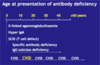
At what age do primary antibody deficiencies present at?
Certain primary antibody deficiencies can present at any age!
What are clinically important Phagocyte deficiencies?
- Disease: Cyclic neutropenia (abnormal low amount of neutrophils) (periodicity – every 3/4 weeks); Defect: unknown
- Disease: Leukocyte Adhesion Deficiency (Lack of CD18 protein on phagocyte); Defect: Adhesion to vascular endothelium
- Disease: Chronic Granulomatous Disease (CGD) (lack of respiratory burst due to lack of enzymes involved in producing free radicals); Defect: intracellular killing
- Disease: Chediak-Higashi Syndrome (failure of phagolysosome formation); Defect: Phagocytosis
- NB: Secondary phagocyte deficiency is much more common!!!!

What are clinically important T cell deficiencies?
- Disease: DiGeorge Syndrome (defect in thymus embryogenesis and incomplete development); Defect in: lack of thymus => lack of functional T cells
- Disease: Severe Combined Immunodeficiency (defect in gamma-chain used by many receptors (IL-2, IL-4, IL-7, IL-9) – involves T and B cell abnormalities); Defect: Stem cell defect
- Disease: Severe Combined Immunodeficiency (defect in adenosine deaminase (ADA), purine nucleoside phosphorylase (PNP)); Defect: death of developing thymocytes
- Disease: Severe Combined Immunodeficiency and Omenn’s Syndrome (defect in genes critical for TCR rearrangement, maturation); Defect: Defective T cell development
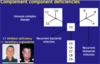
What are Complement Component deficencies?
C1 inhibitor deficiency => hereditary angioedema – risk of airway obstruction => suffocation. If this condition is not properly diagnosed, 30% mortality.
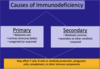
What could Secondary deficiencies be due to?
[*] Secondary Immune deficiencies: decreased production of immune components due to:
- Malnutrition (most common cause in the world)
- Infection e.g. HIV
- Liver diseases
- Lymphoproliferative diseases
- Drug-induced neutropenia – immunosuppressive / cytotoxic
- Splenectomy due to infarction e.g. sickle cell anaemia, trauma, autoimmune haemolytic disease, infiltration (tumour), coeliac disease, congenital
- “Physiological” – age
[*] Secondary Immune Deficiencies due to increased loss or catabolism of immune components
- Protein-losing conditions such as Nephropathy, Enteropathy
- Burns

What factors cause neutropenia and discuss the management of neutropenia
[*] Factors causing neutropenia (abnormal low levels of neutrophils)
- Drugs e.g. phenytoin, chloramphenicol, alcohol (abuse)
- Autoimmune neutropenia
- Infections e.g. infectious mononucleosis, hepatitis B or C, human immunodeficiency virus, cytomegalorvirus infection, typhoid
- Bone marrow infiltration with malignancy
- Aplastic anaemia
- Vitamin B12 or folate or iron deficiency
- Chemotherapy – cytotoxics and immunosuppressants
- Exposure to chemical agents e.g. Benzene, organophosphate, radiotherapy
[*] Management (neutrophil count of <1.0 x10^9)
- Treat suspected neutropenic sepsis as an acute medical emergency and offer empiric antibiotic therapy immediately
- Assess patient’s risk of septic complications
Describe the presentation and management of the Asplenic/Splenectomised patient
[*] The Asplenic/Splenectomised Patient
Presentation:
- Increased susceptibility to encapsulated bacteria (most invasive, difficult to get rid of – can only be phagocytosed if opsonized) e.g. Haemophilus influenzae, Streptococcus pneumoniae and Neisseria Meningitidis
- OPSI (overwhelming post-splenectomy infection) => sepsis and meningitis: 0.25-5% splenectomised individuals per year, 5% lifetime risk; 40-70% mortality (low risk but risk of mortality is high)
Management:
- Penicillin prophylaxis (life-long)
- Immunisation against encapsulated bacteria (at least two weeks before splenectomy if possible)
- Patient information; Medic Alert bracelet
Why is the spleen so important?
- Largest lymphoid organ
- Blood borne pathogens – capable of dealing with encapsulated bacteria!
- Antibody production: (acute response: IgM production which activates complement; long term protection: IgG production (best antibody weapon)
- Splenic macrophages – removal of opsonized microbes and immune complexes
Describe the presentation and management of patients with primary antibody deficiencies
Presentation with primary antibody deficiencies
- Recurrent upper and lower respiratory bacterial infections => bronchiectasis
- GI complications including infections (Giardia)
- Arthropathies (including Mycoplasma/Ureaplasma sp)
- Increased incidence of autoimmune disease
- Increased incidence of lymphoma and gastric carcinomaManagement with primary antibody deficiencies
- Prompt / prophylactic antibiotics – treat acute infection aggressively
- Immunoglobulin replacement therapy
- Goal: Serum IgG > 8g/Litre
- Lifelong treatment
- Different formulations IvIg and ScIg (young patients – once a week)
- Conditions: CIVD, XLA (BRuton’s Disease), Hyper-IgM Syndrome)
- Management of respiratory function
- Avoid unnecessary exposure to radiation
Describe the presentation and management of patients with phagocyte deficiencies
[*] Presentation of patients with phagocyte deficiencies
- Prolonged and reuccrent infections
- Skin and mucous membranes (ulcers)
- Osteomyelitis
- Deep abscesses
- Commonly staphylococcal (catalase +ve)
- Invasive Aspergillosis (fungal opportunistic infection – most common cause of death in patients with a phagocyte defect)
- Inflammatory problems (granuloma)
- Will leave noticeable scars
[*] Management of patients with phagocyte deficiencies
- Prophylactic antibiotics/ anti-fungal agents / Immunization
- Surgical management
- Interferon-gamma (CGD)
- Steroids (CGD)
- Stem cell / bone marrow transplantation (ultimate resort)

Describe the presentation and management of DiGeorge Syndrome
[*] DiGeorge Syndrome Management:
- Neonatal cardiac surgery (depending on nature and severity of cardiac abnormality)
- Supplement to correct hypocalcaemia
- Immune defect variable (only 20% of DiGeorge have immunodeficiency -?) (low T cell number)
If <0.4x10^9/L, Pneumocystis Pneumonia (PCP) prophylaxis with antibiotics
Bone marrow transplantation (severe forms)
- Use only X-irradiated and CMV(-) – blood
- No live vaccines (BCG, MMR, oral polio) – because of the possibility of replication and becoming pathogenic

What is SCID? Describe the presentation and epidemiology of it.
[*] Severe Combined Immunodeficiency (SCID) – effectively a complete absence of adaptive immunity. Something wrong with both T cells and B cells. Primary immunodeficiency
Epidemiology:
- Incidence: 1:50,000 births
- Most severe form of primary immunodeficiency
- At least 10 known causative genes
- X-linked and AR variants
- Sex: 3:1 male/female
- Age at presentation: mean 6.5 months
- Mortality rate: 100% (if undiagnosed)
Presentation
- Failure to thrive
- Protracted diarrhoea
- Long term antibiotic therapy – extreme susceptibility to all types of infection (bacterial, viral, fungal, opportunistic)
- Deep skin and organ abscesses
- Low lymphocyte count (<4.0-10.0 cells/microliters)
- High susceptibility to fungal and viral infections: Pneumocystis pneumonia (PCP), Varicella-zoster virus (VZV) – lies dormant in the body – part of the Herpes family, Cytomegaloviruses (CMV), Epstein `barr virus (EBV)
Describe the management of SCID
Management: fatal if not treated
Short term management:
- No live vaccines
- Only irradiated, CMV- blood products
- Aggressive treatment of infections
- Prevention of new infections: reverse barrier nursing / laminar flow, prophylactic antibiotics and anti-fungals, IV immunoglobulin therapy
Long term management:
- Bone marrow / stem cell transplantation
- Gene therapy
Describe Common Variable Immunodeficiency
- Commonest primary antibody deficiency associated with panhypogammaglobulinaemia (1:15,000)
- Associated with recurrent bacterial infections (especially URTI/LRTI)
- May be associated with autoimmune disease
- Subset of patients has granulomatous disease
- Family history lacking in most cases of CVIS
- Mainstay of treatment is immunoglobulin replacement +/- (prophylactic antibiotics). As immunoglobulins only have a limited lifespan, this immunoglobulin replacement therapy is required forever and so patients needs regular transfusions for the rest of their lives.
What infections can occur in patients with primary immunodeficiencies?
[*] Infections in patients with (primary) immunodeficiencies
T Cells/ Combined
- Systemic Lung / GI etc
- Intracellular: viruses, mycobacteria, fungi (Candida, Aspergillus), protozoa (Cryptosporidium), Opportunistic (Pneumocystis jiroveci)
B Cells/ Antibody
- Sinopulmonary GI (skin/joint)
- Pyogenic bacteria: Pneumococcus, H. influenza type B, Enterovirus (enceph), Mycoplasma (joint)
Complement
- Systemic (bact)
- Neisseria: Mengingococcus
- Pyogenic Bacteria: Pneumococcus, H influenza B
- Cytomegalovirus / Herpes simplex virus
Phagocytes:
- Skin / lung /lymph/GI/liver
- Bacteria: Catalase-positive Staphylococci, Serratia, E coli, Klebsiella, B. cepacia
- Fungi: Candida, Aspergillus
Describe Neutropenic Sepsis
- Secondary immunodeficiency
- A leading cause of morbidity and mortality in cancer patients receiving cytotoxic chemotherapy
- Fever and neutropenia common in patients undergoing myelosuppressive chemotherapy
- Neutropenia (usually) defined as <1.0 x 10^9/L
- Bacteraemia (positive blood cultures) in up to 30% of these
- Early onset antimicrobial treatment (within 1st hour of sepsis; “door to needle” target time) reduces mortality, and each hour of delayed treatment worsens survival.
Describe Aspergillus infection in an immunocompromised host
[*] Aspergillosis is caused by several species of the genus Aspergillus but primarily by Aspergillus fumigatus.
[*] Aspergillus is rarely pathogenic in the normal host but can produce disease in immune-suppressed individuals and patients treated with broad-spectrum antibiotics.
[*] Aspergilli reside in dust soil and decomposing organic matter.
[*] Aspergillosis manifests itself in several forms, depending in part on the patient’s immunologic status.
[*] Acute aspergillus infections:
- The most severe, and often fatal, form of aspergillosis is acute invasive infection of the lung, from which the infection can be disseminated to the brain, GI tract, and other organs.
- A less severe, noninvasive lung infection gives rise to a fungus ball (aspergilloma), a mass of hyphal tissue that can form in lung cavities derived from prior diseases such as tuberculosis.
- Although the lung is the most common primary site of infection, the eye, ear, nasal sinuses and skin can also be primary sites.
