Session 3 Flashcards
(33 cards)
What is the primary intestinal loop?
The midgut elongates enormously.
Runs out of space – Makes a loop that:
- has the SMA as its axis
- is connected to the yolk sac by the vitelline duct
- has cranial and caudal limbs

Why in the 6th of foetal growth week do we see herniation?
- During 6th week, growth of the primary intestinal loop is very rapid – elongation
- Liver is also growing rapidly – abdominal cavity is too small to accommodate both so intestines herniate into the umbilical cord.
Describe midgut rotation and descent of the caecal bud.
As midgut loop herniates through umbilicus there is a single 90 degree counter-clockwise turn around the superior mesenteric artery.
There’s then a further 90 degree rotation which inverts the loop so cranial portion is now caudal in position.
As herniation is resolved there is a final 90 degree rotation. 270 degrees anti clockwise in total.
This ensures the cranial limb is positioned so that it returns to the abdominal cavity first so it and its derivates are pushed to the left hand side. The caudal limb then returns second.
As the loop is herniated, it’s continuing to elongate and develop. After folding there should be complete resolution of the herniation.
The caecel bud descends down along the right hand side of the abdomen to give us the ascending colon.
This process of rotation and descent ensures normal disposition of the abdominal viscera. (Transverse colon should be in front of the duodenum). It also minimises movement of duodenum as its plastered to posterior wall.

How might malrotation occur?
- Incomplete rotation – Midgut loop makes only one 90° rotation resulting in left-sided colon
- Reversed rotation – Midgut loop makes one 90° rotation clockwise resulting in transverse colon passing posterior to the duodenum

What are the risks associated with midgut defects?
- Most complications present in the neonatal period
- Risk of volvulus (an obstruction caused by twisting of the stomach or intestine) which can lead to strangulation and then ischaemia
What happens to the remnants of the yolk stalk after herniation from primary intestinal loop rotation?
• The vitelline duct can persist resulting a number of different abnormalities e.g vitelline cyst
Meckel’s diverticulum - ileal diverticulum, the most common GI anomaly
Vitelline cyst - Vitelline duct forms fibrous strands
Vitelline fistula - Direct communication between the umbilicus and intestinal tract

What is Meckel’s diverticulum?
Ileal diverticulum left after closing of the vitelline duct.
- 2% population affected
- 2 feet from ileocaecal valve
- Usually detected in under 2s
- 2:1 ratio male:female Sometimes can contain ectopic pancreatic or gastric tissue
What is recanalisation in gut development?
- The primitive gut is a simple tube
- In some gut structures, cell growth becomes so rapid that the lumen is partially or completely occluded – affected regions: oesophagus, bile duct, small intestine
- Recanalisation occurs to restore the lumen. This is a normal process (not pathological)
- If recanalisation is wholly or partially unsuccessful, atresia or stenosis of the structure can occur

Describe possible atresias and stenoses of the GI tract
- Atresia – Lumen obliterated
- Stenosis – Lumen narrowed
- Most occur in duodenum – Most likely cause is incomplete canalisation – But “vascular accidents” may also contribute
- Pyloric stenosis – hypertrophy of the circular muscle in the region of the pyloric sphincter – NOT a recanalisation failure. Its a common abnormality of the stomach in infants – narrowing of the exit from the stomach causes characteristic projectile vomiting
Describe development of anal canal and the implications that this has
Proctodeum - A small depression in the caudal most region of the embryo.
• At the cloacal membrane in the proctodeum there’s a junction between two embryonic germ layers (ectoderm and endoderm). This cloacal membrane ruptures which gives an opening for the bladder and an opening for the hindgut. The outer covering of ectoderm at the proctodeum is pushed up into embryo so the exiting distal portion of each tract is lined with ectoderm This explains the differences between above and below the pectinate line. As superior structure is derived from hindgut (endoderm) and inferior lining is derived from ectoderm.
Above pectinate line
- Blood supply from IMA
• Innervation
– pelvic parasympathetics (S2,3 and 4)
– Sympathetic
• Inferior mesenteric ganglion and plexus
- Columnar epithelium
- Lymph. drainage = internal iliac nodes
• Below pectinate line
– Blood supply from Pudendal artery
– somatic innervation - S2,3 and 4 pudendal nerve
– Stratified epithelium
– Lymph. drainage = superficial inguinal nodes
So above the pectinate line the only sensation possible is stretch as it only has visceral sensory afferents, while below the pectinate line the tissue is temperature, touch and pain sensitive as it has somatic innervation

Describe visceral pain of GI
- Visceral pain poorly localised
- The pain pattern reflects the development of the structure
- Foregut and its derivatives – epigastrium pain
- Midgut – periumbilical pain
- Hindgut – suprapubic pain
But remember that the parietal peritoneum receives somatic innervation

Describe some important hindgut abnormalities
- Imperforate anus – Failure of anal membrane to rupture
- Anal / anorectal agenesis
- Hindgut fistulae

Which midgut and hindgut structures have retained/fused mesenteries?
- Mesenteries are retained by: – Jejunum – Ileum – Appendix – Transverse colon – Sigmoid colon
- Structures of midgut/hindgut with fused mesenteries: – Duodenum – Ascending colon – Descending colon – Rectum (no peritoneal covering in distal 1/3)
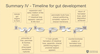
Give an overall timeline for gut development in the foetus
Briefly summarise the mesenteries?
Ventral mesentery
Lesser omentum - foregut to liver
Falciform ligament attaches liver to ventral body wall
Dorsal mesentery
– Greater omentum
– Gastrolienal ligament attaches stomach to spleen
– Lienorenal ligament attaches spleen to kidney
– Mesocolon supplies ascending and proximal 2/3 transverse colon
– Mesentery proper supplies Jejunal and ileal loops
Describe the composition of saliva
- Mostly water
- Hypotonic (depending on flow rate)
- Rich in potassium and bicarbonate (pH ranges from slightly acidic to ~8) (Don’t want to be too acidic as teeth have enamel)
- Mucins help with lubrication
- Amylase (secreted by salivary glands)
- Lingual lipase (secreted by lingual glands)
- Contains a diversity of immune proteins (e.g. IgA, lysozyme, lactoferrin(sequesters iron to prevent bacterial growth))
What are the three phases of swallowing and roughly how long does each one last?
Oral preparatory phase (0-7.4)
Pharyngeal phase (7.4-7.6)
Oesophageal phase (7.6 onwards)
Describe the oral preparatory phase of swallowing
- Oral preparatory phase (0-7.4)
- Voluntary
- Pushes bolus towards pharynx
- Once bolus touches pharyngeal wall, pharyngeal phase begins
Describe the pharyngeal phase of swallowing
- Pharyngeal phase (7.4-7.6)
- Involuntary
- Soft palate seals off nasopharynx
- Pharyngeal constrictors push bolus downwards
- Larynx elevates, closing epiglottis (action of suprahyoids)
- Vocal cords adduct (protecting airway) and breathing temporarily ceases
- Opening of the upper oesophageal sphincter
Describe the oesophageal phase of swallowing
- Oesophageal phase (7.6 onwards)
- Involuntary
- Closure of the upper oesophageal sphincter
- Peristaltic wave carries bolus downwards into oesophagus
Describe the neural control of swallowing and the gag reflex
Reflex arc for swallowing and gag reflex: Mechanoreceptors in pharyngeal wall detect presence of bolus, when these are activated, impulse is sent along glossopharyngeal nerve to medulla which sends an efferent response down the vagus nerve to pharynx which stimulates pharyngeal constrictors. After 6 months hyper active gag reflex moves back in the mouth to allow eating of solid food. Before this baby would just gag it up.

Describe prevention of gastro-oesophageal reflux at the gastro-oesophageal junction
- Lower oesophageal sphincter - Functional sphincter formed from smooth muscle of distal oesophagus
- Diaphragm envelopes the oesophagus and acts as a physiological sphincter - Intra-abdominal oesophagus gets compressed when intraabdominal pressure rises
- Mucosal ‘rosette’ at cardia of stomach (increases surface area)
- Acute oblique angle of entry of oesophagus which forms a flap valve which can close when stomach pressure rises
How does umbilical hernia differ from omphalocoele?
Umbilical hernias have covering of both skin and subcutaneous. Physiological resolution of the primary intestinal loop herniation is resolved, only a weakness left behind
What are the functions of saliva?
Hydrate food to be able to form a bolus
Enables speech through mouth lubrication
Teeth health (pH and immune proteins)
Solvent to dissolve food so we can taste it
Digestion
Prevent transmission of disease


