SM 106a - Heart and Mediastinum Anatomy (Incl. Lab Objectives) Flashcards
(219 cards)
Which part of the heart is cut open in this picture?
How do you know?

Left ventricle and Left Atrium
Ventricle has thick muscular walls, no opening for coronary sinus to drain into
Left atrium sits on top of left ventricle

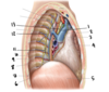
What structure is labeled by #8?

Costal Pleura

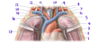
What structure is labeled by #1?

Ascending Aorta

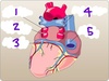
Which structure is labeled by #16

Right ventricle

Which structure is labeled by #13?

Pectinate muscles (in the left auricle)

Which structure is labeled by #10?

Left atrium

Which structure is labeled by #7?

Opening of inferior vena cava and valve

Which of the three major coronary arteries supplies blood to the left ventricle?
Front and bottom of LV: Anterior interventricular artery (aka Left anterior descending artery)
Back and side of LV: Circumflex artery
Where is the Aortic Semi-Lunar Valve?
Where do you place your stethoscope to listen?
Valve: Between the left ventricle and the aorta
Stethoscope: In the second intercostal space to the right of the patient’s sternum
(Circled A)

Which structure is labeled by #14?

Right coronary artery

What is cardiac tamponade?
Cardiac tamponade = fluid in the pericardial cavity that compresses the heart
Parietal pericardium has low compliance; increased volume in the sac puts pressure on the heart, rather than expanding the pericardial sac.
Describe the pathway of sympathetic innervation of the heart
- Presynaptic neuron
- Spinal cord
- Lower cervical or uppor thoracic sympathetic trunk
- Synapse in sympathetic trunk
- Cell body is here in the stellate ganglion
- Postsynaptic neuron
- Sympathetic trunk
- Cardiopulmonary splanchnic nerves
- Plexus at the bifurcation of the trachea
What structure is labeled by #12?

Right Ventricle

What structure is labeled by #9?

Left Internal Jugular Vein

Which structure is lableled by #7?

Right coronary artery

Which coronary artery suplies the left atrium?
Circumflex artery
Why does angina sometimes radiate down a person’s arm?
The left ventricle is supplied by nerves from T1
Nerves from T1 also supply the brachial plexus
Therefore, anginal pain can be referred to nerves in the arm
Which structure is labeled by #5?

Interventricular septum

Which structure is labeled by #1?

Ligamentum Arteriosum

What structure is labeled by #16?

Right Brachiocephalic Vein

Which part of the lung does the heart interface on the…
Left side?
Right side?
Right side = middle lobe of the right lung
Left side = lingula of the upper lobe of the left lung
What structure is labeled by G?

Left subclavian artery

Which structure is labeled by #3?

Opening of the left coronary artery
List the differences between the left and right ventricles
- Smooth top of chamber
- Left is called the aortic vestibule
- Right is called the conus arteriosus
- Left is round with thicker walls
- Right is bellow-shaped with thinner walls