Soft Tissue / MFR 2 Flashcards
(18 cards)
Thoracic
Direct/Indirect Thoracolumbar MFR
Patient: prone
Physician: stands beside the patient
- Place both hands palm down on the thoracolumbar junction B/L, fingers spread out slightly
- Engage tissues with a ventral force
- Move tissues inferiorly and superiorly, left and right, and clockwise/counterclockwise, noting in which direction there is ease of motion and restriction of motion
- Either treat the direct or indirect barrier
- Consider utilizing REMs to enhance release

Thoracic Longitudinal & Lateral MFR
Patient: lateral recumbent
Physician: stands facing patient
- Caudad forearm contacts the iliac crest, cephalad forearm contacts axilla, fingers contact medial aspect of erector spinae muscles
- Spread elbows apart while applying lateral traction on paraspinal muscles
- Have patient breathe deep for activating force

Seated Paraspinal Lumbar MFR
Patient: seated
Physician: seated next to patient
- Palm on medial aspect erector spinae muscle group, other hand across patient’s chest grasping contralateral shoulder
- In repetitive fluid motion, apply force anteriorly and laterally while depressing and translating erector spinae laterally until tissue release

Direct/Indirect Thoracic MFR, prone
Patient: prone
Physician: stands beside patient
- Hands on bilateral sides of thoracic spine
- Engage tissues with a ventral force
- Move tissues inferiorly and superiorly, left and right, and clockwise/counterclockwise, noting in which direction there is ease of motion and restriction of motion
- Either treat the direct or indirect barrier
- Consider utilizing REMs to enhance release

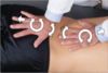
Lumbosacral MFR
Patient: prone
Physician: stands beside patient
- Place one hand over the inferior lumbar segment and the other hand over the superior sacral segment
- Monitor inferior and superior glide, left and right motion, and clockwise/counterclockwise motion, noting the direction of ease of motion or restriction of motion
- Treat indirect or direct barrier, consider utilizing REMs.
Prone I-Sacral Release
Patient: prone
Physician: standing next to patient
- Place bottom hand over sacrum with heel over base and fingers over apex. Place other hand on top in opposite direction.
- Evaluate pattern of restriction by rocking sacrum into multiplanar direction, noting laxity and restriction.
- Treat indirect or direct barrier by stacking dysfunction; consider utilizing REMs

Upper Limb & Shoulder MFR
Patient: prone with arm dangling from the table
Physician: seated on the side of the involved upper limb
- Grasp the humeral head of the patient with both hands and monitor the tissues for tissue texture response to the following motions introduced through the humeral head: flexion/extension, IR/ER of the humerus, adduction/abduction of the humerus, protraction/retraction of the scapular, superior/inferior scapular motion, traction/compression
- Engage either for direct or indirect MFR. Follow the release until there is no more tissue creep.

Lateral Stretch, Rhomboid Region
Upper Extremity
Patient: lateral recumbent
Physician: Standing, facing patient
- Caudal hand loops beneath axilla and grasps inferior portion of medial scapular border.
- Cephalad hand grasps superior border of the medial scapula.
- Apply lateral traction to scapula for 1-2 seconds in repetitive, rhythmic manner

Elbow MFR
Patient: seated or supine
Physician: on side of involved upper limb
- Hold the patient’s hand with one hand and the proximal radius and ulna with the other hand
- Test elbow flexion/extension and forearm supination/pronation to determine directions of laxity and restriction
- Indirect: gently and slowly move the elbow to its position of laxity, and follow any tissue release until it is completed.
- Direct: slowly move the elbow into its restriction and apply steady force until tissue give is completed.
- Slowly return elbow to neutral and retest motion

Still’s Wrist MFR
Patient: seated
Physician: standing, facing patient
- Grasp carpal bones between thenar eminences
- Test flexion/extension, ulnar/radial deviation for restriction/laxity
- Stack restrictive barriers and instruct patient to make a fist and/or spread fingers widely for 5 seconds and then relax hand
- Engage next restrictive barrier and repeat until now new restrictive barriers are encountered

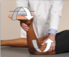
Hamstring Hypertonicity ME/MFR Tx
Patient: supine
Physician: Standing on side to be treated
- With knee extended and contralateral ASIS stabilized, flex patient’s hip until fascial barrier is met
- Patient is then instructed to push leg downward toward table while physician resists for 3-5 seconds
- Engage the next restrictive barrier and repeat until motion is restored
*alternate technique with knee flexed can be done for gluteus maximus hypertonicity

Iliotibial Band, Prone (Fascia Lata)
Patient: prone
Physician: stand on side opposite IT band being treated
- Use caudad hand to grab foot or ankle, flex knee to 90*.
- Cephalad hand will contact lateral thigh
- Push foot and lower leg out laterally while simultaneously engaging the IT Band by compressing cephalad hand into patient’s IT band and pulling posteromedially.
Iliotibial Band, Lateral Recumbent
Patient: lateral recumbent
Physician: stand facing the front of the patient
- Stabilize patient by placing cephalad hand on the posterolateral aspect of the iliac crest.
- Make a fist with caudad hand and place the flat portion of the proximal phalanges over the distal lateral thigh
- Engage tissue giving a slight downward pressure into IT band and slide fist proximally towards the greater trochanter region. Then move proximal to distal.
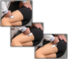
Knee MFR/INR
Patient: supine
Physician: standing on same side of knee being treated
- With superior (cephalad) hand, grasp distal femur to stabilize, with inferior (caudad) hand grasp tibia/fibula and use it as lever to examine for three-dimensional laxity and restriction
- Assess in full extension followed by flexion, IR/ER, Ab/Adduction
- Passively move LE to treat either direct/indirect
Knee MFR
Patient: supine or seated
Physician: standing on same side of knee being treated
- Grasp proximal leg with both thumbs on tibial plateau between knees
- Move tibia into anterior/posterior, medial/lateral glide, and IR/ER to determine position of laxity and restriction
- Treat restrictive barrier directly or indirectly

Sprained Ankle Indirect MFR
Patient: supine
Physician: at foot of table
- Monitor facial milieu or other individual ligaments with cephalad hand
- Use caudal hand on forefoot to introduce: inversion/eversion, plantarflexion/dorsiflexion, IR/ER. Then engage as many indirect barriers as possible and use the inherent mechanisms to release the fascia. Follow release until there is no more tissue creep.

Plantar Fascia (Longitudinal Stretch) MFR
Patient: supine
Physician: at foot of table
- Stabilize foot by placing hand over dorsum of foot
- Make a closed fist with your other hand and contact sole of patient’s foot just proximal to metatarsal heads
- Exert moderate pressure and move fist distal to proximal towards the calcaneus along the plantar fascia

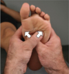
Plantar Fascia “X” MFR
Patient: supine
Physician: at foot of table
- Cross thumbs to make an “X” and place thumb pads over the area of concern at the plantar fascia. Impart an inward force that is vectored distal and lateral and continue this pressure until barrier is met, and further until release is palpated.
- Repeat with foot alternately attempting plantar/flexion and dorsiflexion.