Sport's Medicine Flashcards

1
Q

Specific Injuries based on anatomic site

  1. Shoulder? 4
  2. Elbow? 2
  3. Knee? 4
  4. Foot and Ankle sprains? 1
A
  1. Shoulder
    - Rotator cuff disease
    - Degeneration
    - Instability
    - Biceps and SLAP Lesions
  2. Elbow
    - Medial pain issues
    - Lateral pain Issues
  3. Knee
    - ACL
    - Meniscal Injuries
    - Articular Cartilage
    - Anterior Knee pain
  4. Foot and Ankle
    - sprains
2
Q

Who’s at risk for rotator cuff injuries? 2

A
  1. Trauma
  2. Repetitive overuse
3
Q
  1. What kind of population sufferes from degenerative tenon?
  2. Describe the process?
A
  1. Many sports played competitively into 60’s, 70’s and 80’s

2.

  • Tendons undergo normal aging and degeneration
  • Sport further stress on tendon complex
4
Q

Describe whats going on in the pictures below?

A
  1. Supraspinatus- No sign of white, which is good. no fat
  2. More white streaks, pretty substantial tear in a younger individual. fatty white streaks
  3. Probably not reconstructable and have to start thinking replacable.
5
Q

What is our goal for nonoperative treatment?

What are these? 6

A
  1. Reduce Inflammation

2.

  • Time
  • Activity shutdown
  • NSAID’s
  • Subacromial injection
  • Modalities
  • PT: ROM and Strength
6
Q

Supraspinatus exercise?

A
7
Q

Infra/Teres Strengthening?

A
8
Q

Subscapularis Strengthening?

A
9
Q

What is the surgical option for a Full thickness rotato cuff tear?

3

A
  1. Open Repair
  2. Mini-Open Repair
  3. Arthroscopic Repair
10
Q

Describe the Open Repair for RC

A
11
Q

Describe Mini-Open Repair for RC?

A
12
Q

Describe the Arthroscopic repair for RC?

A
13
Q

Post-op Course for RC surgery?

6

A
  1. Sling for 6 weeks
  2. Rehab for 3 mos
  3. Golf 4 to 5 mos
  4. Tennis 6 mos
  5. Swimming 7 to 8 mos
  6. Full recovery 1 year
14
Q

Shoulder Arthritis:
1. Early/Moderate Tx? 5

  1. Severe? 1
A

1.

  • Activity Modification
  • NSAIDS
  • Steroid Injections
  • PT
  • Arthroscopy
    2. Shoulder Replacement
15
Q

What is going on in the following pictures?

A

Osteophyte off the humerus itself. They almost always wear out posteriorly. This is best treated with a replacement.

16
Q

TSR results

  1. Failure rate?
  2. Advantages? 2
A
  1. 3% failure

2.

  • Predictable pain relief
  • Excellent function
17
Q

What is often a sign of shoulder instability?

A
18
Q

Treatment of 1st Dislocation

  1. Have to determine what?
  2. Tx Options? 3
  3. Whats your biggest issue with stability?
A
  1. Anterior vs. Posterior

2.

  • Reduction: X-ray
  • Immediate: ER Brace
  • Surgical
    3. Labral tear
19
Q

Instability Treatment
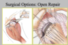
Open Repair

  1. Pros? 3
  2. Cons? 2
A
  1. Pro’s:
    - Higher success rate
    - Better in ligamentously lax
    - Glenoid reconstruciton possible
  2. Con’s:
    - Risk of over tightening
    - Painful post-op
20
Q

What does neer test?

A

Biceps Disease
Neer: Outlet Impingement

21
Q

What contributes to bicep dz?

3

A
  1. Acromion shape and slope
  2. AC joint enlargement
  3. Cuff and biceps problems
22
Q

Biceps Degeneration/Tendonitis

How would you treat the following:

  1. Isolated?
  2. W/ Rotator Cuff Tendonitis?
  3. W/ Rotator Cuff Tear?
A
  1. Non-Operative Management: if failure then surgical tx
  2. Non-Operative Management: if failure then surgical Tx
  3. Surgical Tx
23
Q

What tests would we do for Bicep pathology?

4

A
24
Q

Tests for Subscap?

2

A

Stomach compression test

Lift off test

25
Q

Diagnostic Imaging: for shoulder path?

2

A
  1. MRI:

Moderate accuracy for biceps disease: Gadolinium recommended

  • Up to 97% assoc. with RCT
    2. Ultrasound- Injection guide
26
Q

Non-operative Management: Bicep injury

Spontaneous Rupture treated how?

4

A

Spontaneous Rupture

Treated non-operatively

Non-operative Management

  1. Rest
  2. NSAIDS
  3. Physical Therapy- Rotator cuff strengthening
  4. Injections- Intra-articular
27
Q

Surgical Indications:
Tenotomy or Tenodesis

5

A
  1. Subluxation or dislocation of biceps
  2. > 25 % tear
  3. Significant inflammation, atrophy, hypertrophy
  4. Routine during TSR and HHR (esp. fracture/stiffness)
  5. Irreparable rotator cuff tear
28
Q

Tenotomy 3 vs. Tenodesis 2?

OUr indications?

A

Evolving with new techniques

  1. Tenotomy:
    - Elderly, cosmesis less of concern
    - Easier rehab
    - Revision
  2. Tenodesis:
    - less than 50 years,
    - cosmesis/strength IS a concern
29
Q

SLAP means what?

A

Superior Labral tear from Anterior to Posterior

30
Q

What is Obriens test for?

A

Labral Tear

318 PATIENTS, 56 WITH POSITIVE TEST POSITIVES CONFIRMED SURGICALLY

SENS - 100%

SPEC - 98%

AC JOINT

31
Q

What is the crank test?

A

For labral tear

32
Q

SLAP Lesion Non-operative management

4

A
  1. Non-operative Management
    - Rest
    - NSAIDS
    - Physical Therapy X 3 mos
    - Throwing Program
33
Q

SLAP lesions PT includes?

3

A
  1. Rotator cuff strengthening- Instability
  2. Scapula strengthening
  3. Posterior Capsular Stretching- Internal Impingement
34
Q

Slap Repairs: Indications

5

A
  1. Young patient (<40)
  2. Mechanical symptoms
  3. Associated Instability
  4. Associated internal impingement
  5. Acute rotator cuff tear
35
Q

What indicates instability with a SLAP lesion?

4

A
  1. “Drive-Through” sign
  2. Esp. if glenohumeral ligaments attached
  3. Pagnani test
  4. Rodofsky test
36
Q

Slap Repairs: Contraindications

4

A
  1. Elderly- Consider tenotomy
  2. Frozen shoulder
  3. Anatomic variant (No exposed cartilage and Doesn’t match symptoms)
  4. Chronic rotator cuff tear
37
Q

Medial elbow Pain DDx

6

A
  1. Medial epicondylitis
  2. Ulnar Neuropathy
  3. Flexor Pronator strain
  4. Zpronator sundrome – entrapment of AIN
  5. Ulnar or Medial Collateral Ligament**
  6. Olecrenon Stress Fracture**
38
Q

Lateral Elbow Pain DDx

4

A
  1. Lateral Epicondylitis
  2. Radial Tunnel Syndrome – entrapment of PIN
  3. Lateral ulnar Collateral ligament
  4. Capitellar OCD
39
Q

Lateral Epicondylitis

  1. AKA?
  2. PP?
  3. Cause?
  4. Histology?
  5. Tx?
A
  1. Tennis Elbow
  2. Overuse injury involving eccentric overload at origin of common extensor tendons
  3. Repetitive pronation/dupination with elbow extended
  4. Microtear of ECRB
  5. Non-operative TX 95% successful
40
Q

Lateral Epicondylitis

  1. Presentation: Pain with what? 3
  2. Tx? 2
A
  1. Presentation
    - Pain with resisted wrist extension, gripping
    - Pain at ECRB insertion
    - Pain with resisted wrist extension with elbow extended
  2. Treatment
    - Ice, NSAID, rest, ultrasound
    - Larger raquet grip
41
Q

Lateral Epicondylitis

More severe tx? 2

A
  1. Injections
    - Some benefit from corticosteroid injection
    - No indication for effectiveness of PRP
  2. Rarely requires release and debridement of ECRB
42
Q

37 y/o RHD Pitcher

no previous elbow problems

felt pop / pain with fastball

could not continue pitching

Dx?

A

Elbow MCL

43
Q

Physical Exam for elbow MCL

  1. Where is the pain?
  2. To find location, which tests would you do? 2
  3. Where will you have mild pain, weakness? 2
A
  1. pain - medial elbow @ AMCL insertion
  2. Location, Location, Location
    - Jobe
    - Milking
  3. mild pain, weakness
    - wrist flexion
    - forearm pronation
44
Q

What are the following showing?

A

1, Normal

  1. Partial tear
  2. Complete Tear
45
Q

Medial Collateral Ligament Complex

What is included in this? 3

A
46
Q

Elbow MCL treatment?

5

A

Rehab?

  1. Standard x 6 wks
  2. 2 wks - acute inflammation
  3. Rest, ice, modalities (phono, ionto, contrast pools)
  4. Then active/passive c/ modalaties
  5. Throwing Program x 3 mos: Short toss >> Long toss >> Mound
    - If fails- repeat
47
Q

Surgical Indication for elbow MCL

4

A

1, no problems with ADL

  1. no problems with warm up
  2. pain at 70-100% effort
  3. pain / tingling ulnar distribution

Shoulder ??

48
Q

ACL Injury happens with what movements? 6

A
  1. Sudden deceleration
  2. Twist
  3. Pivot
  4. Cut
  5. Clipping/pile-up
  6. Backward fall skiing
49
Q

Rehab time for ACL?

A

4-6 months

50
Q

Surgical Facts for ACL?

3

A
  1. Ligament does not heal
  2. Ligament does not do well with primary repair
    - Sewing the ends together
    - Studies have shown success rates of 40% or less
  3. Only way to restore is through reconstruction
51
Q

ACL: Only way to restire is through resconstruction?

2

A
  1. Probably due to synovial environment
  2. Unlike MCL which is outside joint and can heal without surgery
52
Q

Surgical Options for ACL:

  1. Reconstruction?3
  2. Allograft? 3
A
  1. Reconstruction
    - Patella tendon
    - Hamstring tendon
    - Quadriceps tendon
  2. Allograft tendon
    - Patella tendon
    - Achilles tendon
    - Tibial tendon
53
Q

Anterior Knee Pain

DDX? 3

A
  1. Patello-femoral pain- Cartilage changes, maltracking
  2. Quadricep or Patellar Tendon Pain
  3. Osgood Schlatters’ Disease
54
Q

Anterior Knee Pain

  1. Very common, especially in who?
  2. Tracking problem?
  3. Conservative tx? 2

Then surgery

A
  1. Very common Esp females
  2. Tracking problem- Q angle
  3. Conservative treatment
    - Rehab
    - Brace

Surgery

55
Q

Osgood Schlatters

  1. AKA?
  2. More common in who and at what age?
  3. Cause?
  4. Prognosis?
A
  1. Tibial Tubercle Apophysitis
  2. More common in males

Boys – 12-15

Girls 8-12

  1. Stress from extensor mechanism
  2. Self-limiting, dependant on growth plate closure
56
Q

Osgood Schlatters Presentation

3

A
  1. Pain at anterior aspect, worse with kneeling
  2. Tender over enlarged tubercle
  3. Worse with resisted extension
57
Q

Osgood Schlatters TX? 4

Prognosis?

A
  1. NSAID’s, rest, ice, activity modification
  2. Quad plus HAMSTING strengthening
  3. For severe symptoms – cast
  4. Rarely requires ossicle excision
    - Skeletally mature with symptoms

90% resolve – time limited

58
Q

FOOT AND ANKLE

  1. Examine patient while in what positions?
  2. Assess ROM of what? 3
  3. Midfoot is examined with what? 2
  4. What exam important in all patients?
  5. Key to any exam? 3
A
  1. sitting and standing
  2. ankle, hindfoot and forefoot
  3. pronation and abduction stresses
  4. Neurovascular
  5. Key to exam
    - WHERE DOES IT HURT?
    - DISCRETE PALPATION
    - MANIPULATION
59
Q

What are the most common eating disorders in sports?

A

Ankle ligament injuries are the most common injuries in sports

60
Q

Lateral ankle ligaments are the most common injured structures

What direction are they being overextended?

Which ligaments? 2

A

Inversion

  1. Anterior Talofibular
  2. Calcaneofibular Ligaments
61
Q

Differential Diagnosis of Twisting Injuries

A
  1. Lateral Ligament Structures
  2. Syndesmosis
  3. Articular Cartilage of the Talus: Osteochondral Dessicans
  4. Peroneal Tendons
  5. Base of the 5th metatarsal
  6. Subtalar Joint
  7. Fractures about the ankle: Growth plate injuries
  8. Lateral and Anterior Process of the Talus Fractures
  9. Stieda process or Os Trigonum
  10. Superficial Peroneal or Sural Nerves
  11. Calcaneal-cuboid and Lisfranc joints
62
Q

Anatomy of the Ankle-
Ligaments

  1. Primary Ligamentous Support- Lateral Ligament Complex includes? 3
A

1.

  • ATFL - Anterior Talofibular Ligament
  • CFL - Calcaneofibular Ligament
  • PTFL - Posterior Talofibular Ligament
63
Q

Anatomy of the Ankle Muscles

  1. Peroneal Tendons function? 2
  2. Posterior Tibialis Muscle function? 2
A

1.

  • Laterally provide resistance to inversion injuries
  • Nerves of proprioception in the ankle joint and ligaments signal these to contract to protect the joint from plantar flexion and inversion

2.

  • Antagonist to the peroneals
  • Provides resistance to Eversion stresses
64
Q

Medial ligament injuries

  1. Medial structures are less likely to be injured because?
  2. Results from?
A
  1. of the more prominent bony barriers to eversion
  2. Result from ankle external rotation and eversion
65
Q

Ligament sprains tend to occur when the joint is in a position that provides little bony stability. In the ankle this is what? 2

Describe the mechanism of injury?

A
  1. Plantar Flexion and Inversion

2.

Talus is narrower posteriorly than anteriorly and in dorsiflexion the wider talus engages the mortise with a tighter fit

Inversion is less stable because of the lengths of medial and lateral malleoli differ

Lateral malleolus extends further distally and blocks lateral talar movement

In this position ligament injuries will occur

66
Q

Risk factors for Inversion Injuries

Biomechanical factors may cause excessive forces to the lateral ankle and place the ankle at risk for inversion injuries such as?

A
67
Q

Lateral Ankle Injuries
Physical Exam

  1. Inspection? 3
  2. Palpation? 4
  3. Ligaments ? 5
  4. Tendons? 2
A
  1. Inspection

Identify

  • swelling,
  • ecchymosis,
  • deformity
    2. Palpation: Must palpate bony structures
  • Tenderness over bone or growth plates bring high suspicion for fractures
  • Proximal Fibula: Maissnouve
  • Base of 5th Metatarsal
  • Lateral Talar Process
    3. Ligaments: Syndesmosis or interosseous injuries
  • ATFL,
  • CFL,
  • PTFL,
  • anterior tibiofibular ligament,
  • deltoid
    4. Tendons
  • Peroneal
  • Achilles
68
Q
  1. What is the anterior drawer test specific to?
  2. How do you do it?
  3. Talar tilt test: Ankle in neutral to place stress on what?
  4. Apply stress where?
  5. Must block what?
A
  1. Anterior Drawer Test: Specific to the ATFL

2.

  • Ankle in neutral position with the heel and tibia stabilized
  • Forward stress placed on heel
    3. Talar Tilt Test: Ankle in neutral to place stress on CFL
    4. Apply a varus stress
    5. Must block subtalar motion
69
Q

Lateral Ankle Injuries
Diagnostic Tests

  1. X-Ray evaluation is warranted when what?
    - Not all acute ankle injuries need X-rays
  2. Which views? 3
  3. Look for what? 4
  4. MRI has a valuable role in the evaluation of what and not what?
A
  1. palpable pain is present on bony areas
  2. AP, lateral and mortise views

3.

  • fractures,
  • displacement of mortise,
  • widening of growth plates (fibular),
  • loose bodies or OCD
    4. chronic ankle pain, but has no role in the acute situation
70
Q

Chronic Ankle Instability

Reconstruction may be indicated after failure of rehab program of what? 3

A
  1. proprioception,
  2. muscle strengthening and

3, Achilles stretching

71
Q

Syndesmotic Injuries
High Ankle Sprains

  1. Three ligaments unite the distal tib-fib? 3
  2. MOI?
  3. Rehab time?
A

1.

  • Anterior tibiofibular
  • Posterior tibiofibular
  • Interosseous
    2. Mechanism of injury is EXTERNAL ROTATION and DORSIFLEXION
    3. Often have a longer rehab time with more long-term disability than lateral ankle sprains
72
Q

Syndesmotic Injuries

Dx?

5

A
  1. Tender over anterior syndesmosis
  2. Common to have deltoid tenderness
  3. Maisonneuve fracture will have tenderness at proximal fibula plus syndesmosis
  4. Compression Squeeze Test- Squeeze at mid-calf will cause pain at ant. Syndesmosis
  5. External Rotation Test- Done with knee at 90 degrees
73
Q

Radiographic Evaluation in Syndesmotic Injuries

  1. 50% will show what?
  2. Tibiofibular clear space what size?
  3. HOw reliable is this test?
  4. Tibiofibular overlap an AP of what?
  5. Tibiofibular overlap on Mortise of what?
  6. What kind of views?
A
  1. 50% will show a bone avulsion fracture from the anterior or posterior tubercle of the tibia
  2. less than 6 mm
  3. Most reliable test
  4. AP > 6mm
  5. >1 mm
  6. External Rotation stress views
74
Q

Treatment of Syndesmotic Injuries

  1. Partial injury? 2
  2. If widening is noted on plain or stress view – surgical repair is indicated? 4
  3. May develop what kind of pain on push off?
A
  1. Partial injury should be
    - treated with boot or walking cast for 2-4 weeks followed by rehab
    - Return to play is usually twice as long as for severe lateral ankle sprain – 4.5 to 6 wks
  2. If widening is noted on plain or stress view – surgical repair is indicated
    - Syndesmosis screw placed 1-2 cm proximal to joint line with ankle in max dorsiflexion
    - Non-weight bearing cast - 4 wks
    - Weight bearing immobilization - 4 wks
    - Screw removal at 10-12 wks
  3. May develop heterotopic ossification with pain on push-off
75
Q

Assessment of Persistently Painful
Sprained Ankles

What is highest on your diff for this?

A

INCOMPLETE REHABILITATION

76
Q

Stress Fractures

  1. What is it?
  2. Result of?
  3. Most often occur with?
  4. Where most often?
  5. Can also be associated with?
A
  1. Fatigue induced fracture of the bone caused by repeated stress over time
  2. Result of accumulated trauma from sub- maximal loading
  3. Most often occur in weight-bearing bones

4.

  • Tibia,
  • metatarsals,
  • navicular,
  • femoral neck
    5. May occur associated with growth plates
77
Q

Stress fractures - etiology

3

A
  1. Bones are constantly remodeling, with overuse and stress the capacity to do so is exhausted and a weak area develops
  2. Osteoblasts are overwhelmed
  3. Bone and muscles serve as shock absorbers to stress - with muscle fatigue the bone may be taxed
78
Q

Stress Fractures - Etiology

What is the female triad? 3

A

FEMALE TRIAD - commonly associated with recurrent stress fractures

  1. Amenorrhea
  2. Disordered eating
  3. Osteoporosis
79
Q

FEMALE TRIAD - commonly associated with recurrent stress fractures

  1. Amenorrhea caused by?
  2. Lead to?
A
  1. Caused by hormonal abnormalities

  1. Lead to demineralization and stress fractures

3.

80
Q
  1. Stress Fractures - Presentation?
  2. Usually resides with?
  3. Tenderness and swelling?
A
  1. Pain with weight bearing that increases with exercise or activity
  2. Usually resides with rest
  3. May have area of localized tenderness on or near the bone and generalized swelling
81
Q

Stress Fracture - Diagnosis

2

A
  1. X-rays- Acutely may not show evidence – may take 10-14 days before bone remodeling is present
  2. MRI or bone scan is more sensitive
82
Q

Stress Fractures - Treatment

3

A
  1. Rest combined with unloading of stress area to the time when pain is not present- Walking boots, crutches
  2. Gradual return to activities that caused issues- 10 % increase per week
  3. For fractures that don’t respond or have significant risk to not heal - fixation
83
Q

Stress Fractures - Prevention

5

A
  1. Allow for gradual ramp up of loading activities – allow bone to adapt to increased stresses
  2. Strengthening of muscles – calf and shin
  3. Replace shoes every 300-700 miles
  4. Increase calcium and Vit D
  5. Address issues associated with female triad
84
Q

Tibial Stress fractures

  1. Pain where?
  2. Tx? 2
A
  1. Pain directly over fracture

2 Treatment

  • Activity restriction with protected weight bearing
  • Rarely IM nail
85
Q

Femoral neck Stress Fracture

  1. Compression side where?
  2. Tension side where?
  3. Study of choice?
  4. Which side needs surgical tx?
A
  1. Compression side
    - Inferior medial neck
  2. Tension side
    - Superior-lateral
  3. MRI study of choice
  4. Tension side need surgical treatment