What is a TIA?
An ischaemic usually embolic neurological event with symptoms lasting less than 24h. This is what makes it transient.
Without intervention more than 1 in 12 patients will go on to have a stroke within a week.
Signs of TIA.
The signs will be specific to the artierial territory and what part of the brain that artery supplies.
E.g. amaurosis fugax can occur when the retinal artery is occluded. This causes unilateral progressive vision loss like curtain descending.
Global events like syncope and dizziness are rare but can happen.
Attack may occur in singularity but can also be many.
What do multiple highly stereotyped attacks aka crescendo TIAs suggest?
Critical intracranial stenosis commonly in the superior division of the MCA.
Causes of TIA.
Atherothromboembolism of the carotid is the main cause - listen for bruits.
Cardioembolism such as a mural thrombi post-MI or in AF, valve disease or in prosthetic valve.
Hyperviscosity - polycythaemia, sickle cell anaemia and myeloma.
Vasculitis is a non-embolic cause.
Differentials of TIA.
Hypoglycaemia
Migraine aura
Focal epilepsy
Hyperventilation
Retinal bleeds
Rare mimics such as;
Malignant hypertension
MS
Intracranial tumours
Peripheral neuropathy
Phaeochromocytoma
Somatisation
Investigation in TIA.
Bloods - FBC, U&Es, ESR, Glucose, Lipids,
CXR
ECG
Carotid doppler
CT and MRI
Echocardiogram
Treatment of TIA.
Similar to stroke patient should be given Aspirin 300mg OD for 2 weeks and then be switched onto clopidogrel 75mg OD.
If this is CId then give aspirin 75mg OD combined with slow-release dipyridamole.
Control cardiovascular risk factors
Anticoagulation indications if need be
Carotid endarterectomy.
Explain control of cardiovascular risk factors in TIA.
Optimise BP ( < 140/85 mmHg)
Hyperlipidaemia
DM
Help stop smoking
When should a carotid endarterectomy be done?
Perform within 2 weeks of first presentation if 70-99% stenosis and operative risk is acceptable.
When should antiocoagulation be done in TIA?
If there is a cardiac source of emboli.
How should people with crescendo TIA (two or more TIAs in a week) be treated?
As being at high risk of stroke.
What is the risk assessment tool designed to improve the prediction of short-term risk of a stroke after a TIA?
ABCD2 score.
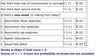
Explain the ABCD2 score.
Age > 60 (1)
BP >140/90 (1)
Clinical features
Unilateral weakness (2)
Speech disturbance without weakness (1)
Duration of symptoms
> 1h (2)
10-59 min (1)
Diabetes (1)
What do different scores in ABCD2 tell you?
4 or more indicates that the patient is at high risk of an early stroke and must be assessed by a specialist within 24h.
A score of 6 or more strongly predicts a stroke (8.1% within 2 weeks and 35.5% in the next week)
Driving and TIA.
Prohibited from driving for at least 1 month.
What is a stroke?
A sudden onset of a focal neurological deficit lasting more than 24 hours or with imaging evidence of brain damge due to either infarction or haemorrhage.
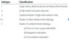
Causes of stroke.
Small vessel occlusion/cerebral microangiopathy or thrombosis in situ.
Cardiac emboli
Atherothromboembolism form e.g. carotids.
CNS bleeds
Carotid artery dissection
Vasculitis
SAH
Venous sinus thrombosis
Antiphospholipid syndrome
Thrombophilia
Fabry disease
CADASIL
Differentials of strokes.
Head injury
Hypo/hyperglycaemia
Subdural haemorrhage
Intracranial tumours
Hemiplegic migraine
Post-ictal
CNS lymphoma
Wernicke's encephalopathy
Hepatic encephalopathy
Encephalitis
Toxoplasmosis
Cerebral abscesses
Mycotic aneurysm
Drug overdose
What can be used to help medical staff distinguish between a stroke and a stroke mimic?
ROSIER scale.
Explain ROSIER scale.
Stands for Rule Out Stroke In the Emergency Room

Risk factors of stroke.
HTN
Smoking
DM
Heart disease
Peripheral vascular disease
Increased PCV
Carotid bruit
COCP
Dyslipidaemia
Alcohol use
Increased clotting
Low antithrombin III
etc...
A stroke can be infarct or haemorrhagic.
What points to haemorrhagic?
Remember this is unreliable.
Meningism
Severe headache
Coma
Pointers to ischaemia.
Remember this is unreliable.
Carotid bruit
AF
Past TIA
IHD
Different types of strokes.
Total anterior circulation stroke (TACS) (Worse prognosis)
Partial anterior circulation stroke (PACS)
Lacunar stroke (LAC)
Posteiror circulation stroke (POCS)
Define TACS.
All three of the following need to be present for a diagnosis of a TACS:
Unilateral weakness (and/or sensory deficit) of the face, arm and leg
Homonymous hemianopia
Higher cerebral dysfunction (dysphasia, visuospatial disorder)
Define PACS.
Two of the following need to be present for a diagnosis of a PACS:
Unilateral weakness (and/or sensory deficit) of the face, arm and leg
Homonymous hemianopia
Higher cerebral dysfunction (dysphasia, visuospatial disorder)
Define POCS
One of the following need to be present for a diagnosis of a POCS:
Cranial nerve palsy and a contralateral motor/sensory deficit
Bilateral motor/sensory deficit
Conjugate eye movement disorder (e.g. horizontal gaze palsy)
Cerebellar dysfunction (e.g. vertigo, nystagmus, ataxia)
Isolated homonymous hemianopia
Define LACS.
One of the following needs to be present for a diagnosis of a LACS:
Pure sensory stroke
Pure motor stroke
Senori-motor stroke
Ataxic hemiparesis
Define LACS.
One of the following needs to be present for a diagnosis of a LACS:
Pure sensory stroke
Pure motor stroke
Senori-motor stroke
Ataxic hemiparesis
What is TOAST classification of stroke?
Classification of subtype.

What is a rapid assessment tool of suspected stroke?
FAST
ROSIER
Explain FAST.
Face (facial drooping)
Arm (weakness)
Speech (slurred)
Time (time to call 999)
This is a public health tool.
What is a clinical stroke assessment tool to evaluate and document neurological status in acute stroke patients?
NIH stroke scale (NIHSS)
Explain NIH stroke scale.
Scores you on each of the following and subsequent severity;
1. Level of consciousness
2. Best gaze
3. Visual
4. Facial palsy
5. Motor arm
6. Motor leg
7. Limb ataxia
8. Sensory
9. Best language
10. Dysarthria
11. Extinction and inattention
Acute management of stroke.
Protect airways.
Maintain homeostasis.
Do a screen swallow and make the patient nil by mouth until this is done.
CT/MRI within 1h. This is essential to assess if thrombolysis can be given or not. Should not be given in haemorrhagic stroke. Diffusion weighted MRI is most sensitive for an acute infarct, but CT helps rule out primary haemorrhage.
Once haemorrhagic is excluded give aspirin 300mg and continue for 2 weeks then switch to long-term antithrombotic treatment.
Thrombolysis (will be discussed in next flashcard)
Explain when thrombolysis should be done in stroke.
This should be considered as soon as haemorrhagic stroke has been excluded and provided the onset of symptoms was less than 4.5h ago.
Benefits of thrombolysis outweigh the risks within this window.
Alteplase is the agent of choice and must be given by trained staff.
Always do a CT 24h post-lysis to identify any bleeds.
Contraindications to thrombolysis.
Haemorrhage
Mild/non-disabling deficit
Recent surgery, trauma or artery/vein puncture at uncompressible site
PRevious CNS bleed
Aneurysm or AVM
Severe liver disease, varices or portal HTN
Seizures at presentaiton
Hypo/hyperglycaemia
GI or urinary tract haemorrhage in last 21 days
Known clotting disorder
Low platetelts
INR > 1.7
History of intracranial neoplasms
and the list goes on
When might thrombectomy be considered?
In large artery occlusion in the proximal anterior circulation.
Primary prevention of stroke (before any stroke occurs)
Treat HTN
DM
Dyslipidaemia
Cardiac disease
Help quit smoking
Exercise help
Liflong anticoagulation in AF and prosthetic valves and prevention post TIA
Secondary prevention of stroke (preventing any further stroke)
Control risk factors like in primary prevention
Antiplatelet agents after stroke. If there is no primary haemorrhage give 2 weeks of aspirin 300mg and then switch to long-term clopidogrel monotherapy. If this is not tolerated or contra-indicated then give low dose apsirin plus slow-release dipyridamole.
Should also give anticoagulation after stroke from AF.
Tests to determine risk factors for further strokes.
HTN - check for retinopathy, nephropathy or cardiomegaly on CXR
Cardiac source of emboli - 24h ECG for AF, CXR, ECho
Carotid artery stenosis - Carotid dopple US +/- CT/MRI angiography.
Hypo/hyperglycaemia
Dyslipidaemia
Vasculitis - ESR, ANCA, VDRL to loof for active untreated syphilis
Platelet and bleeding study
Hyperviscosity
Genetic tests for CADASIL and Fabry disease
Driving and stroke.
Not permitted to drive for one month.
Driving and recurring TIAs.
Not permitted to drive for 3 months and you must be assessed by a doctor prior to resumption of driving.
What should patients with stable neurological symptoms from their stroke or TIA with carotid stenosis of 50-99% according to NASCET and 70-99% according to ECST be assessed and referred to?
Carotid endarterectomy within 1 week of onset of stroke or TIA symptoms.
When should the carotid endarterectomy be performed?
Within a maximum of 2 weeks of onset of stroke or TIA symptoms.
There is a small risk of stroke during the surgery
What can people with severe middle cerebral artery infarction be at risk of?
Malignant MCA syndrome.
What should patients with severe middle cerebral artery infarction be considered for?
Decrompressive hemicraniectomy if any deteriorationin their clincal condition occurs.
They should be referred within 24 hours of onset of symptoms and treated within a max of 48h.
Management of cardiac disease that can result in stroke.
CHA2DS2VASc score should be done in order to determine if someone is suitable for anticoagulation if they are in AF and at risk of stroke.
This should be done along with HASBLED score.

Assessments for re-enablement after stroke.
Assess swallowing - if difficult make nil by mouth
Minimise fall risk
Ensure bladder and bowel movements are OK
Minimise spasticity
Monitor the progress and resolution of symptoms and signs
Monitor mood
Assess need for DNAR
If the patient has issues swallowing and doesn't seem to recover from it, what should be consdiered?
Enteral feeding via NG and PEG.
Indications of enteral feeding.
Usually indicated due to poor swallowing and risk of aspiration.
Risks of enteral feeding.
The decision to feed is often based on patient and family preference.
Even with a PEG or NG tube aspiration can occur, and some patients may never tolerate oral feeding again.
Requirements for decompressive hemicraniectomy in severe middle cerebral artery infarction.
Within 48 hours of symptoms.
Under 60 yo
CT infarct of at least 50% MCA territory and an NIHSS score of above 15.
How should the aspirin be given after a stroke if the patient is dyspagic?
Rectally or by enteral tube still 300mg though.