Structure & function Flashcards

1
Q

What is the largest vital organ in the body?

A

The skin

2
Q

Systemic disease can manifest on the skin. T/F

A

True

3
Q

The skin can be divided into two layers. What are these?

A

Epidermis and dermis

4
Q

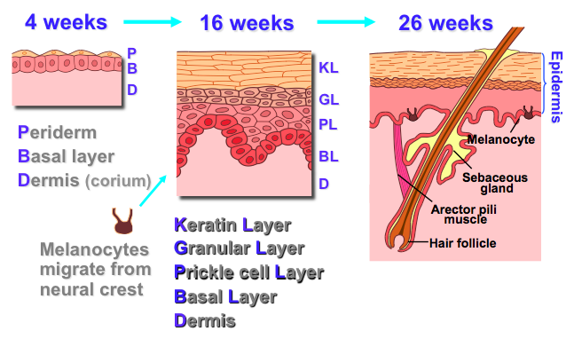
What are the embrological origins of the epidermis, dermis and melanocytes?

A

Epidermis - ectoderm. Dermis - mesoderm. Melanocytes - migrate from neural crest.

5
Q

How developed is the skin during gastrulation? (7-10 days)

A

There is cellular organisation into the germ layers

6
Q

How long does complete skin development take?

A

26 weeks gestation

7
Q

Describe the embryological development of the skin

A
8
Q

What name is given to the patten in which skin develops? What structures do they follow?

A

Blaschko’s lines. They don’t follow any structures

9
Q

What are appendages? (in relation to the skin)

A

Nails, hair, glands, mucosae

10
Q

What are the epidermal cell layers (from top to bottom)?

A

Keratin, granular, prickle cell and basal

11
Q

From which epidermal cell layer do new cells differentiate?

A

Basal layer

12
Q

There are marked differences between the epithelium at different sites of the body (e.g between the sole of the foot and the armpit). T/F

A

True

13
Q

Epithelial cell turnover is regulated by which factors?

A

Growth factors, hormones and cell death

14
Q

How long does it take for a keratinocyte to migrate from the basement membrane to the keratin layer?

A

28

15
Q

Describe the features of the basal layer

A

Small cuboidal cells forming a single layer. Possesses lots of intermediate filamints of keratin. The most metabolically active layer of the epidermis

16
Q

Describe the features of the prickle cell layer

A

Large polyhedral cells connected by many desmosomes and possessing lots of intermediate filaments.

17
Q

What happens when epithelial desmosomes are burst (intraepithelial blistering)? How might this occur?

A

Water escapes giving the skin a wet and glistening appearance. This may happen during inflammation where there is increased water.

18
Q

Describe the features of the granular layer

A

Two/three layers of flatter cells possessing odland (lamellar) bodies and keratohyalin granules (containing filggrin & involucrin). High lipid content. One of it’s functions is to remove the nuclei from keratinocytes

19
Q

What do we call keratinocytes which have lost their nuclei?

A

Corneocytes

20
Q

Describe the features of the keratin layer

A

Mostly an insoluble cornified envelope consisting of mostly ketain and filaggrin. Also possesses lamellar granules which release lipid. Acts as a waterproof barrier

21
Q

What virus can cause warts? Which does the virus do to the cells?

A

Human papilloma virus. It causes over proliferation of keratinocytes.

22
Q

Are mucosal surfaces typically affected by skin disease?

A

They can be. Most commonly seen in severe skin disease

23
Q

What is the most common epidermal cell?

A

Keratinocytes

24
Q

What are melanocytes

A

Pigment producing dendritic cells

25
Where are melanocytes found?
Basally & suprabasally
26
What is the function of melanocytes? Describe this process
Converts tyrosine to melanin. The melanin is transferred from melanocytes to keratinocytes via dendrites. The melanin forms a protective "cap" over the DNA of the cells, to protect from UV radiation.
27
From which organelle do melanocytes originate?
golgi apparatus
28
What is vitiligo?
an autoimmune disease which causes the depigmentation of areas of skin (due to loss of melanocytes)
29
What is albinism?
a genetic condition which results in loss of pigmentation in the skin
30
What is Nelson's syndrome?
hyperpigmentation of the skin due to an overactive thyroid producing more melanin stimulating hormone
31
Where do Langerhans cells originate?
bone marrow (like many other immune cells)
32
Where are Langerhan's cells found?
suprabasally in the epidermis, dermis, lymph nodes
33
What is the function of langerhan's cells?
They are immune cells involved in antigen presentation
34
What special feature do Langerhan's cells possess?
Birbeck granules/racket organelles
35
What is the function of Merkel cells?
Mechanoreceptors
36
Where are Merkel cells found?
Basally - between keratinocytes and nerve fibres
37
Merkel cell tumours are usuallu benign.T/F
False - they are rare malignant tumours with an extremely high mortality rate (caused by a virus)
38
Hair follicles are also known as what?
Pilosebacous units
39
What cells control our hair pigmentation?
Melanocytes (only those located above the dermal papilla)
40
List the components of a pilosebacious unit
Hair shaft, medulla, cortex, external and internal root sheath, papilla of hair follicle, matrix, arrector pilli muscle, sebaceous gland
41
What is hair composed of?
Specialised keratins
42
List the phases of hair growth and what occurs during each phase.
Anagen (growing), Catagen (involuting) and Telogen (resting)
43
What are the different types of hair?
Lanugo (in utero), vellus (fine, downy skin hair) and terminal (pubic, underarm, head hair)
44
What is the time scale for each of the phases of hair growth?
Anagen - several years Catagen - 3/4 weeks Telogen - everyday shedding occurs (50-100 hairs)
45
When may hair loss in humans be asynchronus? How will the hair appear when it first grows back?
Under times of huge stress or hormonal changes (e.g pregnancy). It will first appear white/grey until pigment is added
46
What is virilisation? What causes this?
The apperance of typically male features (e.g facial hair) on a female. Excess androgen.
47
What is alopecia areata?
Autoimmune hair loss
48
Nails are more biochemically similar to hair than to bone. T/F
True
49
What are nails made of?
Specialised keratins
50
Name some parts of the nail
Lunula, cuticle, nail plate, hyponychium, nail bed, nail matrix
51
What are the two main causes of a nail which appears crumbly?
Fungal infection Small repeated traumas & so poor differentiation over a period of time (e.g handball players)
52
Damage to the stem cells in the nail matrix may cause unusual nail growth permenantly. T/F
True
53
What are some of the functions of the dermo-epidermal junction?
Support, anchorage, adhesion, growth & differentiation of epidermal cells
54
What type of barrier is the dermo-epidermal junction?
Semi-permeable membrane
55
Name the two sections that the dermo-epidermal junction is divided into
lamina lucida & lamina densa
56
Define (and give an example of ) inherited and autoimmune conditions of the dermo-epidermal junction
Inherited - mutations in the proteins of the dermo-epidermal junction resulting in skin fragility (e.g epidermolysis bullosa) Autoimmune - antibodies formed against proteins in the dermo-epidermal junction (e.g dermatitis herpetiformis)
57
What are the symptoms of epidermolysis bullosa? What may occur in severe conditions?
Skin blistering, tearing, inability to be touched or held due to skin fragility. Epidermolysis bullosa dystropica of the hands.
58
What is bullous pemphigoid? Which test may diagnose it?
An autoimmune blistering disorder due to defects of skin tethering (i.e problems within the dermo-epidermal junction). Immunofluorescent histology
59
List the components of the dermis
Cells (*fibroblasts,* macrophages, mast cells, langerhan's cells), fibres (collagen, elastin), ground substance, blood vessels, lymphatics & nerves
60
What is the function of fibroblasts in the dermis?
Secretion of collagen
61
What is ground substance composed of?
Mucopolysaccharides, glycosaminoglycans
62
What are the two main extrinsic factors which contribute to photo aging? What happens to give the apperance of aged skin?
UV light & smoking. Less well formed collagen fibres, less elastin (solaris elastosis) and less nutrients being directed to the dermis
63
What is the function of horizontal plexuses?
They connect deep and superficial vessels
64
What is angioma? What is a haemangioma?
They are basically interchangeable terms used to describe a benign tumour of the blood vessels
65
What is another name for an angioma? What are the main problems with angiomas?
Port wine stains. They may sometimes bleed or look unsightly
66
What is the characteristic histology of an angioma?
dilated vascular spaces
67
What things may cause chronic lymphoedema?
Reccurent infection causing permenant damage to the lymphatics Lymph node blockage
68
What are the functions of pacinian and meissners corpuscles respectively?
Pacinian - pressure Meissner - vibration
69
Give an example of a disease which affects the nerve fibres in the skin
Neurofibromatosis
70
What is hyperhydrosis?
Abnormal excess sweating
71
Where are sebaceous glands found?
Mostly on the skin, scalp and chest but found all over the body apart from the soles of the feet & palms of the hands
72
What are the main functions of sebaceous glands? When do they become active?
Prevention of moisture loss/moisturation of the skin & protection against bacterial/fungal infections. Post-puberty
73
What do sebaceous glands produce? What is this composed of?
Sebum - squalene, wax esters, triglycerides & free fatty acids
74
What causes/contributes to acne? When is it usually triggered?
increased sebum, blocked sebaceous ducts & bacteria. During puberty
75
Where are apocrine sweat glands found?
Armpits and perineum
76
What is the function of apocrine sweat glands?
Androgen dependent scent glands
77
Where are eccrine sweat glands found?
The whole skin surface but particularly the palms and soles and armpits
78
What stimulates eccrine sweat glands?
Mental state, temperature and gustatory sensations
79
What are the functions of eccrine sweat glands?
cooling by evaporation and moistens palms & soles to increase grip
80
What is erythroderma?
Erythema on over 80% of the body
81
What is toxic epidermal necrolysis?
A severe skin reaction, usually triggered by drugs, which causes the epidermis to lift from the dermis
82
What are the conseqeunces of skin failure and how does this occur?
Dehydration - fluid loss Hypoalbuminaemia - protein loss Infection - loss of barrier protection Hypothermia - heat loss Spread of infection - loss of immune defense Disordered thyroxine metabolism Stigma - psychological impact Pain - sensation not lost with skin
83
What does the skin barrier protect against?
Trauma, UV radiation, irritants, toxins, allergens & pathogens
84
What is icthyosis? Give an example of what causes it
Scaly skin due to abnormal keratin layer. Steroid-sulphatase deficiency/X-linked
85
What is the typical pattern of irritant hand dermatitis caused by hand washing?
Most severe on finger tips or in between fingers
86
The skin is involved in thyroid hormone AND vitamin D metabolism. T/F
True
87
How is vitamin D stored in the liver and kidney respectively?
Liver: hydroxycholecalciferol Kidney: 1,25 - dihydroxycholecalciferol
88
Where do humans get vitamin D from?
Oily food in the diet & UV light
89
What proportion of thyroid hormone metabolism occurs in the thyroid gland? Where does the rest occur? What is it that gets metabolised?
20%. Peripheral tissues e.g the skin. Thyroxine (T4) is converted to Triiodothryonine (T3)
90
What receptors in the skin allow it to detect body temperature? What are the changes that modify body temperature?
Thermoreceptors. Blood vessel constriction/relaxation, behavioural, sweating, hair arrector muscles, shivering, etc
91
How is tissue lost due to frostbite?
Strong vasoconstriction cuts off the blood supply to tissue causing it to die
92
Give an example of a non-specific immune response involving the skin
Scratching to remove a parasite
93
Crusted scabies only occur in patients with weakened immune systems and represent a strong immune response to few mites. T/F
False - there will be many mites in a patient with crusted scabies (and no itch!)
94
What is Hansen's disease?
Leprosy (i.e a chronic granulomatous infection)
95
What is eczema herpiticum?
Eczema infected with herpes simplex virus
96
What are the communication functions of the skin?
Important visually, for giving off scent and in sociosexual behaviour
97
Acne vulgaris is associated with unemployment. T/F
True
98
What are the different types of nerve receptors within the skin?
99
What are neuropathic ulcers? Give two examples of diseases which might lead to the formation of these ulcers
Ulcers that form due to a loss of nerve sensation preventing the patient from feeling/noticing ulceration of the skin. Diabetes and leprosy
100