TEMPOROMANDIBULAR JOINT Flashcards
(42 cards)
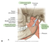
Ligaments of the TMJ

- Lateral TMJ (late phase biomechanics)
- Sphenomandibular
- Stylomandibular
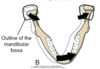
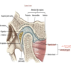
TMJ articular disc regions attachments:
- Posterior
- Intermediate
- Anterior
Posterior region of the TMJ articular disc attaches to the following:
- Collagen rich inferior retrodiscal lamina
- Elastin rich superior retrodiscal lamina

Anterior region of the TMJ disc attaches to the following:
- Tendon of the superior head of the lateral pterygoid muscle
- Temporal bone anterior to articular eminence
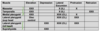
Masseter muscle:

- Origin: inferior zygomatic bone and arch
- Insertion: external surface of the mandible between the angle and coronoid process
- Action: bilaterally elevates and protrudes the mandible unilaterally ipsilateral lateral excursion
- Innervation: mandibular nerve, branch of CN V – trigeminal nerve
Temporalis muscle

- Origin: temporal fossa
- Insertion: coronoid process and ramus of the mandible
- Action: bilaterally elevates and protrudes the mandible; unilaterally ipsilateral lateral excursion
- Innervation: mandibular nerve, branch of CN V – trigeminal nerve
Medial pterygoid:
- Origin: lateral pterygoid plate
- Insertion: internal surface of the mandible between the angle and mandibular foramen (anterior to ramus)
- Action: bilaterally elevates and protrudes the mandible; unilaterally contralateral lateral excursion
- Innervation: mandibular nerve, branch of CN V – trigeminal nerve
Lateral pterygoid (superior head):

- Origin: greater wing
- Insertion: medial capsule, TMJ, disc, and pterygoid fossa
- Action: bilaterally eccentrically controls the disc during closing; unilaterally contralateral lateral excursion
- Innervation: mandibular nerve, branch of CN V – trigeminal nerve)
Lateral pterygoid (inferior head):

- Origin: lateral pterygoid plate
- Insertion: pterygoid fossa and neck of mandible
- Action: bilaterally DEPRESSES and protrudes the mandible; unilaterally contralateral lateral excursion
- Innervation: mandibular nerve branch of CN V Trigeminal
what three muscles close the mouth (elevation of the mandible)
- Masseter
- Temporalis
- Medial pterygoid
what two muscles open the mouth (depression of madible)
- Lateral pterygoid (inf head)
- Suprahyoid
3 lateral excursion muscles
- Medial pterygoid
- Lateral pterygoid (Inf and superior heads)
- All contralateral excursion
protrusion muscles
lateral pterygoid (inferior and superior heads)
retrusion muscles
temporalis
What are the secondary muscles of mastication?
- infrahyoids
- suprahyoids

In the adult the mouth can be opened an average of_____ as measured between the incisal edges of the upper and lower front teeth
50 mm
Maximal opening of the mouth typically occurs during actions such as…
yawning and singing
The interincisal opening is typically large enough to fit ______ adult “knuckles” (proximal interphalangeal joints)
three
Functional movement (depression) for eating is about ___ mm or ____ knuckles
18 mm
one knuckle
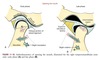
The early phase of depression of the mandible, constituting the first 35% to 50% of the range of motion, involves primarily ______ of the mandible relative to the cranium.
rotation
the condyle rolls posteriorly

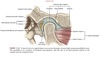
The late phase of opening the mouth consists of the final 50% to 65% of the total range of motion. This phase is marked by a gradual transition from primary rotation to primary
translation.
The transition can be readily appreciated by palpating the condyle of the mandible during the full opening of the mouth.
During the translation the condyle and disc slide together in a forward and inferior direction against the slope of the articular eminence
The early phase of elevation of the mandible involves primarily ______ of the mandible relative to the cranium.
translation
in a posterior and superior direction
The late phase of closing (elevation) the mouth is marked by a
rotation
condyle roll anteriorly
protrusion normal ROM
3 mm