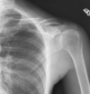
Thursday [02/09/2021] Flashcards

(100 cards)

1
Q

What is a central line?

A

Central venous catheter:

  • critically ill patients or those requiring prolonged IV therapies, for more reliable IV access
  • normally in the neck [internal jugular vein], chest [subclavian artery], groin [femoral vein], or through veins in the arms [PICC line]
2
Q

Why are PICC lines sometimes preferred?

A

Alternative to centra lines in major veins such as the subclavian or internal jugular as may result in pneumothorax
- generally used when patient expecting more than 2w of IV therapy

3
Q

What is caput medusea?

A

Caput medusae is the appearance of distended and engorged superficial epigastric veins, which are seen radiating from the umbilicus across the abdomen. The name caput medusae (Latin for “head of Medusa”) originates from the apparent similarity to Medusa’s head, which had venomous snakes in place of hair. It is caused by dilation of the paraumbilical veins, which carry oxygenated blood from mother to fetus in utero and normally close within one week of birth, becoming re-canalised due to portal hypertension caused by liver failure.

4
Q

Hypothyroid signs

A

Common symptoms include:

tiredness
being sensitive to cold
weight gain
constipation
depression
slow movements and thoughts
muscle aches and weakness
muscle cramps
dry and scaly skin
brittle hair and nails
loss of libido (sex drive)
pain, numbness and a tingling sensation in the hand and fingers (carpal tunnel syndrome)
irregular periods or heavy periods

5
Q

Hypothyroid Dx

A

A blood test measuring your hormone levels is the only accurate way to find out whether there’s a problem.

The test, called a thyroid function test, looks at levels of thyroid-stimulating hormone (TSH) and thyroxine (T4) in the blood.

Doctors may refer to this as “free” T4 (FT4).

A high level of TSH and a low level of T4 in the blood could mean you have an underactive thyroid.
Suspect a diagnosis of primary hypothyroidism if TSH levels are above the normal reference range (usually above 10 mU/L) and FT4 is below the normal reference range.

6
Q

Causes of mottled skin abdomen

A
  • Lupus
  • poor circulation
  • RA
  • antiphospholipid syndrome
  • hypothyroidism
  • pancreatitis
  • shock
  • end of lilfe
7
Q

What is colloid?

A

A colloid is a mixture in which one substance of microscopically dispersed insoluble particles are suspended throughout another substance. However, some definitions specify that the particles must be dispersed in a liquid,[1] and others extend the definition to include substances like aerosols and gels

8
Q

When is colloid albumin used?

A

Acute or sub-acute loss of plasma volume e.g. in burns, pancreatitis, trauma, and complications of surgery (with isotonic solutions),
Plasma exchange (with isotonic solutions),
Severe hypoalbuminaemia associated with low plasma volume and generalised oedema where salt and water restriction with plasma volume expansion are required (with concentrated solutions 20%),
Paracentesis of large volume ascites associated with portal hypertension (with concentrated solutions 20%)

9
Q

What is urea?

A

Urea, also known as carbamide, is an organic compound with chemical formula CO(NH2)2. This amide has two –NH2 groups joined by a carbonyl (C=O) functional group.,
Body uses mainly in nitrogen excretion. Liver forms it by combining two ammonia molecules [NH3] with CO2 in the urea cycle.

10
Q

Causes of high urea

A

A urea test is done to:

See if your kidneys are working normally.
See if your kidney disease is getting worse.
See if treatment of your kidney disease is working.
Check for severe dehydration. Dehydration generally causes urea levels to rise more than creatinine levels. This causes a high urea-to-creatinine ratio. Kidney disease or blockage of the flow of urine from your kidney causes both urea and creatinine levels to go up.

11
Q

Signs of liver disease?

A

Liver disease doesn’t always cause noticeable signs and symptoms. If signs and symptoms of liver disease do occur, the may include:

Skin and eyes that appear yellowish (jaundice)
Abdominal pain and swelling
Swelling in the legs and ankles
Itchy skin
Dark urine color
Pale stool color
Chronic fatigue
Nausea or vomiting
Loss of appetite
Tendency to bruise easily

12
Q

Compensated liver disease

A

Compensated cirrhosis means the liver is scarred but still able to perform most its basic functions at some level. The stage or grade of scarring depends on how well the liver is able to function. If the cause for damage is not eliminated, like having the Hepatitis C virus, or drinking alcohol, drug use, etc… liver damage will continue to progress and the patient will begin to experience more severe break down in liver function.

With compensated cirrhosis, the pressure in the portal vein is not too high and the liver still has enough healthy cells to perform its function.

13
Q

Signs of compensated liver disease

A

Patients can live for years without being aware of liver damage with little to no symptoms. Not all patients experience these symptoms but common symptoms of compensated cirrhosis are:

itching
fatigue
loss of appetite
stomach upset
weight loss
bruising
swelling/retaining fluid in legs or abdominal area
confusion (brain fog)
Loss of muscle mass

14
Q

Which diaphragm side should be higher?

A

Right due to the liver

15
Q

How to analyse an CXR?

A

RIPE: rotation, inspiration, penetration,
ABCDE

16
Q

a

A

a

17
Q

What is RSI?

A

a

18
Q

Tricks for inserting good cannula

A

a

19
Q

Signs of AKI?

A

Anuria

20
Q

Dx of AKI

A

a

21
Q

Types of pressure sores

A

a

22
Q

What slows wound healing in patients?

A

a

23
Q

Why is cricoid pressure used?

A

a

24
Q

Shoulder anatomy

A

a

25
What is carpal tunnel syndrome?
a
26
When is nerve block used?
a
27
What is MAC?
a
28
What should the MAC be in an unconscious patient?
like above 1 I think
29
Three says assessing patient intubated correctly?
1. Chest rising 2. Misting of tube 3. CO2 up and down witjhbreathing
30
What's the aqntidote to rocuranium?
Summagammex or something
31
What's the aqntidote to rocuranium?
Summagammex or something
32
Horner's syndrome signs
a
33
Bells' palsy signs
a
34
CN Bell's palsy
a
35
CN Horner's
a
36
What are central lines used for?
Administer medication or fluids unable to be taken by mouth or would harm a smaller peripheral vein, obtain blood tests [specifically central venous oxygen saturation], administer fluid or blood products for large volume resuscitation, measure central venous pressure
37
What can be used to PICC line in correct place?
US or CXR or the use of fluoroscopy
38
What's a Hickman line?
Or central venous catheter - administration of chemotherapy or other medications as well as withdrawal of blood for analysis - some types mainly used for the purpoise of apheresis or dialysis - used for long-term IV access
39
Where is a Hickman line inserted into?
surgeon makes two incisions -\> one at the jugular vein or nearby vein/groove, one thoracic wall catheter then inserted into SVC, preferably near the junction of it and RA entrnace then sututred XR and US used to position catheter during procedure
40
What are ports?
In medicine, a port is a small medical appliance that is installed beneath the skin. A catheter (plastic tube) connects the port to a vein. Under the skin, the port has a septum (a silicone membrane) through which drugs can be injected and blood samples can be drawn many times, usually with less discomfort for the patient (and clinician) than a more typical "needle stick".
41
When are ports used?
Ports are used mostly to treat hematology and oncology patients. Ports were previously adapted for use in hemodialysis patients, but were found to be associated with increased rate of infections and are no longer available in the US
42
Where are ports inserted to in the patient?
The port is usually inserted in the upper chest (known as a "chest port"), just below the clavicle or collar bone, with the catheter inserted into the jugular vein.
43
Differences between Hickman, PICC and POrtacaths
Portacath under the skin, Hickmann and Picc lines access above the skin - less chance of infection POrtacath - bigger scar left - Hickman and PICC lines require flushing with saline and Heparain to avoid blood clotting [though think Portacath the same too?]
44
Cause of caput medusae
It is also a sign of portal hypertension due to liver failure
45
How to determine the direction of flow in the veins below the umbilicus in caput medusae?
Determine the direction of flow in the veins below the umbilicus. After pushing down on the prominent vein, blood will: flow toward the legs → caput medusae flow toward the head → inferior vena cava obstruction.
46
What is postpartum thyroiditis?
``` Postpartum thyroiditis (PPT) The hypothyroid phase of PPT usually occurs between 3–8 months (most often at 6 months) postpartum and typically lasts 4–6 months. ```
47
What is livedo reticularis?
Mottled skin abdomen I think
48
What is antiphospholipid syndrome?
Authoimmune hypercoagulable state caused by antiphopholipid antibodies - provokes blood clots [thrombosis] in both arteries and veins as well as pregnancy/related Cx liek miscarriage
49
Dx of APS
One clinical event [thrombosis or pregnancy Cx] and two positive blood test results spaced at least 3m apart that detect lupus anticoagulatn, anti-apolipoprotein antibodes or anti-cardiolipin antibodies
50
Types of APS
Antiphospholipid syndrome can be primary or secondary. Primary antiphospholipid syndrome occurs in the absence of any other related disease. Secondary antiphospholipid syndrome occurs with other autoimmune diseases, such as systemic lupus erythematosus (SLE). In rare cases, APS leads to rapid organ failure due to generalised thrombosis; this is termed "catastrophic antiphospholipid syndrome" (CAPS or Asherson syndrome) and is associated with a high risk of death.
51
Does using colloids or crystalloids have a difference on patient mortality?
Using colloids (starches; dextrans; or albumin or FFP) compared to crystalloids for fluid replacement probably makes little or no difference to the number of critically ill people who die. It may make little or no difference to the number of people who die if gelatins or crystalloids are used for fluid replacement. Starches probably increase the need for blood transfusion and renal replacement therapy slightly. Using albumin or FFP may make little or no difference to the need for renal replacement therapy. We are uncertain whether using dextrans, albumin or FFP, or crystalloids affects the need for blood transfusion. Similarly, we are uncertain if colloids or crystalloids increase the number of adverse events. Results from ongoing studies may increase our confidence in the evidence in future.
52
WHat's the difference between crystalloids and colloids?
Crystalloids are low-cost salt solutions (e.g. saline) with small molecules, which can move around easily when injected into the body. Colloids can be man-made (e.g. starches, dextrans, or gelatins), or naturally occurring (e.g. albumin or fresh frozen plasma (FFP)), and have bigger molecules, so stay in the blood for longer before passing to other parts of the body. Colloids are more expensive than crystalloids. We are uncertain whether they are better than crystalloids at reducing death, need for blood transfusion or need for renal replacement therapy (filtering the blood, with or without dialysis machines, if kidneys fail) when given to critically ill people who need fluid replacement.
53
Advantages of CVC
The Advantages of a CVC Immediate access: A CVC can be used as soon as it is placed, so it’s a good choice if urgent or emergent dialysis is needed. Long-term access: A CVC can remain in place for days, weeks, and even months, so it’s useful for patients undergoing long-term treatment. No needles: No needles are required for access, whether you are receiving medicine with your CVC or utilizing it for dialysis treatment. The tubes from the catheter connect directly to IV tubing or the tubes on the dialysis machine. Blood can also be drawn from the CVC, negating the need for needles during routine or recurring blood tests.
54
Disadvantages of CVC
Serious infection from CVC: Infections can be localized (to the area of the insertion site) or systemic (affecting the whole body). Infection is responsible for the removal of about 30%–60% of CVCs and hospitalization rates are higher in CVC patients than patients with other types of dialysis accesses. The most dangerous infectious complication is catheter-related bloodstream infection (CRBSI), associated with high rates of morbidity and mortality. (ii) Risk of Clots: Blood clots can form on and/or in the internal opening of the catheter and slow or entirely block the blood flow. When this occurs, the clots need to be dissolved, usually with a type of medicine, before care can continue. Damage to Blood Vessels: Over time, a CVC can cause a stenosis, or narrowing, of the vein(s) where it’s placed. This can cause damage to the walls of the vein and may prevent blood from flowing adequately.
55
Advantages of PICC
No Needles: A PICC can spare your veins multiple needle sticks needing for giving medications or drawing blood, improving overall patient satisfaction. More comfortable: Sometimes, certain medications or chemotherapies can have irritating effects when administered through a traditional IV. A PICC line spares your veins from this effect during treatment
56
Disadvantages of PICC
Risk of Infection: Like any line or device introduced into the skin, a risk of infection can occur if the area isn’t kept clean. Malfunction: A PICC can move out of position in the vein if not well-secured or completely covered and may need to be repositioned or removed. A blood clot can also form which may need medication to clear it
57
Hormones involved with urea
The cycling of and excretion of urea by the kidneys is a vital part of mammalian metabolism. Besides its role as carrier of waste nitrogen, urea also plays a role in the countercurrent exchange system of the nephrons, that allows for re-absorption of water and critical ions from the excreted urine. Urea is reabsorbed in the inner medullary collecting ducts of the nephrons,[32] thus raising the osmolarity in the medullary interstitium surrounding the thin descending limb of the loop of Henle, which makes the water reabsorb. By action of the urea transporter 2, some of this reabsorbed urea eventually flows back into the thin descending limb of the tubule,[33] through the collecting ducts, and into the excreted urine. The body uses this mechanism, which is controlled by the antidiuretic hormone, to create hyperosmotic urine—i.e., urine with a higher concentration of dissolved substances than the blood plasma. This mechanism is important to prevent the loss of water, maintain blood pressure, and maintain a suitable concentration of sodium ions in the blood plasma. The equivalent nitrogen content (in gram) of urea (in mmol) can be estimated by the conversion factor 0.028 g/mmol.[34] Furthermore, 1 gram of nitrogen is roughly equivalent to 6.25 grams of protein, and 1 gram of protein is roughly equivalent to 5 grams of muscle tissue. In situations such as muscle wasting, 1 mmol of excessive urea in the urine (as measured by urine volume in litres multiplied by urea concentration in mmol/l) roughly corresponds to a muscle loss of 0.67 gram.
58
Types of diuretics
1. Thiazides 2. Loop diuretics 3. Potassium-sparing diuretics 4. Aldosterone antagonists 5. Other [like osmotic diuretic, mercurial diuretics]
59
Thiazide duiretic PP
Thiazides and related compounds are moderately potent diuretics; they inhibit sodium reabsorption at the beginning of the distal convoluted tubule. They act within 1 to 2 hours of oral administration and most have a duration of action of 12 to 24 hours; they are usually administered early in the day so that the diuresis does not interfere with sleep. Examples: bendroflumethiazide, chlortalidone, indapamine, metolazone
60
Use of thiazide diuretics
Thiazides are used to relieve oedema due to chronic heart failure and, in lower doses, to reduce blood pressure.
61
Use of loop diuretics
Loop diuretics are used in pulmonary oedema due to left ventricular failure; intravenous administration produces relief of breathlessness and reduces pre-load sooner than would be expected from the time of onset of diuresis. Loop diuretics are also used in patients with chronic heart failure. Diuretic-resistant oedema (except lymphoedema and oedema due to peripheral venous stasis or calcium-channel blockers) can be treated with a loop diuretic combined with a thiazide or related diuretic (e.g. bendroflumethiazide or metolazone).
62
Example loop diuretics
Furosemide and bumetanide are similar in activity; both act within 1 hour of oral administration and diuresis is complete within 6 hours so that, if necessary, they can be given twice in one day without interfering with sleep. Following intravenous administration furosemide has a peak effect within 30 minutes. The diuresis associated with these drugs is dose related. Torasemide has properties similar to those of furosemide and bumetanide, and is indicated for oedema and for hypertension.
63
Potassiums sparing diuretics
Amiloride hydrochloride and triamterene on their own are weak diuretics. They cause retention of potassium and are therefore given with thiazide or loop diuretics as a more effective alternative to potassium supplements. See compound preparations with thiazides or loop diuretics.
64
What must not be given with potassium sparing diuretics
Potassium supplements must not be given with potassium- sparing diuretics. Administration of a potassium sparing diuretic to a patient receiving an ACE inhibitor or an angiotensin-II receptor antagonist can also cause severe hyperkalaemia.
65
Action and examples of aldosterone antagonists
Spironolactone potentiates thiazide or loop diuretics by antagonising aldosterone; it is a potassium-sparing diuretic. Spironolactone is of value in the treatment of oedema and ascites caused by cirrhosis of the liver; furosemide can be used as an adjunct. Low doses of spironolactone are beneficial in moderate to severe heart failure and when used in resistant hypertension [unlicensed indication]. Spironolactone is also used in primary hyperaldosteronism (Conn’s syndrome). It is given before surgery or if surgery is not appropriate, in the lowest effective dose for maintenance. Eplerenone is licensed for use as an adjunct in left ventricular dysfunction with evidence of heart failure after a myocardial infarction; it is also licensed as an adjunct in chronic mild heart failure with left ventricular systolic dysfunction. Potassium supplements must not be given with aldosterone antagonists.
66
What is mannitol used for?
Mannitol is an osmotic diuretic that can be used to treat cerebral oedema and raised intra-ocular pressure.
67
Causes of liver disease
Infection - hep A, hep B, hep C Immune system abnormality -Autoimmune hepatitis - Primary biliary cholangitis - Primary sclerosing cholangitis Genetics - Hemochromatosis - Wilson's disease - Alpha-1 antitrypsin deficiency Ca and other growths - Liver cancer - Bile duct cancer - Liver adenoma Other - Chronic alcohol abuse - Fat accumulation in the liver (nonalcoholic fatty liver disease) - Certain prescription or over-the-counter medications - Certain herbal compounds
68
Decompensated liver disease
Liver damage and severe scarring can progress to the point where the liver can no longer function properly and the patient will begin to experience more severe symptoms. Liver function will continue to decrease with the patient being more at risk for more severe complications like internal bleeding, liver cancer and liver failure and the need for liver transplant.
69
Sx of decompensated liver disease
Internal bleeding from large blood vessels in the esophagus (varices) Fluid buildup in the abdomen (ascites) Swelling in legs Hepatic Encephalopathy with confusion, slurred speech, disoriented or brain fog Yellowing of eyes and skin (jaundice) Severe fatigue Loss of appetite Continued weight loss or muscle mass Nausea Redness in the palms of hands Bruising
70
D is ABCDE
Common causes of unconsciousness - review patietns ABC - check patients drug chart for drug-induced causes depressed concioscuness - examine the pupils [size, equlaity, reaction to light] - AVPU - measure blood glucose
71
What should do first to every patient who are unconscious?
15l non-rebreathe mask -\> do for every patient
72
Why don't give 4l non-rebreathe mask to patients?
won;t work, not high enough
73
Which drugs that are antihypertensives should you stop pre-operatively?
ACE inhibitors and ARBs
74
Which patients should you worry about giving too high an oxygen amount?
COPD patients
75
Where should aim to get blood from patient who's hypovolaemic? What size?
Try ACF first -\> aim for 18, 16 if can't. The bigger the better.
76
Which and how much fluid should you give to resuscitate the patient?
- either hartmanns or saline, Hartman more preferred - give her a bolus 250/500 -\> give her 250 then check for response, then 250 again up to 1-2l
77
How to get additional information about a patient?
Previous A and E records, collateral history, call Gp to get records
78
Ix for patient unconscious?
Blood glucose, A and E, electrolytes, ABG, lactate, head CT, temperature, tox screen with paracetamol and salyclic levels
79
Immediate treatment for patient with hypoglycaemia
10% glucose 5ml/kg standard infusion
80
Why not higher than 10% glucose periopherally?
20-50 will wreck veins if given, 5% too low
81
Causes of hypoglycaemia
RE-EXPLAIN - renal failure - exogenous - pituitary - liver failure - infection - neoplasm
82
Why don't give benzos when patient agitated after waking up from hypoglycaemic episode
Body will use all the dextrose it's been given and she'll go into a hypoglycaemic state
83
Give 2 antiemetics used in anaesthetics
Ondansetron and dexamethasone
84
Airway obstructed how to manage patient if by self?
- Call anesthetist - open airway with jaw thrust and head tilt chin lift
85
What next after- open airway with jaw thrust and head tilt chin lift
Get guedel in
86
Why try to get guedel in first?
Less likely trauma to nose, more readily available on the ward
87
Why try to get in nasal airway in?
If patient gags geudel, get it out immediately!
88
How to size naso airway?
With finger, put lube in, try larger nostril first as each nose has a slgith deviation
89
What to do if 15l not working?
use non-rebreathe 15l
90
What is on this CXR?
PICC line
91
What is on this CXR?
tracheostomy
92
Go through the urea cycle
93
Label
94
What type of fracture?
Correct! A comminuted fracture occurs when a bone breaks into multiple segments. In this patient borh the tibia and fibula are broken into multiple segments
95
Leg XR
Correct! This is a spiral fracture in which the femur is broken into 3 segments (also a comminuted fracture).
96
This patient presented to the ER with shoulder pain after a bicycle accident.
Fracture of the clavicle * * * Correct! This patient has fractured the left clavicle in the middle third of the bone. This is a very painful injury.
97
This patient has fractured several bones in the foot and subsequently had hardware placed for fixation. 1. Can you identify which bones have had hardware placed?
Cuboid, medial cuneiform, calcaneus
98
CXR equipment
Cnetral line!
99
CXR
Pacemaker
100
Drugs used for induction of anaesthesia
Propofol, midazolam etc.