Tuesday [31/08/2021] Flashcards

(102 cards)

1
Q

DDx for chest pain side of chest

A
  • Pulmonary embolism - Infection: viral [most commonly], bacterial [pneumonia or TB] - Injury or trauma [causing a fracture/bruising] - Lung Ca near the pleural surface [with smoking 20 a day] - Autoimmune disorder like RA or lupus - Pneumothorax - Pleural effusion - Pericarditis - Heart problem - GORD - Anxiety - Costochondritis
2
Q

RFs for PE

A
  • Recent surgery, fractures, immobility - Personal/FHx clotting disorders - Obesity, malignancy, infection, pregnancy - COVID/vaccine - combined pill [oestrogen] - female [oestrogen] - smoking
3
Q

Which scan to test for DVT?

A

Doppler scan

4
Q

Blood test for PE? How good is it?

A

D-dimer -> sensitive but not specific

5
Q

When can D-dimer also be raised?

A

During surgery/pregnancy

6
Q

Ix for PE [12]

A
  • USS of the leg: using Doppler, if positive for DVT then can assume PE, if not then PE not r/o - Blood test for D-dimer: sensitive but not specific [as may be high if had recent surgery/pregnancy]. If negative, then cannot also be used to r/o PE either. - USS of the heart or echocardiography: can be used for massive PE - Isotope scan [or V/Q] and CTPA scan: both involve XR, though CTPA more accurate. V/Q used if allergic to dye, or if have kidney disease. - General tests: heart, lung and blood usually done. ECG to look at if any strain on the heart [incl. AF which can occur due to a PE], blood tests to look at signs of a heart attack, infection or inflammation, test for ABG check O2 in blood, CXR for a pneumonia - S1Q3T3 -> PE [sign of right heart strain] - Blood test: hypercoagulable, anaemia - ABG for hypoxia/lactate, bicarb - Troponin - Clotting to see if liver disease bad - Clotting -> fibrin degradation - CTPA -> see if kidneys can take dye - CXR -> r/o other causes - Could do bedside echo
7
Q

ECG changes in PE

A

Sinus tachycardia Also, S1Q3T3 sign for right heart strain [though it’s actually quite rare]

8
Q

Pre-test probablility PE

A

Well’s score

9
Q

What is D-dimer?

A
  • One of the protein fragments produced when a blood clot gets dissolved in the body. Normally undetectable unless the body is forming/breaking down blood clots. Then, its level can significantly rise. One for the final degradation products of fibrin from a blood clot.
10
Q

Most suitable test to give for patient serious suspicion PE? [e.g. if had positive D-dimer and clinical signs]

A

CTPA [computerised tomographic pulmonary angiogram]

11
Q

Which tests suitable for investigating PE in pregnant lady?

A
  • V/Q scan - Technically should do m but no one does it. Really non-specific. - Should do CTPA
12
Q

What is started immediately whilst awaiting result for PE? Give example drugs?

A
  • Anticoagulation is usually started immediately in order to prevent clot worsening, whilst awaiting results - Either apixaban/rivaroxaban offered most people with a PE, or LMWH [5d] -> dabigatran/edoxaban tablets. - Alternatively, LMWH offered same time as warfarin for at least 5d until the INR is stable
13
Q

What type of drug is warfarin, what type of drug is rivaoxaban?

A
  • Both are anticoagulants: warfarin is a vitamin K antagonist [VKA] and rivaroxaban is a direct oral anticoagulant [DOAC]
14
Q

most common SE of anticoagulants?

A
  • Most common SE of anticoagulants is bleeding; DOACs do not require INR monitoring however regular follow-up is required
15
Q

What is the HASBLED scoring system?

A

HAS-BLED is a scoring system developed to assess 1-year risk of major bleeding in people taking anticoagulants for atrial fibrillation

16
Q

How long should warfarin be taken for for a patient with DVT/PE? What is the INR range

A

For people with deep vein thrombosis (DVT) and pulmonary embolism (PE): - Duration of treatment will vary for each person, depending on a variety of factors. Experts are not unanimous on the optimal duration of warfarin treatment, but usually it should be continued for at least: o Six weeks in people with distal DVT (calf vein thrombosis). o Three months in people with proximal DVT or PE where there are known temporary risk factors and there is considered to be a low risk of recurrence. o Six months in people with proximal DVT due to an unknown cause (idiopathic). o Long term if there have been recurrent DVTs or PEs. - Warfarin can be stopped abruptly without harm when the duration of treatment is completed - Should be within INR of 2-3 - Generally treated for 3m

17
Q

What is INR?

A

The international normalized ratio (INR) is a calculation based on results of a PT and is used to monitor individuals who are being treated with the blood-thinning medication (anticoagulant) warfarin (Coumadin® Evaluate extrinsic pathway and common pathway of clotting.

18
Q

What is thrombophilia?

A

Condition that increases the risk of blood clots, usually treated with anticoagulants

19
Q

5 most common types of thrombophilia

A

There are many types of thrombophilia. Some types run in families and others develop later on in life. Common types of thrombophilia include: - factor V Leiden - protein C deficiency - protein S deficiency - antithrombin deficiency - antiphospholipid syndrome

20
Q

Which biochemical test do you want ot know within the first few minutes of a patient arriving semi-conscious/unconscious?

A

Blood glucose -> see if patient hypoglycaemic

21
Q

`Standard infusion for patient in hospital if hypoglycaemic?

A

10% glucose 5ml/kg standard infusion

22
Q

Out of hospital what can do if patient known diabetic and hypoglycaemic?

A

Shove chocolate into mouth [paramedics can give glucagon, but takes minutes to work]

23
Q

What should be part of initial assessment patient with head injury trauma?

A

Primary review cABCDE Immobilise cervical spine traumtic injury possible

24
Q

initial Ix for patietnt unconsicous?

A

Measure capillary glucose, check pupil size and reactivity, calculate GCS and coma score
25
Why is temperature important observation to look for unconscious patietn?
Temperature: - sign of infecftion - also, mainly hypothermic could be patient
26
Levels of hypothermia
below 35 mild hypothermia Moderate hypothermia 32 severe hypothermia below 28
27
How to warm a patient with hypothermia?
Passively warm: blankets, room, heating Actively warm: fluids/oxygen warmed, can give NG tube and warmed IV fluids Extracorporeal membrane oxygenation in extreme circumstances
28
GCS eyes
1- no response 2- to pain 3 - to voice 4 - open
29
GCS verbal
1 - no response 2 - moans, intelligabl 3 - nonsensical speach 4 - disorientated 5 - orientated and alert
30
GCS motor
1 - no response 2 - decerebrate extension 3 - decorticate flexion 4 - withdraws from pain 5 - localises to pain 6 - follows commands
31
Describe 3 methods for applying painful stimulus
- Typically, trapezius squeeze - Applying supraorbital or rubbing the sternum - Mandibular pressure often favoured
32
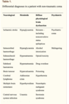
DDx for patient with decreased level of conssciousness?
picture basically 1. Neurological 2. Metabolic 3. Physiological 4. Pshcyatric 5. Traumatic
33
Ix for unconscious patient?
- FBC - blood glucose - U and E - calcium and bone prfile - LFT - clotting screen - toxicology screen [incl. paracetamol, salicylate and blood alcohol level ECG] - ECG - ABG [inlc. CO concentration] - blood cultures for patietns signs infection - CT head - ECG for arrhythmias/cardiac arrest
34
Which drug antidote opioid overdose?
Naloxone
35
What can urine dip look at?
Common causes of taoxins -\> though not sensitive
36
What can patients get like with raised CO2 in repsriatory acidosis?
sleepy and acidotic
37
Why is O2 not given in COPD patient/
Can get acidotic
38
How to give naloxone hcl in OD?
Naloxone Hydrochloride for acute opioid overdose-high-dose regimen [for adult IVI] - Initially 400 micrograms, then 800 micrograms for up to 2 doses at 1 minute intervals if no response to preceding dose, then increased to 2 mg for 1 dose if still no response (4 mg dose may be required in seriously poisoned patients), then review diagnosis; further doses may be required if respiratory function deteriorates following initial response, intravenous administration has more rapid onset of action, doses may be given by intramuscular route but only if intravenous route is not feasible
39
How to give naloxone if can't get IV access?
IV -\> IM [but can take 15m] -\> interosseous
40
Examination finding IVDU?
- Person pale - Marks on arms of IVDU - Blue tint to the lips/fingertips
41
Assuming naloxone Tx successful what is the risk?
- Risk of acute withdrawal -\> headaches, changes BP, rapid HR, sweating, nausea, vomiting, tremors - Can give 200mcg slowly can give in increments if patient okay -\> so don’t get up really quickly - Patient will likely be respiratory acidosis
42
5 head injury pathologoes
Skull fracture Extradural haemorrhage [young person] Subdural haemorrhage [older] COning Laport strcutrre basal skull fracture
43
Where is a common place for extradural haemorrhage?
Middle meningeal artery
44
What interval seen in extradural haemorrhage?
Lucid interval
45
What can be seen in patients with coning on CT?
pressure causing midlines shift
46
Tx for extradural haemorrhage?
Cranioplasty is done -\> vein clampled etc.
47
Subdural haemorrhage Tx?
Burr hole
48
Why in older peoople SdH
reccurence common in chronic subdrual, old and frail - small brain not realised that there's damage
49
What high risk patients SdH?
Cone if on anticoagulants
50
Why can people with SdH not have prominent findings on CT?
Long-term SdH -\> blood not lysed and so it's not white
51
What can subluxation of cervical spine cause?
Quadriplegia -\> driving without seatbelt,
52
Which level of the spine are there breahting problems present if below?
C3/4 -\> Chrostpher Reeve
53
Signs of basal skull fracture
o Blood and CSF leak o Ear o Nose o Panda eyes o Battle sign bleeding middle cranial fossa
54
Which patients do not need CT head injuey?
Very few, basically give to every patient - loads of RFs including GCS of 13 or below, any suspected focal nuerologcial deficit, if on antiocoaulgaiton
55
Which area of the brain hernaties throughin coning?
- Uncal area of temporal lobe -\> herniates through tentorium and that causes dilated pupils - Basal skull fracture
56
Whihc particualr precaution should take all patient head injury?
Full cervical spine immobilization should be attempted for people who have sustained a head injury and any risk factors for cervical spinal injury for example: • Glasgow Coma Scale (GCS) less than 15 on initial assessment. • Neck pain or tenderness. • Focal neurological deficit. • Paraesthesia in the extremities
57
Which Ix should all paitents basically have head injury?
- CT head - CT spine - Blood tests -\> group and save/cross-match if SDH seems likely, in anticipation of operative intervention
58
Patient vomited CT scan how to clear airway?
- Using a suction device to remove vomit from airway - Log roll patient in the CT scanner - Further aspiration can be prevented by intubating patient
59
Indications for intubating patients with head injury?
Indications for intubation - Deterioration LOC - Facial injuries: bleeding into airways - Ventilationary insuffienacy [O2 below 10, CO2 6] - Multiple fits - agitation - GCS below 8
60
What is needed to facilitate intubation?
- RSI [including anaesthetics, muscle relaxants] - Anaesthetists and help
61
What will need to do to ensure safe transfer of patient?
- Should be accompanied by a doctor with appropriate training and experience in transfer patients with acute brain injury, should have dedicated and trained assistant and all equipment needed o Several steps o Multiple check-list - Intubate patient with GCS of 8 or below - Yes, until imaging has confirmed spinal stability - Don’t use hard collar anymore actually
62
Benefits and risks of cervical spine collar?
- Benefits: to spine - Risks: venous outflow obstruction and are a nociceptive stimulus, which might elevate intracranial pressure, uncomfortable
63
DDx for patient collpasing, tingling, lips swelling, hot, feeling anxious/nasueated?
● Anaphylactic reaction to a food allergen ● Syncope ● Stroke ● Seizure ● Panic attack ● Septic shock [don’t have to be hot, can be hypothermic] ● Life-threatening asthma attack For each cause consider, in this case, is it likely, possible or unlikely? ● Anaphylaxis - very likely, especially considering she had just started eating a meal ● Syncope - possible, syncope can really happen whenever. But the prodrome doesn't really seem like syncope ● Stroke - unlikely ● Seizure ● Panic attack - possible, especially because she felt anxious and nauseous before collapsing ● Life-threatening asthma attack - this is a differential in children
64
Name of the rash typically seen allergies
● Urticaria (hives) - maculopapular rash ● Raised, erythematous rash with a pale centre ● Intensely itchy
65
histyory questions to ask in allergy?
● What was the trigger? ● Have they ever had an anaphylactic reaction before? -\> what has that been like when they’ve had it Has the patient been in intensive care before? FH of atopy
66
Systems affected anaphylaxis and waht to expect to see?
● Respiratory - airway smooth muscle constriction, causing stridor (due to laryngeal swelling) and eventually the airways close ○ Also, bronchospasms, giving tachypnoea and SOB, hypoxia, wheezing ● Dermatology - urticaria (not present in 20%) ● Cardiac - hypotension and tachycardia ● GI - inflamed mucus membranes, nausea and vomiting ● Neuro - agitation, confusion and anxiety
67
Mechaniism for anaphylaxis reactions?
● Type 1 hypersensitivity reaction mediated by IgE -\> affecting all systems [ABCDE] ● IgE is released by mast cells and peripheral basophils, causing inflammation
68
Difference between anaplhylactic and anaphylactoid reactions? Does the difference matter in these circumstances?
● An anaphylactic reaction is always IgE-mediated ● An anaphylactoid reaction mimics an anaphylactic reaction but is not IgE-mediatedNo - both are life-threatening conditions which need emergency management with Adrenaline
69
ABCDE apprahc to patient with anaphlyaxis
○ A - ensure they're nursed lying flat so the airway stays open. Remove any food blocking the airway too ○ B - check O2 sats and correct with supplementary O2 if needed ○ C - obtain IV access to give fluids (normal saline) to correct any drop in BP ● Give antihistamines (chlorphenamine) first - Don’t use steroids ● Call for help ● Lay the patient flat ● Raise the patient's legs - this redirects blood flow to the essential organs
70
When is adrenaline indicated?
- First-line treatment for anaphylaxis. Give IM adrenaline early in the anterolateral thigh for airway/breathing/circulation problems: - single dose IM adrenaline well-tolerated and poses minimal risk to an individual having an allergic reaction. If in doubt, give IM adrenaline. - repeat IM adrenaline after 5 minutes if A/B/C persist
71
patient continues to deteriorate what likley cause/
Anaphylactic shock now
72
Route and dose of adrealine for patients?
● \_\_\_\_\_\_\_\_\_\_\_\_\_\_500mcg IM ● This dose can be repeated at 5min intervals ● The preferred site of injection is the anterolateral aspect of the thigh
73
What other tx can be helpful aptients with anaplhyalxis?
● High flow O2 - establish airway ● IV fluid challenge ● Chlorphenamine - salbutamol and bronchodilator - hydroperitonea Don’t give steroids in anaphylaxis
74
Roel of adrenaline IV?
Intravenous (IV) adrenaline must be used only in certain specialist settings, and only by those skilled and experienced in its use. o IV adrenaline infusions form the basis of treatment for refractory anaphylaxis: seek expert help early in patients who’s respiratory and/or cardiovascular problems persist despite 2 doses of IM adrenaline - IV adrenaline without infusion can kill - Seniors: slow dilution
75
Risks of adrenaline?
● The adrenaline needs to be the correct titration, or it will be too much - stroke - VT or tachycardia [serious CV problems]
76
Who should be administering adrenaline by this route?
● Only a specialist. Following the second dose of adrenaline her vital signs stabilise. Within 30 minutes she has returned to normal. Sarah is keen to return home now but her parents are still concerned.
77
What need to consider patients safe discharge anaphlaxis?
Needs to be monitored for 16-24 hours so that you can see if she's having a biphasic reaction. Rebound phenomenon. - though very rare, not seen happen once Mr K
78
Adfvice given to patient upon discharge?
- Give an epi pen - Give advice on how to do use it - Allergy challenge: mast cell tryptase - Refer to allergy clinic
79
Any tests available for Dx of anaphylaxis?
Mast cell triptase When should it be taken?
80
narrow complex tahycardia casues?
Narrow complex tachycardia 1. Exercise 2. SVT: cardiac monitor, valsaval maneavour, carotid massage [young people] -\> adenosine [works on AVN, blocks AVN 30-40s] 3. AF 4. other
81
Commonly used caculation for maximum HR?
220-age
82
broad complex tachycardia causes?
Broad complex tachycardia: - ventricular tachycardia -\> chemical cardioversion [amiodarone], electric cardioversion [defib.]
83
What can lead sodium lead to brain patient? How low does it have to be?
hyponatreamia -\> can be fitting if below 120
84
HSS occurs in whom mianly? Dx of it, and Sx
T2DM Sx Altered level of consciousness Neurologic signs including: blurred vision, headaches, focal seizures, myoclonic jerking, reversible paralysis[5] Motor abnormalities including flaccidity, depressed reflexes, tremors or fasciculations Hyperviscosity and increased risk of blood clot formation Dehydration[5] Weight loss[5] Nausea, vomiting, and abdominal pain[5] Weakness[5] Low blood pressure with standing[5]Dx Plasma glucose level \>30 mmol/L (\>600 mg/dL) Serum osmolality \>320 mOsm/kg Profound dehydration, up to an average of 9L (and therefore substantial thirst (polydipsia)) Serum pH \>7.30[8] Bicarbonate \>15 mEq/L Small ketonuria (~+ on dipstick) and absent-to-low ketonemia (\<3 mmol/L) Some alteration in consciousness BUN \> 30 mg/dL (increased)[5] Creatinine \> 1.5 mg/dL (increased
85
Hypernatreamia numbers?
over 150, not common, can be iatrogenic
86
What to give patients with over 5 potassium?
- Give calcium -\> salbutamol -\> insulin/dextrose
87
Nice guidelines for coma
88
Monro-Kellie homeostasis principle
89
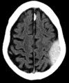
type of haemorrhage?
There is a large crescent-shaped density in the left cerebral hemisphere. It is dark in colour. Crescent = Subdural Dark = Chronic
90
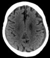
What's the main pathology?
There is a hypoattenuating area in the right frontal lobe (around the middle of the right cortical surface). There is loss of grey-white matter differentiation, and the density is only slightly lower than the surrounding brain tissue, indicating that this is most likely an acute infarct. The ventricles appear asymmetrical because the patient’s head was slightly tilted in the scanner.
91
Whats' the main pathology?
There is a degree of small vessel disease, most clearly seen next to the right lateral ventricle at the top of the image as an area of low attenuation. There is cerebral atrophy, evident by the widened sulci (the gaps between gyri) and the dilated ventricles. The bright areas within the ventricles are the choroid plexus, which have calcifications. This is a very common normal finding. An intraventricular bleed would generally settle at the dependent part of the ventricles. Ventricular enlargement due to hydrocephalus is generally associated with narrow sulci.
92
What's the main patohlogy?
There is a crush fracture of L1 vertebral body, with cortical break and significant loss of vertebral height. The spinal cord does not currently appear to be compressed, as the bright CSF signal on MRI T2 is still visible between the affected vertebral body and the grey cord immediately posterior to it. There is some high signal in the cord however, which could indicate cord oedema from injury to the cord.
93
Non-ctrast Ct scan, history of HTN and sudden onset left facial droop. CT show?
Intracerebral haemorrhage ## Footnote The image shows a large area of high density in the right frontal lobe due to acute bleeding. The CT was non-contrast and so this can’t be an enhancing mass. Its position indicates it is in the brain (intra-axial) rather than extra-axial, so subarachnoid and subdural haemorrhage are ruled out. The focus of high density in the posterior horn of the right lateral ventricle is the calcified choroid plexus, a normal finding.
94
Poor historian with increasing fonusion over severla weeks, what doe simage show?
Bilateral subdural collections ## Footnote The left-sided subdural collection is easy to see. There is also a thin subdural collection on the right. If you see one abnormality, don’t forget to continue viewing the scan systematically.
95
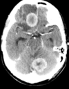
Post contrast CT brain: patient with colon cancer, what is most likely Dx?
Two advancing mets ## Footnote In a patient with a known malignancy a mass in the brain is most likely due to metastatic disease. Two primary lesions occurring simultaneously, benign or malignant, is rare. The image shows enhancement of normal vessels as well as the enhancing masses. There is no evidence of subarachnoid haemorrhage
96
Non-contrasrt CT brian: atnicoagulated patient with sudden onset of confusion and right hemiparesis\> What does image show?
Intraventircular haemoeehage ## Footnote High density material fills the left lateral ventricle. This is blood which extended from an intracerebral haemorrhage of the adjacent peri-ventricular white matter.
97
What doe sthis image show?
Depressed skull fracture ## Footnote This CT brain image on bone window settings shows a depressed skull fracture. Remember to look at both the brain windows and the bone windows on all CT images of the brain.
98
sudden onset severe headache, what does this show?
Subdural haemorrhage ## Footnote Increased density material seen in the subarachnoid space indicates acute haemorrhage. Although subarachnoid bleeding can extend into the ventricles, the ventricles do not contain blood in this image.
99
Alcoholic found unscoius, what does image show?
Extradural haemorrhage ## Footnote This image shows lentiform increased density material indicating acute blood in the extra-axial space (not in the brain). These are the typical appearances of an extradural haematoma.
100
2 CT brian images, pre and post contrast, patient with worsening headache. Which finding corect?
Maas with entral enlargement ## Footnote A large mass in the left cerebral hemisphere is causing mass effect with effacement of the sulci and left lateral ventricle (**asterisk**). Low density surrounding the mass indicates vasogenic oedema of white matter. The mass enhances peripherally but not centrally. The central portion of the mass is avascular due to cell necrosis. **Note:** Abscess is a differential diagnosis
101
2 CT images, pre- and post contrast: patient with worseninfg postrual headacha ena dcerebrellar signs, most likely Dx?
Movement effect ## Footnote This elderly patient presented with confusion and he could not co-operate with instructions to keep still in the CT scanner. The images have been acquired while the patient is shaking his head. Image quality is non-diagnostic. A repeat scan later showed no acute abnormality. The confusion was due to a urinary infection, a much more common cause of confusion in elderly patients than intracranial pathology.
102
2 CT brian images, brain and bone window setting: what does do these images show?
Posterior fossa meningioma ## Footnote These are typical appearances of a meningioma. This enhancing mass arises from the dura mater; in this case from the tentorium cerebelli, a sheet of dura mater which separates the posterior fossa structures from the rest of the brain. The mass compresses the adjacent cerebellum.