Tubulointerstitial Dz Pathology Flashcards
(35 cards)
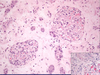
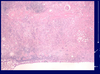
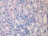
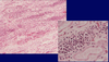
Acute TID; characterized by active inflammation; see eosinophils and lymphocytes (left) and neutrophils (right)

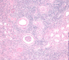
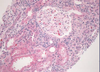
Normal kidney tubules and glomerulus

Tubular necrosis; necrotic cells slough off into the tubular lumen; seen in Acute TID

Edema; seen in interstitium as foamy/vacuoles between tubules. Characteristic of Acute TID

Interstitial fibrosis; seen in Chronic TID; fibrinous tissue is very pale and separates the renal tubules
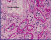
Tubular atrophy; tubules shrink in diameter, the epithelium simplifies and the basement membrane thickens; seen in Chronic TID where tubules atrophy in response to slow ischemia

Chronic inflammation; uniform small lymphocytes which are remnants of a previous active inflammation. Seen in Chronic TID.
Acute TID; active inflammatory cells in interstitium: lymphocytes, neutrophils, plasma cells, macrophages

Chronic TID; fibrosis of interstitium; tubular atrophy
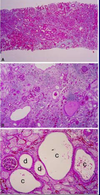
Interstitial Nephritis; inflammation localized to interstitum primarily, with minor involvement of the tubules

Acute interstitial nephritis; infiltration of plasma cells, eosinophils, lymphyocytes, macrophages; also see edema
Acute interstitial nephritis; diffuse infiltration of inflammatory cells; tubules relatively unaffected.
Acute allograft rejection; form of acute interstitial nephritis; where lymphyocytes actually infiltrate the tubules

Acute Tubular Necrosis (main photo); normal photo in bottom Right corner; ATN most commonly affects the Prox. CT and can be caused by ischemia or toxins

Ischemic ATN; caused by shock, hypovolemia, and hypoxemia; usually milder than toxic ATN, the tubular epi. cells fall off individually.

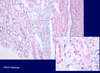
ATN urine sediment; contains epithelial cells and necrotic debris

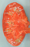
Ascending Acute Pyelonephritis; with white-pus streaks located in the renal medulla.

Acute Ascending Pyelonephritis; with pus-streaks within the medulla; also see papillary necrosis

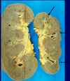
Hematogenous pyelonephritis; has “flea-bitten” appearance
Acute pyelonephritis; neutrophil infiltration

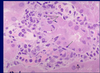
Bacteria in Acute Pyelonephritis; look like pale blue smudges

Fungus (arrow) in Acute Pyelonephritis

WBC casts in the medulla in acute pyelonephritis

Chronic interstitial nephritis; with fibrosis, atrophy and chronic inflammation