Vertebral Column Flashcards
(98 cards)
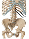
Vertebral Column
Skeleton of neck and back. Functions to support weight, protect spinal cord, serve as axis and pivot, aid posture/movement

What are the differentiated segments of the vertebral column?
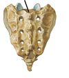
7 Cervical vertebrae, 12 thoracic vertebrae, 5 lumbar vertebrae, 5 sacral vertebrae, variable coccygeal vertebrae (typically 4)
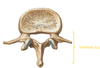
Vertebral Body
Anterior most structure of the vertebral column. Major weight bearing component of the structure. Size of vertebrae varies (increases) as you go down the vertebral column (weight borne on the vertebra significantly increases as you go down the spine)

Vertebral (Neural) Arch
Paired pedicle and lamina of a vertebra.
Pedicle
Paired structures on either side of vertebral foramen. joins vertebral arch and body
Lamina
Paired structures on either side. Flat plates contacting pedicles and spinous process, bound the vertebral foramen.

Vertebral foramen (Canal)
Midline of vertebral column. Houses the spinal cord. Formed by pedicles and lamina.

Superior vertebral notch
Sit above the vertebral arch, immediately superior to the pedicle just posterior to the body. When the vertebrae are articulated, these notches correspond with their superjacent inferior counterpart to form the intervertebral foramen for the spinal nerve

Inferior vertebral notch
deep incisure placed inferior to the more superiorly situated pedicle. Its anterior border is the back of the vertebral body and its posterior wall is the inferior articular process. When the vertebrae are articulated, these notches correspond with their subjacent superior counterpart to form the intervertebral foramen for the spinal nerve

Intervertebral foramen
Formed by superior and inferior vertebral notch; where the spinal nerve roots exit

Transverse processes
Processes (2 per vertebrae) projecting horizontally bilaterally from intersection of lamina and pedicles. Serve as a point of attachment for muscles and ligaments.

Spinous process
Process (1 per vertebra EXCEPT C1) projecting posteriorly and inferiorly from the junction of the laminae. Serves as an attachment for muscles and ligaments.

Zygapophysis/Articular process
4 per vertebra, 2 superior (pre-zygapophyses) and 2 inferior (post-zygapophoses). Act to link vertebrae adjacent together. Spring from junction of pedicles and lamina. superior processes or prezygapophysis project upward from a lower vertebra, and their articular surfaces are directed more or less backward. inferior processes or postzygapophysis project downward from a higher vertebra, and their articular surfaces are directed more or less forward and outward

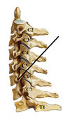
Typical Cervical Vertebra: Transverse Foramen
Located on either side of the body. Vertebral arteries run C2-C6, enter through foramen magnum to reach the brain (become basillar artery). C7 has transverse foramen, but the artery does not run through it! (Runs along it)

Typical Cervical Vertebra: Anterior and Posterior tubercles
The transverse processes are divided into an anterior and posterior portion, termed the anterior and posterior tubercles. Anterior portion is analagous to the rib, posterior portion is more analagous to “true” transverse process

Carotid Tubercle
Anterior tubercle of C6. Carotid artery runs alongside it. Carotid artery can be compressed at this point easily to occlude bloodflow.
Typical Cervical Vertebra: Vertebral Foramina
large for C3 – C7 due to cervical enlargement of spinal cord
Typical Cervical Vertebra: Uncinate Process/Uncovertebral joint
Raised margins of superior border of body. Form saddle-shaped joint on C3-C7 and T1. Prevents a vertebra from sliding backwards off the vertebra below

Spinous Process of C7
Vertebra prominens; very long and prominent.

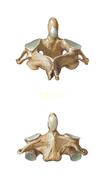
Atlas
C1. Articulating surfaces for articulation with the occipital condyles (superior articulating surfaces) and Axis (Inferior articulating surface). LACKS A VERTEBRAL BODY; instead has an anterior and posterior arch (appears as a ring). Groove for vertebral artery on superior surface

Axis
C2. Superior articulating surface (articulation with C1). Dens/odontoid process: former body of C1, allows pivoting for skull
Dens
Odontoid process. Allows for pivoting of skull. Enlarged spinous process of C2. Articulates with C1.

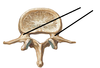
Typical Thoracic Vertebrae: Superior Costal Facet
Location where rib forms articulation with the top of a vertebra. Superior costal facet is located on the inferior thoracic vertebrae; inferior costal facet is located on the superior vertebrae. While these terms may be confusing, it helps to know that the costal facets are named for their position on the vertebral body itself, NOT for the part of the rib that they articulate with

Typical Thoracic Vertebrae: Inferior Costal Facet
Location where rib forms articulation with the top of a vertebra. Inferior costal facet is located on the superior vertebrae; superior costal facet is located on the inferior thoracic vertebrae;. While these terms may be confusing, it helps to know that the costal facets are named for their position on the vertebral body itself, NOT for the part of the rib that they articulate with