VIVA – Anatomy – Eyes Flashcards
(11 cards)
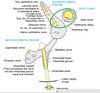
Draw the layers of the upper eyelid

Draw and label the superior orbital fissure
What are the attachments of the orbital septum?
- Laterally, the septum is attached to the orbital margin, 1.5 mm in front of the lateral orbital tubercle attachment of the lateral palpebral ligament à Eisler fat pocket separates the lateral palpebral ligament from the orbital septum
- from there, the septum continues along the superior orbital rim at the arcus marginalis
- superomedially, the septum bridges the supraorbital groove, passes inferomedially anterior to the trochlea, and then follows the posterior lacrimal crest. As it runs down the posterior lacrimal crest, it lies anterior to the medial check ligament and posterior to the Horner muscle (and hence, behind the lacrimal sac).
- line of attachment crosses the lacrimal sac fascia to reach the anterior lacrimal crest at the level of the lacrimal tubercle
- from there, it passes inferiorly down the anterior lacrimal crest and laterally along the inferior orbital rim
- a few millimeters lateral to the zygomaticomaxillary suture, the attachment leaves the rim and lies several millimeters from it on the facial aspect of the zygomatic bone, thus forming the fat-filled premarginal recess of Eisler. The line of attachment then continues to again reach the lateral orbital rim, just below the level of the Whitnall ligament.
- blends with some fibres of levator palpebrae superioris superiorly

What are thickenings of orbital septum?
- Thickened at lid margins as tarsal plate
- Dense fibrous tissue
- Crescent shaped that curve with the eyeball
- Eyelashes attach to tarsal plates
- Contain Meibomian Glands
- Modified sebaceous glands
- Secrete oily substance minimising tear evaporation
- Palpebral conjunctiva densely adherent to tarsal plates
- Orbicularis oculi is anterior to the tarsal plate
- Attaches to anterior lacrimal crest and margins of orbit
- Septum thickens here as the medial palpebral ligament
- Anchors tarsal plates to anterior lacrimal crest
- Lateral palpebral ligament is thinner and fuses with raphe of orbicularis oculi
- Attaches to marginal tubercle of Zygomatic bone
- Palpebral fissure sits between the lids
Draw the visual pathway

What visual field defects are associated with lesions at each level?

Where do the parasympathetic and sympathetic fibres run within CN III?
- Superior division carries sympathetic fibres from the cavernous plexus to the visceral-muscle part of the levator
- Inferior division gives off nerve to inferior oblique which gives off parasympathetic root to ciliary ganglion à cells bodies in Edinger Westphal nuclei
How do you test each extraocular muscle?
The initial clinical examination of the extraoccular eye muscles is done by examining the movement of the globe of the eye through the six cardinal eye movements.
When the eye is turned in (nasally) and horizontally, the function of the medial rectus muscle is being tested.
When it is turned out (temporally) and horizontally, the function of the lateral rectus muscle is tested.
When turning the eye down and out, the inferior rectus is contracting.
Turning the eye up and out relies on the superior rectus.
Paradoxically, turning the eye up and in uses the inferior oblique muscle, and turning it down and in uses the superior oblique.
All of these six movements can be tested by drawing a large “H” in the air with a finger or other object in front of a patient’s face and having them follow the tip of the finger or object with their eyes without moving their head.
Having them focus on the object as it is moved in toward their face in the midline will test convergence, or the eyes’ ability to turn inward simultaneously to focus on a near object
In isolation, what movements do the following muscles produce?
Superior oblique?
Down and out
In isolation, what movements do the following muscles produce?
Superior rectus?
Up and in
