VIVA – Anatomy – Neck Flashcards
(266 cards)
What are the branches of the ECA?
Superior thyroid (ant)
Ascending pharyngeal (medial)
Lingual (ant)
Facial (ant)
Occipital (post)
Post-auricular (posterior)
Superficial temporal and maxillary are terminal
What is your technique for ligation?
Horizontal skin incision at level of hyoid/SMG in skin crease – post 1/3 over SCM
Subplatysmal flaps
Identify ant border of SCM and retract posteriorly
Carotid sheath identified
Vascular loop around common carotid
Identify hypoglossal crossing
ECA usually anterior and superficial to ICA
ICA does not branch in the neck à identify 2 branches of ECA
Ligate ECA between superior thyroid and lingual branches – if ligate lower may cause clot that can migrate proximally
?Need to ligate facial vein
What structures pass between ECA and ICA?
Glossopharyngeal nerve
Pharyngeal branch of vagus
Stylopharyngeus muscle
?Styloglossus
Styloid + stylohyoid ligament
Deep lobe of parotid
Branchial fistula tract
What are the course and branches of Superior Thyroid Artery (Ant)
- Arises at commencement ECA from anterior surface
- Can arise from CCA
- Usually the first branch
- Runs nearly vertically downwards and forward with associated vein
- Enters upper pole thyroid
- External laryngeal nerve closely associated
- Branches
- Infrahyoid
- Superior laryngeal artery
- Pierces thyrohyoid membrane with internal laryngeal nerve
- Branch to SCM
- Supplies
- Adjacent muscles
- Larynx
- Thyroid gland
What are the course and branches of Ascending Pharyngeal (Post)
- Arises just above commencement ECA
- Smallest branch of ECA
- Arises from posterior surface
- In 14% arises from the occipital artery
- Ascends vertically anteromedial to the ICA along side wall pharynx
- Anterior to prevertebral fascia
- Supplies pharyngeal wall, soft palate, tonsil, inferior tympanic branch
- Meningeal branches to foramen lacerum, jugular foramen, hypoglossal
- Branches
- Posterior Meningeal
- Pharyngeal
- Inferior tympanic
What are the course and branches of Lingual Artery (Ant)
- Arises above superior thyroid
- Usually at or above the level of the hyoid
- Runs up then along the upper border greater horn hyoid (or above)
- Deep to digastric and stylohyoid
- Lies against lateral wall of pharynx to pass medial to the posterior border hyoglossus
- Runs with deep lingual vein to tip of tongue
- Crossed by hypoglossal nerve and facial vein
- Branches
- Suprahyoid
- Sublingual
- Dorsal Lingual
- Deep Lingual
What are the course and branches of Facial Artery (Ant)
- Arises above lingual
- Sometimes via a common linguofacial trunk
- At approximate level of digastric / angle of mandible
- Runs upwards and forwards on superior constrictor
- Deep to digastric and stylohyoid muscles but then hooks around posterior belly of digastric to reach upper surface submandibular gland
- Makes an “S” bend curling down over submandibular gland and up over mandible at anterior border of masseter
- Anterior to the facial vein
- Runs upwards and medially to the side of the nose as the angular artery towards the medial canthus
- Deep to platysma / Risorius / Zygomaticus major and minor
- Superficial to buccinators
- Landmarks for FAMM flap
- Based anterior to stensons duct
- Use a doppler
- Branches
- Cervical Branches
- Ascending Palatine
- Tonsillar – given off as lies on superior constrictor à tonsil and soft palate
- Branches to SMG
- Submental artery à given off just before crosses inf border of mandible à runs along inf surface of mylohyoid, between it and digastric à ant belly digastric, mylohyoid and sublingual/FOM
- Muscular branches to muscles of mastication
- Facial Branches
- Superior and Inferior Labial Arteries
- Each divides into two branches
- Both run beneath vermillion margin of lip
- Anastomose end to end at the midline of the lip
- In or behind the deep fibres of orbicularis oris
- Hence fairly superficial on labial side
- Tortuous
- Angular Artery
- Terminal portion of facial artery
- Gives off a lateral nasal artery which can anastomose with the dorsal nasal branch (ophthalmic –ICA)
- Multiple anastomoses with STA / IMA / Ophthalmic / Contralateral Facial
- Superior and Inferior Labial Arteries
- Cervical Branches
What are the course and branches of Occipital Artery (Post)
- Arises from posterior ECA at same level as facial artery
- Crossed at origin by hypoglossal nerve
- SCM branch passes anterior to nerve and “holds it down”
- Passes along lower border posterior belly digastric
- Grooves base of skull at occipitomastoid suture
- Passes back through apex of posterior triangle
- Runs with greater occipital nerve
- Supplies back of scalp
- Branches
- 2 to SCM
- Upper is a guide to accessory nerve
- Mastoid
- Auricular
- Meningeal
- Occipital
- Descending
- 2 to SCM
What are the course and branches of Posterior Auricular Artery (Post)
- Arises above level of digastric
- Can arise within parotid
- Superficial to styloid process
- Passes along upper border posterior belly digastric
- Crosses surface of mastoid
- Runs with lesser occipital nerve
- Branches
- Auricular to pinna and ear
- Stylomastoid branch
- Supplies facial nerve
- Gives off stapedial artery
- Posterior tympanic – Mastoid and stapedial
- Occipital
What are the course and branches of Superficial Temporal Artery
- Terminal branch of ECA
- Runs upwards deep to TMJ
- Crosses posterior root of zygomatic arch
- Branches
- Parotid
- Transverse facial artery near commencement
- Runs just above parotid duct
- Anterior auricular
- Zygomatico-orbital
- Middle temporal artery
- Runs vertically deep to temporalis
- Causes groove in squamous temporal bone
- Anastomoses with deep temporal branches of Internal maxillary artery
- Frontal
- Parietal
What are the landmarks for finding the facial artery for a FAMM flap?
?
What is the blood supply to the SCM?
Upper 1/3 – branches from occipital artery
Middle 1/3 – branches from superior thyroid or EJV itself or both
Lower 1/3 – suprascapular
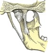
Describe the course and segments (7) and branches of the ICA
- Course
- Lateral to ECA initially
- Slopes up and posterior to move to a medial position
- No branches
- Enters carotid canal at base of skull
- Curves upwards from foramen lacerum to enter posterior aspect cavernous sinus
- Between Sphenoid endosteum and inner layer of dura
- Arches up then forwards in medial wall cavernous sinus
- Pierces roof of sinus medial to anterior clinoid process
- Accompanied by SNS fibres (Internal Carotid Nerve)
- Curves backwards along roof of cavernous sinus
- Then curves upwards lateral to the optic chiasma
- Divides into terminal branches at anterior perforated substance
- ACA
- Passes forwards above the optic nerve
- Anterior Communicating Artery
- Lies in chiasmatic cistern
- ACA
Other branch = ophthalmic – commences as ICA emerges from roof of cavernous sinus
Segments
- cervical
- petrous
- lacerum
- cavernous
- clinoid
- ophthalmic
- communicating
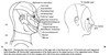
What is the relationship of the petrous segment to the cochlea?
The cochlea lies posterior and superior to the petrous carotid in the ant part of the otic capsule
How do you manage tumour involving the cervical ICA?
- Carotid Artery involvement
- Pre-op imaging
- encased carotid defined is on radiology tumour encircles >270 degrees - unresectable
- concern for involvement if wrapping around more than 180, loss of fascial plane between tumour and carotid on MRI, less than 1.8mm of separation
- 50% of those between 180-270 degrees resectable
- balloon occlusion test – if performed without neuro deficit, can go on to resect carotid segment
- resection still has significant morbidity and mortality associated – 60% neurological and up to 40% mortality
If unexpected involvement
- Abandon
- Shave tumour from vessel
- Sacrifice
- See if vascular surgeon available
- Clamp carotid
- Measure stump pressure via transducer (with art line)
- Pressure > 70 resect without reconstruction
- 55-70 resect + reconstruct
- <50 reconstruct + temporary shunt
- Sacrifice as close to skull base as possible (reduce clot propagation)
- Post op heparin SC
- Reconstruction options
- Saphenous vein
- Gortex or Dacron graft
If the ICA is injured during surgery what is your management? Cervical (neck surgery)
ABC - Alert anaesthetist etc
Control proximally with vessel-loop – is there pulsatile back flow from cranial end suggesting intact circle of Willis (90%)?
Options – ligate, repair, temporary shunt
Do not ligate without preop testing (30% stroke)
Call for help (senior surgeon, vascular)
Gain proximal + distal control + vessel loops +/- reconstruction +/- covering with muscle flap, esp. if irradiated
Strict BP maintenance above 110 mmHg (Note: 90% mortality if not maintained)
10% stroke
Often need to sacrifice vagus (+/- permanent trachy + PEG)
Repair if possible
?Javert shunt + ring clamps
Need heparin
Vascular surgeon
Great saphenous vein is good size match
Resection
Incisions above and below vessel
Clamp vessel above and below
Resect medial clavicle if need to get further proximal
Ligate with 0 silk
Post op S/C heparin
Stroke 50%, death 25%
If the ICA is injured during surgery what is your management? Cavernous (FESS)
- Obliterates visualization
- ABC’s
- Inform anaesthetist
- 2 large bore IVC and Immediate fluid resus
- G&H / Crossmatch and order blood
- Lower BP if possible for 1-2min
- Call for help / 2 surgeon technique
- Use the posterior septum to shield the endoscope from the jet of blood
- Use lens cleaning system
- Large calibre suction (12Fr or greater) x2 in contralateral nostril
- Minimise / Control Bleeding
- Head Up
- Ipsilateral CCA compression to slow
- Vascular clamps (wormald) for atraumatic clamp of injury
- Maximise exposure if bleeding controlled with clamps
- “U clip” anastomotic device
- Pack and Pressure
- ? Material
- PJ recommends muscle from SCM in his work (10x10mm)
- Crushed and placed over injury site
- Floseal / surgiflo useful only for venous bleeding
- Intervential radiology / Vascular surgery / Tie off ICA?
- Follow-up
- Needs an angiogram in the flowing months to assess for aneurysm formation
When can the ICA be safely ligated?
Never completely safely – Rutherford vascular surgery says ligation results in 45% mortality and should be reserved only for injuries at BOS that are not amenable to recon
Safer in younger pts and passing balloon occlusion test à Inflated for 20 min, monitor for any change in neuro function, if no changes deflate +/- additional imaging (SPECT, single photon emission CT)
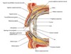
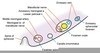
Describe the constrictions of the esophagus?
15cm – cricopharyngeus
23cm – arch of aorta
27cm – L main bronchus
38cm – diaphragmatic hiatus
Blood supply esophagus?
- Inferior thyroid arteries = Upper oesophagus
- Bronchial arteries from Aorta
- Oesophageal branches (L) Gastric artery
- Venous Drainage
- Upper part to brachiocephalic veins
- Middle part to azygos system
- Lower part to (L) Gastric vein à portal vein
- Anastomosis between portal and systemic circulation
Nerve supply esophagus? Extrinsic
- Upper Oesophagus
- RLN
- Middle ganglia cervical SNS ganglia within inferior thyroid arteries
- Middle and Lower Oesophagus
- Thoracic sympathetic trunks
- Greater Sphlancnic nerves
- Vagus (PNS)
- Plexus forms over oesophagus
- Develops into anterior and posterior vagal trunks in lower oesophagus
- Anterior is mainly (L) vagus, posterior (R)
- Motor supply
- Vagus
- Nucleus ambiguous (Upper striated part)
- Dorsal motor nucleus (Lower part)
- Provides secretomotor innervation
- Vagus
Nerve supply esophagus? Intrinsic
Lower 2/3 smooth muscle
- preganglionic = vagus
- postganglionic = myenteric plexus (Auerbach’s) – between inner circular and outer longitudinal
- VIP neurons relax (NO), cholinergic contract
What is Achalasia?
Distal oesophageal flaccidity associated with failure of LOS relaxation
Primary – idiopathic degeneration of ganglion cells in Auerbach’s plexus à loss of post-ganglionic inhibitory neurons à unopposed cholinergic activity
Secondary – carcinoma, CVA, Chaga’s disease, post vagotomy, diabetic autonomic neuropathy, infiltrative disorders
Achalasia - What does it look like on Ba Swallow?
Classic bird beak’s deformity, failure of peristalsis, air-fluid level in upright position