Week 1 Flashcards
(183 cards)
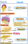
<p><p><p><p><p>Free nerve ending stimuli, rapid or slow</p></p></p></p></p>
<p><p><p><p><p>Pain (fast and slow) Crude touch Pressure Heat and cold Rapid</p></p></p></p></p>
<p><p><p><p><p>Markel cells stimuli, rapid or slow</p></p></p></p></p>
<p><p><p><p><p>In hairless skin Stimuli is pressure (touch)
| Slow</p></p></p></p></p>
<p><p><p><p><p>Hair follicle receptor stimuli, rapid or slow</p></p></p></p></p>
<p><p><p><p><p>Touch
| Rapid</p></p></p></p></p>
<p><p><p><p><p>Meissner corpuscle stimuli, rapid or slow</p></p></p></p></p>
<p><p><p><p><p>Light flutter (touch, feather)
| Rapid</p></p></p></p></p>
<p><p><p><p><p>Pacinian corpuscle stimuli, rapid or slow</p></p></p></p></p>
<p><p><p><p><p>Vibration
| Rapid</p></p></p></p></p>
<p><p><p><p><p>Ruffini corpuscle stimuli, rapid or slow</p></p></p></p></p>
<p><p><p><p><p>Stretch your leg ورفسيني slowly
| Stretch</p></p></p></p></p>
<p><p><p><p><p>Forebrain consists of</p></p></p></p></p>
<p><p><p><p><p>cerebrum, Diencephalon</p></p></p></p></p>
<p><p><p><p><p><p><p><p><p>Hindbrain consists of</p></p></p></p></p></p></p></p></p>
<p><p><p><p><p><p><p><p><p>pons,
Medulla oblongata,
cerebellum</p></p></p></p></p></p></p></p></p>
<p><p><p><p><p><p><p><p><p>Brainstem consists of</p></p></p></p></p></p></p></p></p>
<p><p><p><p><p><p><p><p><p>Midbrain,
pons,
Medulla oblongata</p></p></p></p></p></p></p></p></p>
<p><p><p><p><p><p><p><p><p>Central sulcus separates which lobes?</p></p></p></p></p></p></p></p></p>
<p><p><p><p><p><p><p><p><p>frontal lobe from parietal lobe</p></p></p></p></p></p></p></p></p>
<p><p><p><p><p><p><p><p><p>lateral sulcus</p></p></p></p></p></p></p></p></p>
<p><p><p><p><p><p><p><p><p>aka sylvian sulcus
| separates frontal and parietal lobes from temporal lobe</p></p></p></p></p></p></p></p></p>
<p><p><p><p><p><p><p><p><p>corpus callosum function</p></p></p></p></p></p></p></p></p>
<p><p><p><p><p><p><p><p><p>connects the two hemispheres together and it's made from white matter</p></p></p></p></p></p></p></p></p>
<p><p><p><p><p><p><p><p><p>Grey matter</p></p></p></p></p></p></p></p></p>
<p><p><p><p><p><p><p><p><p>consists of neuronal cell bodies &amp;amp;amp;amp;amp;amp;amp;amp;amp; forms the cortex</p></p></p></p></p></p></p></p></p>
<p><p><p><p><p><p><p><p><p>White matter</p></p></p></p></p></p></p></p></p>
<p><p><p><p><p><p><p><p><p>consists of the myelinated neuronal fibers (axons)</p></p></p></p></p></p></p></p></p>
<p><p><p><p><p><p><p><p><p>the cortex is separated by</p></p></p></p></p></p></p></p></p>
<p><p><p><p><p><p><p><p><p>fissures (sulci) (depression)</p></p></p></p></p></p></p></p></p>
<p><p><p><p><p><p><p><p><p>What's insula?</p></p></p></p></p></p></p></p></p>
<p><p><p><p><p><p><p><p><p>it's a region of the cerebral cortex located deep within the lateral sulcus
it's made up of grey mater</p></p></p></p></p></p></p></p></p>
<p><p><p><p><p><p><p><p><p>What separated the two hemispheres?</p></p></p></p></p></p></p></p></p>
<p><p><p><p><p><p><p><p><p>longitudinal cerebral fissure</p></p></p></p></p></p></p></p></p>
<p><p><p><p><p><p><p><p><p>what is Diencephalon made up of?</p></p></p></p></p></p></p></p></p>
<p><p><p><p><p><p><p><p><p>1) Thalamus
2) Hypothalamus: (has mammillary bodies)
3) Subthalamus:
4) Epithalamus (has pineal body)</p></p></p></p></p></p></p></p></p>
<p><p><p><p><p><p><p><p><p>grey mater makes what in the hemispheres?</p></p></p></p></p></p></p></p></p>
<p><p><p><p><p><p><p><p><p>ganglion or nucleus
- caudata nucleus
- lentiform nucleus
- the cerebral cortex is also formed by grey mater</p></p></p></p></p></p></p></p></p>
<p><p><p><p><p><p><p><p><p>cavities of the CNS and their locations</p></p></p></p></p></p></p></p></p>
<p><p><p><p><p><p><p><p><p>a) 2 Lateral ventricles: in the cerebral Hemispheres
b) Third ventricle: between the
2 diencephalon
c) Fourth ventricle: between Pons, medulla and cerebellum (in hind brain)
d) Cerebral aqueduct: in midbrain Central canal of spinal cord</p></p></p></p></p></p></p></p></p>
<p><p><p><p><p><p><p><p><p>What's the function of Cerebral aqueduct?</p></p></p></p></p></p></p></p></p>
<p><p><p><p><p><p><p><p><p>connects 4th and 3rd ventricles</p></p></p></p></p></p></p></p></p>
<p><p><p><p><p><p><p><p><p>Where can you find CSF? (cerebrospinal fluid)</p></p></p></p></p></p></p></p></p>
<p><p><p><p><p><p><p><p><p>in CNS cavities and Subarachnoid space</p></p></p></p></p></p></p></p></p>
<p><p><p><p><p><p><p><p><p>What are LMN (lower motor neurons) and where do you find them?</p></p></p></p></p></p></p></p></p>
<p><p><p><p><p><p><p><p><p>nerves that control muscles directly e.g. spinal nerves and cranial motor nerves
In the spinal cord and brain stem
Their axons innervate directly the
striated muscles of the body and head respectively</p></p></p></p></p></p></p></p></p>
<p><p><p><p><p><p><p><p><p>What are UMN (upper motor neurons) and where do you find them?</p></p></p></p></p></p></p></p></p>
<p><p><p><p><p><p><p><p><p>neurons that come from the brain cortex and brain stem to control the LMN
their cell Bodies Lie in the cerebral cortex and brain stem The axons of UMNs form descending motor pathways</p></p></p></p></p></p></p></p></p>
receptors for temperature, pain, itch, Pressure/touch, Position sense:
- temperature: Thermoreceptors - pain: Nociceptors - itch: Chemoreceptors - Pressure/touch: mechanoreceptors (respond to distortions in skin eg. phone vibration) - Position sense: Proprioceptors (obstacle while walking, we can avoid it by moving our legs without looking down)
What are Somatosensation
All modalities other than seeing, hearing, tasting, smelling, and vestibular balance. يعني يكون شي عام and they are scattered all over the body
What's transduction and transmission?
- transduction: encoding of stimuli into electrical signals (translation) - transmission propagation of this electric signals to CNS
What's adequate and inadequate stimuli?
if a receptor is more selective (specific) for a single stimulus energy – its adequate stimulus. Differential sensitivity: receptors have a LOW threshold for the adequate stimulus, and a HIGH or no threshold at all to others (inadequate stimuli) Ex: photo receptors are activated by light, that is their adequate stimulus. When you’re being hit on the eye, you can also see light and this is because the high intensity of the stimulus causes the activation of the photo receptors, that would be the inadequate stimuli.
What's receptor (generator) potential?
Change in membrane potential because of a stimuli that allowed ions to diffuse through channels if stimulus is strong, receptor potential reaches AP threshold, AP is generated
ncreasing amplitude of the generator potential results in?
increases in the frequency of AP
What adapts the receptor or the neuron?
The receptor
What are phasic receptors?
- aka dynamic receptors - alert us to CHANGES in sensory stimuli - responsible for our ability to cease paying attention to constant stimuli (aka sensory adaptation) - like wearing a ring, you only feel the pressure when you put it on (put stimuli) and take it off (stimuli removed which causes another receptor potential) - it's RAPID adaptation - Pacinian’s corpuscle, Meissner’s corpuscle

What are Tonic receptors?
- aka static receptors - SLOW adaptation - don't disappear but decrease with time, it only disappears when you remove the stimuli (Generate AP throughout, but diminish slowly. Give continuous info about stimulus) - Imp for when receptors tell you about muscle movements. Eg: when writing, how your finger is continuously moving with movements. If it was the same position for a while, (static) then AP firing could decrease - Proprioceptors, nociceptors, merkel cells

Receptors and their stimuli
Whats the No adaptation receptor?
- Tonic receptors (static receptor) - eg. some Nociceptors - Imp because you want to constantly know that there’s something damaging to the brain (you don’t want to forget about it)
Stimulus intensity coding

Mechanoreceptors
- Respond to distortion of the membrane eg. stretch - eg. lining of the stomach when full or bladder distention or lung inflation - pressure (membrane stretching) cause opening of NA+ channels causing generator/ receptor potential - Direct pressure on skin and/or high-frequency vibration detected by Pacinian corpuscles.
difference btw direct and indirect activation of mechanoreceptors
- Direct: stretching the channel itself OR through structural proteins that are part of the channel (intra or extra cellular proteins) - Indirect: through membrane structural proteins (protein is NOT related to channel eg. 2nd messengers)
What's the pressure receptor
Markel receptor: - Sustained touch, texture, pressure - sense a touch that lasts longer - SLOW adaptation - eg. Braille (blind people text) - superficial in glabrous skin (skin that doesn't have hair)
What's light touch/ flutter receptor
Meissner Corpuscle: - Changes in light touch, stroke, Flutter hence -> - FAST adapting - superficial in glabrous skin (skin that doesn't have hair)

What's vibration receptor
Pacinian Corpuscle: | - FAST adapting
What's stretch receptor
Ruffini Ending: - skin stretch, sustained pressure - eg. when holding a big object, your hands are being stretched, collagen fibers will stretch, cause afferent that will let your brain know that you’re holding something thats stretching your hand - SLOW adapting

What's another flutter, light touch receptor
Hair follicle: - flutter, light touch - in hairy skin unlike meissner corpuscle - eg. when wind blow on your skin - FAST adapting

How to test Proprioception?
Proprioception = position sense 1. with eyes closed know wether you're moving their fingers/ toes up or down 2. Romberg test: with eyes closed, if their proprioception is not intact, then they will sway, and when they open their eyes, the swaying will stop because vision compensates for proprioceptive loss.
What stimulates muscle spindle?
stimulated by stretch > fibers elongate > sensed by nerve endings A1 and II (2) > fire AP
What do gamma motor and alpha motor control?
- Gamma motor: control spindle sensitivity - alpha motor: muscle contraction of skeletal muscles
What does 1b afferent do and where is it
- in Golgi tendon organ - it signals muscle tension to CNS - the axon of 1b is intertwined with the collagen fascicles - When the Golgi tendon organ is stretched (usually because of contraction of the muscle), the Ib afferent axon is compressed by collagen fibers and its rate of firing increases.
Transduction by Chemoreceptors
For visceral sensations: - PO2 , PCO2 receptors - Hunger: food molecules activate hypothalamic chemoreceptors - Thirst: osmoreceptors ``` For pain: - Lactic acid when exercising open H+ gated ion channels on nociceptive neural endings For itch: - histamine
Thermoreceptors
Transient receptor potential channel (TRP) each one has different sensitivity: - TRPV3 in chilli - TRPV4 in chilli (for both, Adequate stimuli: temperature inadequate stimuli: vanilloid in chilli) - TRPM8 in menthol (nonselective cation channel expressed in small diameter trigeminal and dorsal root ganglion neurons in which cooling and menthol evoke inward depolarizing currents and intracellular calcium rises
Nociceptors
- For pain - FREE nerve endings respond to stimuli and damaged tissue Three types: 1. Thermal nociceptors 2. Mechanical nociceptors 3. polymodal nociception
polymodal nociception:
High intensity mechanical, chemical or thermal (v. hot or v. cold) stimuli UNMYELINATED C axons that conduct more SLOWLY (dull, burning pain, diffusely localised, poorly tolerated). ``` TRPA1 TRPV1 TRPV2 MS ASIC (for acid accumulation)
```Mechanical nociceptors:
ntense pressure to skin thinly myelinated axons stabbing, squeezing, pinching
Thermal nociceptors:
activated by extreme temp (v. high or v. low) in peripheral endings of small diameter, thinly myelinated
somatosensory fibers are fast when
big in diameter and myelinated

Proprioception, mechanoreceptors, nociceptors, thermoreceptors which are myelinated which are not?
myelinated: Proprioception and mechanoreceptors NONmyelinated: nociceptors, thermoreceptors

Lower motor neuron lesions features:
- Decreased (flaccidity) - weakness and w time there will be muscle atrophy - Hyporeflexia (decreased reflex) - Hypotonia (decreased muscle resistance to passive movement) - Fasciculations (muscle contraction seen as skin flickering but not strong enough to move the limb) - found in Ant horn cell - eg. Polyneuropathy, (GBS) gullian barre syndrome - flexors and extensors equally affected - caused by: 1- Poliomyelitis 2- Spinal muscular atrophy 3- Amyotrophic lateral sclerosis (ALS)
Upper motor neuron lesions features:
- Increased tone (spasticity) - Hypertonia - hypereflexia - Above anterior horn cell in spinal cord - eg. stroke, multiple sclerosis - Early UMNL manifest as LMNL ( you don't get stroke right away when there is an UMNL) - extensors are weaker in UPPER limbs (triceps) - flexors are weaker in LOWER limbs - +ve babkinski - +ve clonus - +ve hoffman sign - superficial (abdominal reflexes) are absent (decrease)
Describe Poliomyelitis
- Caused by Polio virus causing anterior horn cells destruction - mostly asymptomatic - few have LMN picture - Diagnosis: RNA of virus in CSF
Describe Spinal muscular atrophy
- Caused by genetics (autosomal recessive) - progressive weakness &amp; atrophy of muscles - Treated by Antisense oligonucleotide
Describe Amyotrophic lateral sclerosis (ALS)
- stephen hawking! - It's progressive, pattern of weakness affects right arm then left arm then left leg then right leg - Affects motor neurons in cerebral cortex and brain stem (UMNL) as well as anterior horn of spinal cord (LMNL)
in Peripheral neuropathy what does each of the following affect? - Neuron-opathy - Plexopathy - Motor neuron disease - Radiculopathy - Peripheral neuropathy
- Neuron-opathy = dorsal root ganglion - Plexopathy = plexus - Motor neuron disease = cell body in anterior horn - Radiculopathy = Root - Peripheral neuropathy = peripheral nerve
Describe Neuronopathy (NOT neuropathy) and an example
- Degeneration of dorsal root ganglia &amp; projections - Diseases affecting the neuron cell body Divides to: - Motor neuron disease (anterior horn affected) - Sensory neuronopathy - Ganglionopthy - eg. herpes zoster (shingles)
Describe Radiculopathy and an example
- affects spinal nerve ROOT - classic presentation is pain, if in the neck it's brachial plexus, or in lower back and goes to lower limbs - weakness in muscles supplied by that root - radiating pain along the root dermatome - DECREASED deep tendon reflex (eg. knee jerk reflex) in corresponding root ONLY - caused by compression eg. herniated disc (nerve root compressed, muscles supplied by that root damaged
Describe plexopathy
- affects plexus (Brachial or Lumbosacra) - affects sensory and motor (shows both symptoms) - weakness and numbness - Caused MOSTly by diabetes mellitus, can be caused by focal mass (pancoast tumor, a lung tumor that affects brachial plexus) - eg. Hornor's syndrome, happens bcz of disturbtion of brachial plexus (C8 - T1)
Describe Peripheral neuropathy
- Causes are either Hereditary or Acquired - Hereditary -> Charcot marie tooth disease (IMPORTANT) - Acquired (MINI-P): Metabolic or medication (diabetes, vit B12 def., chemotherapy) Immune Neoplastic Infections Physical (compression)
Describe Mononeuropathy w two examples
- one peripheral nerve affected - cranial or spinal nerve - caused by trauma, compression or entrapment - loss of function in part supplied (motor, sensory, &amp;/or autonomic) - eg. spinal nerve example, Carpal tunnel syndrome -> numbness in lateral 31⁄2 digits +/- weakness -> examined by phalen sign and tinel sign - eg. Cranial nerve 7 -> bell's palsy -> weakness in face muscle, muscle not moving - treated w/ NSAID, steroids, surgery
Describe Polyneuropathy
- Symmetric - Length dependent - Long fibers affected first (leg fibers first) - Starts at the toes and continues going up until it reaches the hands - example: patient came w/ numbness that started in feet (both sides) toothpicks then started to go to ankles then in both hands
Describe Mononeuropathy multiplex w/ examples
- More than one peripheral nerve affected at the same time or one after the other - ASYMMETRIC - affect cranial or spinal n. - examples: 1. Vasculitis of vasa nervorum 2. Sarcoidosis 3. Diabetes mellitus - Symptoms are motor (weakness) (LMNL features) and sensory (numbness is most imp) divides to Demyelinating and Axonal neuropathy
What are Demyelinating and Axonal neuropathy
- they're both divisions of Polyneuropathy 1. Demyelinating neuropathy: - loss of myelin so signal will be slower - causes: common in DIABETES MELLITUS 2. Axonal neuropathy: - damage occurs to axon itself so signal is not able to pass at all - causes: Guillain-Barre syndrome (GBS) + mostly autoimmune
Which of the following causes of weakness characterized by fasciculations? 1. Stroke 2. Brain tumor 3. Amyotrophic lateral sclerosis 4. Basal ganglia calcifications
3. Amyotrophic lateral sclerosis ALS, cause it's mixed
A 60-year old man previously healthy presented with right sided weakness of 2 hours duration. MRI showed acute stroke. which of the following is NOT expected in this man? 1. Weakness 3/5 in right arm 2. Difficulty speaking 3. Hyperreflexia with spasticity 4. Hypotonia in right leg
3. Hyperreflexia with spasticity cause it doesn't happen that early (2 hrs) remember that early UMNL manifests as LMNL
A 23-year old medical student was diagnosed with carpal tunnel syndrome. Which of the following is NOT characteristic of this condition? 1. Tingling sensation 2. positive hoffman's sign 3. Weakness in thumb abduction 4. Down-going toes on plantar reflex
2. positive hoffman's sign it's an UMNL sign and Note: Down-going toes on plantar reflex is a normal response so it's present even in carpal tunnel syndrome
what does Pia mater form?
1. Filum terminale | 2. Denticulate ligament
What's lumbar cistem?
- it's an enlargement in the subrachnoid mater - contains 3 things: 1) CSF 2) cauda equina 3) filum terminale - we aspirate CSF from here to avoid injuring the spinal cord
about Dura mater?
- Ends with arachnoid at the 2nd sacral vertebrae - extradural space which separates the spinal cord from the vertebra so it's not attached
anterior rami
innervate most of the body and form the brachial and lumbosacral plexus
What's n the intermediate horn? (lateral horn)
visceral motor
Lateral horn in thoracic region
gives rise to "pre-ganglionic sympathetic fibers."
Lateral horn in sacral region
gives rise to "pre-ganglionic parasympathetic fibers."
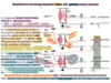
ascending tracts (sensory)
Posterior column: 1) Fasculus gracilis 2) Fasculus cuneatus Anterior column: Spinothalamic tract ``` Lateral column: spinothalamic Posterior spinocerebellar Anterior spinocerebellar Spinotectal Spinoreticular Posterolateral (Lissauer’s)
```descending tracts (motor)
Lateral column: Lateral corticospinal Rubrospinal Lateral reticulospinal ``` Anterior: Anterior corticospinal Tectospinal Vestibulospinal Medial Reticulospinal
```ALL sensory first order neurons are in?
Dorsal root ganglia
ALL sensory first order neurons are in?
Ventroposterior Lateral (VPL) nucleus of thalamus (then it goes to somatosensory cortex)
origin of 2nd order neuron of the Spinothalamic tract
Lamina 1 AND 5 only
end of the first order neurons of the Spinothalamic tract
amina 1 TO 5 (1,2,3,4,5)
Spinothalamic tract 1st order neuron origin end function
Anterolateral pathway 1st order neuron Origin: DRG End: lamina I to V (dorsal root horn) Do one of two things: 1. Terminate at that segment level and synapse with the 2nd order neuron level 2. Join the Lissauer’ tract (posterolateral tract), which will either ascend or descend 1 to 3 segment levels. Then it will terminate and synapse with the 2nd order neuron. Function: - Pain (sharp, prickling and well organized) - Pressure - Temperature
Spinothalamic tract 2nd order neuron origin end
Origin: lamina I and V (dorsal root horn) End: VPL of the thalamus The neuron body of the 2nd order neuron is at either the I OR V lamina. The axon crosses the spinal cord at the anterior commissure, causing the tract to be contralateral. The axon ascends the spinal cord at the anterolateral fasciculus of the white matter. It will terminate at the VPL of the Thalamus.
Spinothalamic tract 3rd order neuron origin end
Origin: VPL of the thalamus | End: Primary somatosensory cortex
Spinotectal tract 1st order neuron origin end function
Lateral pathway Origin: DRG End: lamina I and V (dorsal horn) Function: - Controls pain - Spinovisual reflex: it brings about the movement of the eyes and head towards the source of information
Spinotectal tract 2nd order neuron origin end
Origin: lamina I and V (dorsal horn) End: Superior colliculus The neuron body of the 2nd order neuron is at either the I or V lamina. The axon crosses the spinal cord, causing the tract to be contralateral. The axon ascends the spinal cord at the lateral fasciculus of the white matter. It will terminate at the superior colliculus.
Spinoreticular tract 1st order neuron origin end function
Lateral pathway Origin: DRG End: lamina VI, VII, and VIII (6, 7, 8,) (dorsal horn) Function: - Pain (dull, aching, and poorly localized) - Influence the level of consciousness
Spinoreticular tract 2nd order neuron origin end
Origin: lamina VI, VII, and VIII (dorsal horn) End: reticular formulation of brain stem and intralaminar nuclei of thalamus The neuron body of the 2nd order neuron is at either the VI, VII, or VIII lamina. The axon does not cross the spinal cord, causing the tract to be ipsilateral. The axon ascends the spinal cord at the lateral fasciculus of the white matter. It will give multiple synapse. It synapses contralaterally (it crosses) to the reticular formation of the brain stem, while the axon continues upwards and terminates at the intralaminar nuclei of the thalamus at the same side (ipsilateral).
Visceral sensory tract 1st order neuron origin end function
Lateral pathway Origin: DRG End: dorsal horn ``` Function: Visceral information (ex. distended stomach)
```Visceral sensory tract 2nd order neuron origin end
Origin: dorsal horn End: VPL The neuron body of the 2nd order neuron is at dorsal root horn. The axon travels with the spinothalamic tract. The axon crosses the spinal cord at the anterior commissure, causing the tract to be contralateral. The axon ascends the spinal cord at the anterolateral fasciculus of the white matter. It will terminate at the VPL of the Thalamus.
Visceral sensory tract 3rd order neuron origin end
Origin: VPL | End: somatosensory cortex
Posterior spinocerebellar tract 1st order neuron origin end function
Spinocerebellar pathway (found laterally) Origin: DRG at T1 to L1 or2 (UP) End: Clark’s nucleus (nucleus dorsalis) found at T1 to L1 or 2 Function: Unconscious proprioception
Posterior spinocerebellar tract 2nd order neuron origin end
Origin: Clark’s nucleus (nucleus dorsalis) found at T1 to L1/2 End: cerebellum The neuron body of the 2 order neuron is at Clark’s nucleus (nucleus dorsalis) found at T1 to L1/2. The axon travels upwards and passes through the inferior cerebellar peduncle to terminate at the cerebellum.
Anterior spinocerebellar tract 1st order neuron origin end function
Spinocerebellar pathway (found laterally) Origin: DRG at T12 to L5 (DOWN) End: Spinal border neurons found at T12 -L5 Function: Unconscious proprioception
Anterior spinocerebellar tract 2nd order neuron origin end
Origin: Spinal border neurons found at T12 -L5 End: cerebellum The neuron body of the 2nd order neuron is spinal border neurons found at T12 -L5. The axon crosses the spinal cord, causing the tract to be contralateral. The axon ascends the spinal cord at the lateral fasciculus of the white matter. It then recrosses in the middle cerebellar peduncle, causing the tract to be ipsilateral. It will terminate at the cerebellum.
Cuneocerebellar tract 1st order neuron origin end function
Spinocerebellar pathway (found laterally) Origin: lateral cutaneous (accessory pathway) End: cerebellum The neuron body is found in the lateral cutaneous nucleus in the medulla. This pathway is an accessory pathway as it begins at the middle of another pathway. The axon travels through the inferior cerebellar peduncle to terminate at the cerebellum. Function: Unconscious proprioception
Spinoolivary tract 1st order neuron origin end function
Spinocerebellar pathway (found laterally) Origin: DRG End: dorsal horn Function: Proprioception (the information conveyed is from cutaneous and proprioceptive organs)
Spinoolivary tract 2nd order neuron origin end
Origin: dorsal horn End: inferior olivary nucleus The neuron body of the 2nd order neuron is at the dorsal horn. The axon crosses the spinal cord at the commissure, causing the tract to be contralateral. The axon ascends the spinal cord at the lateral fasciculus of the white matter. It will terminate at the inferior olivary nucleus.
Spinoolivary tract 3rd order neuron origin end
Origin: inferior olivary tract End: cerebellum The neuron body of the 3rd order neuron is at the inferior olivary tract. The axon crosses the inferior cerebellar peduncle (the crossing then recrossing causes the pathway to become ipsilateral) and terminates at the cerebellum
Fasciculus gracilis tract 1st order neuron origin end function
Origin: DRG End: gracilis nucleus in medulla The neuron body is found in the DRG. The central branch of the axon will ascend the spinal cord until it terminates at the gracilis nucleus in the medulla. Function: (important) info from below T6 Conscious proprioception Discriminative touch Vibration Joint movement
Fasciculus gracilis tract 2nd order neuron origin end
Origin: gracilis nucleus in medulla End: VPL The neuron body is at the posterior medulla in the gracilis nucleus. The axon will cross to the other side of the medulla (contralateral) and terminate at the VPL of the thalamus.
Fasciculus gracilis tract 3rd order neuron origin end
Origin: VPL | End: somatosensory cortex
Fasciculus cuneatus tract 1st order neuron origin end function
Function: (important) info from above T6 Conscious proprioception Discriminative touch Vibration Joint movement
Fasciculus cuneatus tract 2nd order neuron origin end
Origin: cuneatus nucleus in medulla End: VPL The neuron body is at the posterior medulla in the cuneatus nucleus. The axon will cross to the other side of the medulla (contralateral) and terminate at the VPL of the thalamus.
Fasciculus cuneatus tract 3rd order neuron origin end
Origin: VPL | End: somatosensory cortex
Anterior Corticospinal origin end function if injured
```Origin (multiple): 1/3 primary cortex, 1/3 supplementary cortex, and 1/3 somatosensory cortex End: anterior horn or interneurons The tract begins in the cerebral cortex (1/3 primary cortex, 1/3 supplementary cortex, and 1/3 somatosensory cortex) and descends to the brain through the cerebral peduncle, then to the Pons (where fibers will move around within the Transverse pontine nucleus). Here the tract divides. The fibers will descend to the medulla into the pyramid WITHOUT CROSSING (15%); these will form the anterior corticospinal tract, which will travel downwards, and few will cross at spinal segments. The fibers terminate at any segment level at the medial group of the anterior horn. Some will synapse with lower motor neurons (3rd order neurons), while others will synapse with interneurons (2nd order neurons). Function: Voluntary control of axial muscles If injured: Injury to any side of the anterior corticospinal tract does not affect muscle innervations because the supply to muscles is bilateral
Lateral Corticospinal
Origin (multiple): 1/3 primary cortex, 1/3 supplementary cortex, and 1/3 somatosensory cortex End: anterior horn or interneurons The tract begins in the cerebral cortex (1/3 primary cortex, 1/3 supplementary cortex, and 1/3 somatosensory cortex) and descends to the brain through the cerebral peduncle, then to the Pons ( where fibers will move around within the Transverse pontine nucleus). Here the tract divides. The fibers will descend to the medulla into the pyramid and CROSS (85% will undergo decussation at medulla); these will form the lateral corticospinal tract, which will travel laterally and downwards. The fibers terminate at any segment level at the lateral group of the anterior horn. Some will synapse with lower motor neurons (3rd order neurons), while others will synapse with interneurons (2nd order neurons). Function: Voluntary control of distal muscles If injured: 1. Injury of the tract (after decussation/ after medulla) will cause ipsilateral deficiency of innervation to the muscles innervated by the tract. Ex. Lesion to left lateral column of spinal cord -> left side of body is paralyzed 2. Injury in the brain (before decussation/ before medulla) will cause contralateral deficiency of innervation to the muscles innervated by that tract. Ex. Lesion to right hemisphere -> left side of body is paralyzed The lateral corticospinal tract is closely related to the rubrospinal tract, so injury in that area will probably affect both tracts
Pyramidal System:
Responsible for voluntary control of face and body muscles
Extrapyramidal system:
responsible for involuntary control + balance + posture
Medial Vestibulospinal origin end function if injured
```Origin: medial vestibular nucleus in medulla End: anterior horn of cervical segments The tract begins at medial vestibular nucleus in medulla and travels through the medial longitudinal fasciculus (fibers which control extra ocular muscle) and terminates at the ventral horn of cervical segments. Some will synapse with lower motor neurons (3rd order neurons), while others will synapse with interneurons (2nd order neurons). Function: Stabilizing head movement Coordinating head movement with eye movement. Ex. When looking up, we move our neck up as well Innervates muscles of head and neck If injured: Injury to any side of the medial vestibulospinal tract does not affect muscle innervations because the supply to muscles is bilateral
Lateral Vestibulospinal origin end function if injured
```Origin: lateral vestibular nucleus in medulla End: anterior horn The tract begins at medial vestibular nucleus in medulla and travels through the lateral fasciculus and terminates at the ventral horn. Some will synapse with lower motor neurons (3rd order neurons), while others will synapse with interneurons (2nd order neurons). ``` Function: Balance Postural changes to compensate for tilts and movements of the body Increase activity of extensor muscles Decrease activity of flexor muscles ``` If injured: Injury of the tract will cause ipsilateral deficiency of innervation to the muscles innervated by the tract. Ex. Lesion to left lateral column of spinal cord -> left side of body is affected
Rubrospinal origin end function if injured
```Origin: red nucleus End: anterior horn, synapsing with the lateral motor neurons Tract begin at red nucleus (found in midbrain). It CROSSES the midbrain and runs laterally with the lateral corticospinal tract and ends at the lateral group of the anterior horn. Some will synapse with lower motor neurons (3rd order neurons), while others will synapse with interneurons (2nd order neurons). Function: Activate flexor muscle If injured: Injury in the tract will cause contralateral deficiency of innervation to the muscles innervated by that tract. Ex. Lesion to right fasciculus -> left side of body is affected
Tectospinal origin end function
Origin: superior colliculus End: anterior horn at cervical level of spinal cord The tract begins at superior colliculus (receives input from optic nerve). The axon descends and crosses in midbrain. They enter spinal cord and they will terminate at the cervical level Function: Reflex Turning head in response to visual stimulus
Mendial Reticulospinal origin end function
Pontine Origin: pontine reticular formation (brainstem) End: anterior horn or interneurons Function: Walking, running Activate extensor muscles
Lateral Reticulospinal origin end function
medullary Origin: medullary reticular formation End: anterior horn or interneuron Function: Walking, running Activates flexor muscles
Lower motor neurons function depends on whether the nucleus of the neuron is found in the lateral group or the medial group within the ventral horn what does each control? (lateral or medial)
- Lateral: control distal limb muscles - Medial: (always bilateral) controls axial/proximal muscles
What are the 1st order neurons in descending pathways?
they are upper motor neurons UMN, they come from the brain. (All the tracts mentioned previously are part of the third order neurons which are lower motor neurons and they send signals directly to the muscles)
Descending autonomic fibers
- Control visceral activities - UMN: o Origin: cerebral cortex, hypothalamus, amygdala, reticular formation o End: lateral horn at T1-L1/2 and S2-S2 o Pathway: originates from cerebral cortex, hypothalamus, amygdala, reticular formation. The axons descend and CROSS at the midbrain. They continue down to the lateral horns in the thoracic or sacral segments. ▪ In thoracic region, the lateral horn in the spinal cord gives rise to preganglionic sympathetic fibers ▪ In sacral region, the lateral horn in the spinal cord gives rise to preganglionic PARAsympathetic fibers
Golgi tendon organ
- Sense Muscle tension
Muscle spindle
- Sense Muscle length (static gamma) - Sense Muscle rate/ speed (dynamic gamma) - has three parts: 1. Specialized intrafusal muscle fibers: contain non-contractile region 2. Sensory fibers (Ia, most myelinated) that terminate in non-contractile region of the intrafusal fibers. - > When the muscle is stretched, the distance between the spirals will increase which will increase the activity of Ia fibers. 3. Motor axons (gamma)
Sensory and motor fibers | gamma or alpha?
- Motor: gamma alpha - Sensory: Ia (1a) II (2)
Two types of motor neurons, alpha (Aa) and gamma (y), what's the difference?
1. Alpha: Aα - Extrafusal innervation Large diameter axons = fast transmission 2. Gamma: γ - Innervates the core of muscle spindle - Intrafusal innervation - two types, dynamic and static - Modulate sensory information in a dynamic or static fashion
linkage btw alpha and gamma motor neurons
2 types of muscle fibers in the Muscle Spindle:
1. Nuclear chain fibers 2. Nuclear bag neurons
Nuclear chain fibers, Nuclear bag neurons, what's the innervation of each?
1. Nuclear chain fibers: innervated by static γ motor neurons only. 2. Nuclear bag neurons: innervated by both static and dynamic γ motor neurons.
two types of muscle fibers, intrafusal and extrafusal, what are they innervated by?
Extrafusal fibers are innervated by α motoneurons, generate force. Intrafusal fibers are innervated by γ motoneurons, adjust sensitivity of muscle spindle
stretch reflex and reciprocal inhibition, which one is mono and which is polysynaptic?
- stretch reflex -> monosynaptic - reciprocal inhibition -> polysynaptic
describe Inhibitory interneurons that are Feedforward inhibition:
- enhances the effect of the active pathway - suppressing the activity of other, opposing, pathways. - Activates one pathway and inhibits all the lateral pathways. - Example: knee jerk reflex.
describe Inhibitory interneurons that are Feedback Inhibition:
- stops the activity within the stimulated pathway and prevents it from exceeding a certain critical maximum. - Inhibits the same Pathway. - Example: autogenic reflex. - Renshaw cells are inhibitory cells that transmit inhibitory signals to the surrounding motor neurons.
What's Flexor Reflex (Flexor-withdrawal reflex)
- stepping on sharp object - receptor is on the skin (not inside the muscle) - polysynaptic - occurs in response to a tactile, painful, or noxious stimulus. Somatosensory and pain afferent fibers initiate a flexion reflex that causes withdrawal of the affected part of the body from the painful or noxious stimulus. - reflex produces flexion on the ipsilateral side & extension on the contralateral side (extensor muscles will contract and flexor muscles relaxes). Extension on the contralateral side is called crossed-extension reflex.
Blood supply for spinal cord? (three)
1. anterior spinal artery for ant. 2/3 2. posterior spinal arteries for post. 1/3 3. great anterior medullary artery of adamkiewicz (from left side of aorta) supplies LOWER part of the spinal cord (IMPORTANT)
pseudounipolar neurons found in:
Dorsal root ganglia
Multipolar neurons found in:
CNS
in neuron regeneration what's the changed in DISTAL end called
Wallerian Degeneration: myelin AND axon will be destroyed HOWEVER Connective tissue especially (endoneurium) AND schwann cells will persist
Role of macrophages in neurons degeneration
- Clean/ digest the debris in Wallerian Degeneration - promotes angiogenesis - type 2 machrophage will produce factors for the regeneration process
Changes of cell body during neuron injury
INJURED: - chromatolysis: Nissl material becomes less in number, fine, granular, dispersed throughout the cytoplasm - nucleus moves towards the periphery (eccentric nucleus) - cell body swells and becomes rounded. - Synaptic stripping: synaptic terminals are seen to withdraw from the surface of the injured neuronal cell body and its dendrites NORMAL: - centrally located nucleus w/ prominent nucleolus. Spread out nissel’s substances
What's nissel’s substances
collection of RER (Rough endoplasmic reticulum) with ribosomes.
in neurons, changes in the cell body and in the proximal segment is called:
retrograde degeneration | (cause going from distal to proximal).
changes in the distal part of neuron is called:
Wallerian Degeneration.
Type 2 MQ will synthesize and stimulate:
1) Anti-inflammation 2) Growth factors 3) Stimulate Schwann cell to differentiate and remeylinate
axon will regrow and go through the Schwann cell but how?
- MQ secret vascular endothelium growth factor that promotes angiogenesis - it will make a connection between the distal and the proximal (bridge) - Along this blood vessel the growing/ proliferating Schwann cell & THEN the axons will be guided till it reaches the distal segment

