Week 10: Musculoskeletal Flashcards
(49 cards)
What are the four key features of the MSK exam?
- Inspect: visually evaluate any signs of deformity, swelling, scars, inflammation or muscle atrophy
- Palpate: use surface anatomy landmarks (bony contours and structures) to localize points of tenderness or fluid collection
- Range of motion: have patient actively move involved joints then move them passively as the examiner
- Special maneuvers: perform stress maneuvers if indicated to evaluate joint stability and integrity of ligaments, tendons and bursae particularly if pain or trauma is present
How is joint pain classified? (3)
- # of joints involved - monoarticular, oligoarticular/pauciarticular or polyarticular
- articular or extra-articular
- acute (days to weeks) vs. chronic (months to years)
Define monoarticular, oligoarticular/pauciarticular and polyarticular
Monoarticular: 1 joint (localized)
Oligoarticular or pauciarticular: 2-4 joints
Polyarticular: more than 4 (diffuse)
Define articular vs. extra-articular. How do these present on exam?
Articular: includes the joint capsule & articular cartilage, synovium & synovial fluid
- Presentation: usually involves swelling & tenderness of the entire joint
Extra-articular: includes peri-articular ligaments, tendons, bursae, muscle, fascia & overlying skin
- Presentation: involves point or focal tenderness in regions adjacent to the articular structure
Which are some examples of monoarticular (7) disease processes? Polyarticular? (8)
- · Monoarticular
- Injury
- Monoarticular arthritis
- Monoarticular osteoarthritis
- Tendinitis
- Bursitis
- Soft tissue injury
- Acute gout
- Polyarticular
- Rheumatic fever
- Rheumatoid arthritis
- Connective tissue disease
- Osteoarthritis
- systemic lupus erythematosus
- Psoriatic arthritis
- Scleroderma
- Gonococcal arthritis
What is crepitus? What does it indicate?
Audible or palpable crunching during movement of tendons or ligaments over bone or areas of cartilage loss
What are the four cardinal signs of inflammation?
- Redness
- Swelling
- Warmth
- Pain or tenderness
What history and exam findings are consistent with rheumatoid arthritis?
History: chronic inflammation of synovial membranes with secondary erosion of adjacent cartilage and bone and damage to ligaments and tendons
Exam findings: tender, often warm but seldom red swollen joints, hands most often affected but may see symptoms in the feet, wrists, knees, elbows and ankles; stiffness in the morning and after inactivity
What history and exam findings are consistent with osteoarthritis?
History: degeneration and loss of joint cartilage from mechanical stress
Exam findings: tender joints usually knees, hips, hands, cervical and lumbar spine, wrists; heberden’s & bouchard’s nodes
How would the FNP perform a preparticipation sports physical? What would be abnormal findings and what would these finding indicate? (12 steps)
Step 1: stand straight facing forward, note any asymmetry or joint swelling
Step 2: move neck in all directions, note any loss of range of motion
Step 3: shrug shoulders against resistance, note for any weakness of shoulder, neck or trapezius muscles
Step 4: hold arms out to the side against resistance and actively raise arms over the head; note any loss of strength in the deltoid muscle
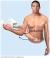
Step 5: hold arms out to the side with elbows bent at 90 degrees, raise and lower arms; note any loss of external rotation and injury of glenohumeral joint
Step 6: hold arms out, completely bend and straighten elbows; note any reduced ROM of the elbow
Step 7: hold arms down, bend elbows 90 degrees and pronate and supinate forearms; note any reduced ROM from prior injury to forearm, elbow or wrist
Step 8: make a fist, clench and then spread fingers; note any protruding knuckle, reduced ROM
Step 9: squat and duck-walk four steps forward; note inability to fully flex knees and difficulty standing up from prior knee injury
Step 10: stand straight with arms at sides facing back, assess for symmetry, leg length and weakness
Step 11: bend forward with knees straight and touch toes; note asymmetry
Step 12: stand on heels and rise to toes; note any wasting of calf muscles
Which muscle groups make up the rotator cuff? (4)
Scapulohumeral group (SITS): supraspinatus, infraspinatus, teres minor & subscapularis
Axioscapular group: trapezius, rhomboids, serratus anterior, levator scapulae
Axiohumeral group: pectoralis major and minor; latissimus dorsi
Biceps & triceps
What exam findings would be consistent with a clavicle fracture in the newborn?
Lumps, tenderness or crepitus along the clavicle
In considering the elbow, what history and exam findings are consistent with lateral epicondylitis?
- pain and tenderness 1cm distal to the lateral epicondyle and sometimes in extensor muscles close by
- can elicit pain when extending the wrist against resistance
In considering the elbow, what history and exam findings are consistent with medial epicondylitis?
- tenderness maximal just lateral and distal to the medial epicondyle
- wrist flexion against resistance increases pain
In considering the elbow, what history and exam findings are consistent with olecranon bursitis?
- swelling and inflammation of the olecranon bursa
- superficial to the olecranon process and may reach 6cm in diameter
What symptoms are consistent with carpal tunnel syndrome? (5)
- noctural hand or arm numbness
- dropping objects
- inability to twist lids off jars
- aching at the wrist and forearm
- numbness of the first 3 digits
What is snuffbox tenderness? What does this indicate?
= tenderness with the wrist in ulnar deviation and pain at the scaphoid tubercle
- indicative of occult scaphoid fracture
What are Dupuytren flexion contractures? Stenosing tenosynovitis? Colles fracture?
- Dupuytren flexion contractures: thickened band overlying flexor tendon of the fourth finger and possible little finger near distal palmar crease; thickened fibrotic cord develops between palm and finger; extension is limited but flexion is normal
- Stenosing tenosynovitis: trigger digits, catching or locking of affected finger
- Colles fracture: tenderness over distal radius after a fall
What are the muscle groups of the hips? (4)
- Flexor group: iliopsoas
- Extensor group: gluteus maximus, hamstring muscles, adductor magnus, gluteus medius
- Adductor group
- Abductor group: gluteus medius and gluteus minimus
How would the FNP perform the Barlow and Ortolani tests. What are positive finding and what do they indicate?
- Barlow: pull the leg forward and adduct with posterior force, feel for any movement of the femur head laterally
- Positive finding: palpable movement of the femur head
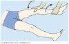
- Ortolani: supine with legs pointing towards you - flex legs to form right angles at the hips and knees, abduct both hips simultaneously until the lateral aspect of each knee touches the exam table
- Positive finding: palpable movement of the femur head back into place
How would the FNP assess for leg shortening? Tibial torsion?
- Leg shortening: inspection of gait, patient supine - assess for symmetry, ,measure distance between anterior superior iliac spine and medial malleolus
- Tibial torsion: have toddler lie prone on the examination table with knees flexed to 90 degrees, assess for internal or external rotation past 10 degrees either way
What is a SCFE? In whom might this occur?
- SCFE: slipped capital femoral epiphysis
- Associated with limp in an obese child
What exam techniques can you use to assess for an effusion? What is a positive result? (3)
- Bulge sign: with knee extended, place left hand above the knee and apply pressure on the suprapatellar recess to displace fluid downward, stroke downward on the medial aspect of the knee and apply pressure to force fluid into the lateral area; tap the knee being the lateral margin of the patella with the R hand
- Positive sign: bulge on medial side between patella and femur
- Balloon sign: place thumb and index finger of the R hand on each side of the patella, with left hand, compress the suprapatellar recess against the femur; palpate for fluid ejected or ballooning into spaces next to the patella and under the right thumb and index finger
- Positive: palpable fluid wave
- Balloting of the patella: compress suprapatellar push and ballotte or push the patella sharply against the femur
- Positive: palpable fluid wave
What are symptoms and exam findings consistent with prepatellar bursitis?
Swelling around the prepatellar bursa - typically due to excessive kneeling