Week 2 Flashcards

(221 cards)

1
Q

Where do mature B-cells reside?

A

Secondary lymphoid organs

2
Q

What is B-cell activation?

A
  • A naiive B-cell has not yet recognized its antigen
  • When a naive B-cell binds to its antigen, it becomes activated
3
Q

What is cross-linking?

A
  • When multiple BCR’s on a single B-cell, all with the same specificity, bind to the antigen
4
Q

How is activation signals conveyed to naive B-cell when it binds its antigen?

A
  • Ig-alpha
  • Ig-beta
  • Non-covalently associated with BCR and they are responsible for signaling cascade within B-cell
5
Q

What is the result of B-cell activation?

A
  • B-cell turns into a plasma cell that secretes antibodies
  • Antibodies have the exact same specificity as the BCR receptor. The only difference between antibodies and BCR is they are secreted instead of tethered to the membrane.
6
Q

Structure of antibodies

A
  • 2 light chains
  • 2 heavy chains
  • Variable regions and constant regions
  • Each antibody has 2 antigen-binding sites (variable regions)
7
Q

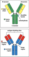
antigen binding site of BCR/antibodies

A
  • There are 2 antigen binding sites
  • Each site is made up of 1 variable region of a light chain and 1 variable region of a heavy chain
8
Q

hypervariable loops of BCR/antibody

A
  • There are 3 hypervariable loops within each V region
  • So there are 6 hypervariable loops per antigen-binding site
  • These are also called “Complementarity determining regions”
  • This is the specific region within the V region that binds the antigen
9
Q

What do the hypervariable loops recognize?

A
  • Epitope - the specific region of an antigen that the hypervariable region binds to
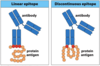
10
Q

linear epitope

A
  • a linear sequence of amino acids recognized by an antibody
11
Q

discontinuous epitope

A
  • a sequence of amino acids within the antigen’s folded shape.
  • recognized by a BCR/antibody
12
Q

multivalent antigen

A
  • When an epitope is present multiple times on a single antigen, we call it a multivalent antigen
  • BCR stimulation is stronger with a multivalent antigen
    *
13
Q

Can multiple antibodies bind to the same antigen?

A
  • Yes
  • BCR A binds to epitope A and BCR B binds to epitope B on the same antigen.
14
Q

polyclonal response

A
  • When multiple different types of B-cells are activated in resopnse to a single antigen
  • Antibodies with the same V-region and antigen specificity can bind to an antigen. But these antibodies can have different C-regions, which dictates the host’s response to the antigen.
15
Q

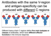
What dictates the immune response to an antibody binding the antigen?

A
  • The C-region of the bound antibody
  • The “class” of antibody bound
16
Q

What are the 5 isotypes/classes of antibodies?

A
  • IgG
  • IgA
  • IgM
  • IgD
  • IgE
17
Q

What is the structure of IgM?

A
  • Pentamer
18
Q

what is the structure of IgA?

A

dimeric

19
Q

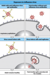
what is the first antibody isotype produced in an immune response? Why?

A
  • IgM
  • Because it is a pentamer, it has 10 antigen binding sites
  • It binds with high avidity and can produce a strong response
20
Q

What are the 3 ways that antibodies participate in immune response?

A
  1. Neutralization - bind pathogen/toxins to prevent the pathogen binding to host cells
  2. Opsonization - bind pathogen directly to mark it for phagocytosis
  3. Activate complement system - results in lysis and phacocytosis
21
Q

Which antibodies participate most in neutralization? Why?

A
  • IgG
  • IgA
  • These are high affinity antibodies
22
Q

Where is IgA found?

A
  • Mucosa (respiratory tract, GI tract)
23
Q

Examples of IgA participating in host defense via neutralization

A
  • Strep
  • Influenza
24
Q

Which antibodies participate in complement fixation?

A
  • IgM - C1q binds to IgM and initiates classical pathway
  • IgG - C1q binds to two or more IgG and initiates classical complement pathway
25
Why would IgA NOT be a part of complement fixation?
* It is found in the mucosa, whereas complement proteins are floating around in the blood
26
Fc receptors
* Are found on innate immune cells (macrophages, neutrophils, NK cells) * Fc receptors recognize and bind the constant region of the antibody (with antigen bound)
27
Which antibody isotypes/classes can Fc receptors on innate cells recognize?
* IgG * IgA * IgE
28
What happens when Fc receptor binds C region of IgG?
* phacocytosis by macrophages * killing by natural killer cells
29
what is a cell's response to Fc receptor binding IgE?
* degranulation of eosinophils * degranulation of mast cells
30
How are monoclonal antibodies produced?
* B-cell hybridoma technology * Inject a pathogen into an animal --\> polyclonal response * Isolate the specific B-cell clone that you want * Fuse the B-cell clone with a myeloma cell --\> "an immortal B-cell" with a single specificity = monoclonal antibody
31
chimeric monoclonal antibodies
* The V-regions are from a mouse * The rest of the antibody is from a human
32
humanized monoclonal antibodies
* The hypervariable regions are from a mouse * The rest is from a human
33
human monoclonal antibodies
* The whole antibody is human but is produced in a mouse * You alter mice antibody genes to produce human antibody genes * This is the least immunogenic of the monoclonal antibodies, but it has ethical considerations
34
ELISA
* Used for DETECTION of antibodies and antigens in the blood * Coat the antigen onto some sort of sticky surface * Direct and indirect ELISA
35
Direct ELISA
* Coat antigen onto a sticky surface * An antibody specific to your antigen of interest is tagged with an enzyme * Incubate antibody with antigen-coated surface * Add enzyme's substrate * If there is a color change --\> your antibody is detecting the antigen it binds to. * The more the color change, the more antibody (and antigen) is present in that sample.
36
Indirect ELISA
* Coat sticky surface with antigen/sample of patient's blood * Add a primary monoclonal antibody specific to antigen of interest that you're testing for * Add a secondary POLYCLONAL antibody that binds to the primary monoclonal antibody. The secondary antibody is the one tagged with an enzyme * Several of these secondary antibodies will bind to the primary enzyme, which increases the sensitivity of the test (more likely to get a positive test if the antigen is present).
37
Sandwich ELISA
* A form of indirect ELISA * Coat sticky surface with a monoclonal antibody * Add the sample * If antigen is present, it will bind monoclonal antibody * Use a primary monoclonal antibody that will bind antigen a second time * Use secondary polyclonal antibody with the enzyme * This increases sensitivity AND specificity over standard direct ELISA
38
What is ELISA used for?
* HIV detection * Viral antigens themselves * Antibodies produced from HIV infection * ELISA has high sensitivity and is used for initial screening for HIV. Diagnosis is confirmed using RT-PCR, which is more specific.
39
What kind of ELISA is used for HIV screening?
* Sandwich ELISA * gp120 is considered the "capture antibody" * The antigen being measured here is the patient's antibodies against HIV viral protein
40
Flow cytometry
* Main purposes is to detect changes or particular subsets of cells in the blood (i.e. allows you to count things) * Main application: you can't see what kind of leukocytes are present under a microscope. This method allows you to figure that out.
41
how does flow cytometry work?
* Use flourophore molecules that have Ab conjugated to them. * The antibodies are specific for proteins on the surface of immune cells. * So Ab-flourophore1 binds to type 1 immune cells. Ab-flourophore2 binds to type 2 immune cells. * Then you send the cell-Ab-flourophore through a detector that spits out results onto a dot plot that you can read to see which immune cells and how many are present.
42
What do we use flow cytometry for?
1. X-linked agammaglobulinemia * This is a disease where you don't produce B-cells * You use flow cytometry to check and can see that basically there are no B-cells in patient's blood 2. CD4 T-cell count in HIV * Used to monitor their T-cell count
43
How is the heavy chain different from the light chain in B-cell genes?
* It has a diversity gene segment * There is only 1 heavy chain locus but 2 light chain loci
44
What is somatic recombination?
* Used by B-cells and T-cells to produce receptors with a wide array of specificity * This is basically DNA splicing
45
Process of Light chain recombination for B-cells
1. V and J segments are selected at random and recombined first. This is the ONLY recombination event for light chains
46
What is the process of heavy chain somatic recombination for BCRs?
1. First recombination is to join a D segment to a J segment 2. Second recombination combes DJ segment to a V segment
47
What is the main difference between light chain and heavy chain recombination in BCRs?
* The light chain undergoes only 1 recombination event (VJ) * The heavy chain undergoes 2 recombination events (DJ then VDJ) * The light chain has no D region
48
What are the enzymes that carry out somatic recombination in BCRs?
recombinases (RAG enzymes)
49
what do recombinase enzymes recognize?
* recombination signal sequences (RSS) in the DNA flanking V,D, and J segments
50
What is the signal joint?
* The DNA that is cleaved out during somatic recombination of BCRs
51
What is the coding joint?
* The area that is joined together to form the recombined VJ segment
52
Non-homologous end joining
* The process by which broken strands of DNA are rejoined during somatic recombination * This rejoining is done by DNA repair enzymes, NOT RAG enzymes
53
What is the major source of diversity in BCRs outside of somatic recombination?
junctional diversity
54
junctional diversity
* The process by which random nucleotides are added between V-J regions (in the light chain) and D-J/V-DJ regions in the heavy chain * A major source of BCR diversity in antigen binding site specificity
55
terminal deoxy-nucleotidyl transferse (TdT)
* The enzyme that adds the nucleotides in junctional diversity process
56
Overview of BCR somatic recombination for both heavy and light chains
57
Stages of B-cell development timed with heavy chain/light chain rearrangement
* Immature B-cell is the stage when the heavy chain and light chains have paired up * This is when you have a functional BCR that can be trafficked to the cell surface * B-cells remain immature as long as they're in the bone marrow
58
what are the earliest identifiable cells of the B-cell lineage?
* early pro-B cells * Heavy chain DJ rearrangement has occurred
59
What cells support B-cell development?
* stromal cells (in the bone marrow)
60
Why is B-cell somatic recombination of the heavy chain inherently imprecise and inefficient?
* Junctional diversity can add nucleotides in such a way that the reading frame of the entire gene is shifted * This frequently results in nonproductive rearrangements * B-cells that have one nonproductive rearrangement can try again on the other chromosome. If both fail, that B-cell dies. * About half of developing B-cells die at the pro-B cell stage (due to nonproductive DJ rearrangement)
61
What is the pre-B cell stage?
* heavy chain rearrangement has occurred successfully (DJ and VDJ rearrangements)
62
What must occur for a pro-B cell to proceed to pre-B cell stage?
* A pro-B cell must have undergone heavy chain rearrangement to form VDJ segment AND * A **surrogate light chain (SLC)** must be made to pair up with the heavy chain while the real light chain is being recombined
63
what is allelic exclusion?
* Expression of only one of two copies of a gene (present on separate chromosomes) is allelic exclusion
64
When do we see allelic exclusion in B-cell development?
* When a functional heavy chain is made and a surrogate light chain pairs with it, you have a pre-B cell. * At this stage, RAG enzymes are turned off to prevent any further rearrangement of the heavy chain on the other chromosome. That is allelic exclusion * This ALSO happens for the light chain
65
why is light chain rearrangement more successful than heavy chain in B-cell development?
* Heavy chain only has 1 locus. Failure at one chromosome gives only one other chance (on the other chromosome). * Light chain has 2 different loci. If rearrangement is nonproductive at light chain locus #1, then the cell keeps trying. * If there are multiple failures, the next locus is tried. * \*\*The light chain can try multiple times at a locus, but the heavy chain only tries once\*\*
66
what is the purpose of heavy chain allelic exclusion vs light chain allelic exclusion?
* Heavy chain allelic exclusion occurs so that heavy chain rearrangement is turned off after a functional heavy chain is made. This ensures only one type of BCR is made. * Light chain allelic exclusion occurs after a fully functional BCR is made. This ensures that each B-cell only expresses one type of BCR. * Together, allelic exclusion at the heavy-chain and then light-chain Ig loci ensures expression and secretion of Igs of a single antigen-specificity in individual B cells.
67
What are the 2 developmental checkpoints in B-cell development?
1. Heavy chain expression inside the B-cell --\> pairing of SLC (and progression to pre-B cell stage). No pairing --\> death. 2. Light chain expression inside the B-cell --\> functional B-cell receptor (and progression to immature B-cell stage).
68
B-cell central tolerance
* The process of making sure B-cells do not attack self * This all occurs in the bone marrow as part of B-cell development
69
What happens if a B-cell is strongly self-reactive in the bone marrow?
* First it's given a chance to rearrange the light chain - this is called "B cell editing" * If that doesn't work, it undergoes apoptosis * These B-cells that are strongly reactive are ones that recognize multivalent self molecules
70
What happens if a B-cell is moderately self-reactive in the bone marrow?
* It is modified to be unreactive but then is still sent to the periphery * An unresponsive B-cell in the periphery is called **anergic**
71
What happens to an immature B-cell that is not reactive towards self in the bone marrow?
* It is sent to the periphery
72
How do autoimmune conditions develop?
* One way is that B-cells can escape central tolerance * B-cells sometimes don't encounter all possible self molecules inside the bone marrow (like insulin). When it is sent to the periphery, it can encounter them and react towards them.
73
Ignorant B-cells
* Self-specific B cells that do not encounter self-antigen in the bone marrow are called ignorant. * These can encounter self antigen outside the bone marrow and be reactive
74
Types of B-cells that can be responsible for autoimmune issues
* Anergic B cells * Ignorant B-cells
75
Overview of B-cell central tolerance
76
What does occupancy depend on?
* Concentration of drug * Affinity of receptor (Kd)
77
What does strength of drug stimulug depend on?
* The number of receptors * The intrinsic efficacy of the drug-receptor complex
78
What does the cell's response to the stimulus depend on?
* Strength of stimulus * Tissue sensitivity
79
Formula to determine fractional occupancy
* Can determine by knowing concentration of the drug and the Kd
80
why would you want to know fractional occupancy?
* Can help you determine the total number of receptors that are occupied (which is important in knowing the stimulus)
81
Definition of Kd
* The drug concentration needed in order to have 50% of receptors occupied
82
Definition of Ec50/Ed50
* Ec50 = Concentration of drug required to generate a half maximal response * Ed50 = dose of drug required to generate a half maximal response * \*\*These two things are different. Dose is what is taken. Concentration is at the site of action.
83
What does Ec50 tell you?
* A measure of the drug's potency * Lower Ec50 = higher potency
84
stimulus
85
what is receptor reserve?
* When Ec50 is reached before Kd * You get a half maximal response before you occupy 50% of receptors
86
What causes receptor reserve?
* The tissue is very sensitive * The intrinsic efficacy of drug-receptor complex is high * You have lots of receptors
87
What determines the potency of the drug?
* Affinity of receptor (Kd) * Total number of receptors * Intrinsic efficacy * Sensitivity
88
What determines the maximum response?
* Intrinsic efficacy * sensitivity * receptor number * NOT Kd
89
What types of antagonism are there?
1. surmountable 2. insurmountable 3. physiologic antagonism
90
what is physiologic antagonism?
* Two different receptors that cause two different responses can antagonize each other
91
5 main cellular responses to stress
1. hypertrophy 2. hyperplasia 3. atrophy 4. metaplasia 5. intracellular accumulation of metabolic products
92
hypertrophy
* definition: cells get bigger * Can be physiologic (ex: workout out) or pathologic (ex: heart gets bigger in response to hypertension)
93
hyperplasia
* cells get bigger AND there is more of them * example of pathologic process: warts from HPV are the result of too much epidermis
94
atrophy
* definition: cells shrink and you have fewer of them * example: 80 year old brain is smaller than a 23 year old brain
95
metaplasia
* definition: a change in cell types * ex: heartburn causes acid to come out of stomach and into distal esophagus. This acid does not just damage the esophageal cells, it turns them from squamous epithelium to columnar epithelium.
96
What are the first changes you see in an injured cell?
* Biochemical changes happen first * Changes visible in light microscopy and organ level lag behind this
97
What are some of the changes you can see in a microscope when a cell has undergone injury?
1. accumulation of water (hydropic changes) 2. accumulation of lipid (steatosis) 3. Glycogen 4. Protein 5. Pigments
98
What is the first change you see to damaged cells under a light microscope? Why?
* hydropic changes (accumulation of water) * Cell injury often disrupts glycolysis, Krebs, and ETS --\> ion balance gets fucked up --\> water inside the cell * You'll see clear vacuoles
99
What is lipid accumulation called?
steatosis
100
What causes steatosis?
* Genetic disorders * alcoholic liver injury * obesity * diabetes * toxins
101
How does steatosis appear in light microscope?
102
What is Fabry's disease? What cellular changes do you see?
* Lysosomal storage disease * See an accumulation of glycolipids --\> appearance of lipid inclusions * Primarily affects heart, kidney, and blood vessels
103
How does glycogen accumulation appear in light microscope?
* Clear vacuoles in cytoplasm
104
what causes glycogen accumulation in cells?
* Glycogen storage disorder * Diabetes
105
what causes protein accumulation in cells?
* Resorption * Excess synthesis of protein * Defects in folding (usually genetic) * Aggregation of abnormal proteins
106
Alpha-1-antitrypsin deficiency
* Normal state: liver secretes anti-trypsin to inhibit trypsin, which is an enzyme that breaks down protein. Anti-trypsin prevents trypsin from degrading elastic layers of the lung. * Pathology: A mutation in anti-trypsin results in accumulation of non-functional (improperly folded) anti-trypsin in the liver. And you lose lung function b/c trypsin is breaking down lung layers. * The anti-trypsin accumulations are seen as big pink globules in the liver.
107
What is alzheimer's an example of re: cell injury?
* accumulation of protein (amyloid proteins)
108
What is amyloidosis?
* Caused by a malignant plasma cell, which results in the accumulation of protein in the cell * specifically, the protein is a build up of light chains
109
What is pathognomonic for amyloidosis?
* Bright luminescence on a congo red stain
110
Intracellular pigments
* Hemosiderin * Lipofuscin * Melanin * Exogenous substances
111
What is hemosiderin? How does it appear under light microscope?
* It is a byproduct of hemoglobin breakdown * Externally it appears as bruising * Under light microscope it appears as brown pigment
112
Lipofuscin - what does it look like under light microscope, what causes it?
* Caused by normal wear and tear - i.e. this is a physiologic change, NOT pathologic * Reddish hue to it
113
anthracosis
* Coal Miner's lung * Example of exonenous substance building up and causing cellular injury
114
Necrosis vs. Apoptosis
* Necrosis is characterized by inflammation; apoptosis is not * Necrosis shows hydropic changes; apoptosis shows cell shrinkage * Necrosis shows autolysis and leakage of intracellular contents; apoptosis shows nuclear fragments and blebbing)
115
What morphologic changes do you see during necrosis?
* Cytosol: cell swelling (hydropic change), eosinophilia * Eosinophilia is a very pink stain of the cells. This is b/c transcription shuts down, and RNA stains blue. But protein stains pink and is more durable than RNA. * Nucleus: karyolysis (chromatin fades), pyknosis (nucleus shrinks), karyorhexis (nucleus fragments)
116
Types of necrosis
1. coagulative necrosis 2. liquefactive necrosis 3. Caseous necrosis 4. Gangrenous necrosis 5. Fibrinoid necrosis 6. Fat necrosis
117
Coagulative necrosis
* "Death in place" * Characterized by ghosts of cells - no nucleus, but the cell architecture remains
118
Most common cause of coagulative necrosis
* Ischemia * Heart attack
119
What is a common finding under light microscope of coagulative necrosis?
* Cells are intact but missing nuclei
120
Liquefactive Necrosis
* The tissue dies and then disintegrates, so you just see a void of tissue
121
Most common cases of liquefactive necrosis
* \*\*Stroke * Abscess (dead tissue is replaced by pus = neutrophils)
122
Caseous necrosis
* Tissue takes on a cheese-like appearance * Dead cells persist but then are surrounded by inflammatory cells
123
Most common causes of caseous necrosis
* Tuberculosis
124
How does caseous necrosis appear under light microscope?
* Central area of dead tissue walled off by inflammatory cells
125
Gangrenous necrosis
* Tissue dies and then is accompanied by bacterial infection
126
Most common cause of gangrenous necrosis
* Diabetes
127
Fibrinoid necrosis
* An accumulation of fibrin in?around the arteriole * Only seen in 1 particular setting: vasculitis
128
Fat necrosis
* This is necrosis specifically of adipose tissue * Most commonly caused by pancreatic injury --\> leakage of digestive enzymes
129
What is the cause of myocardial rupture?
* After a heart attack, the damaged tissue begins remodeling * The tissue is much weaker during this time and can result in rupture * Often occurs 5-9 days after the heart attack during remodeling phase
130
What is the fate of dead tissue?
1. Eaten up by macrophages and neutrophils 2. Scarring 3. Calcifications
131
Chronic Granulomatous Disease cause
* NADPH oxidase disorder
132
Result of defective NADPH oxidase
* Cannot generate reactive oxygen species in phagolysosomes
133
What type of organism are CGD patients most susceptible to?
* catalase positive organisms * Because they can break down hydrogen peroxide * Results in recurrent fungal infections
134
What is an NBT test?
* Measures the ability of phagocytes to generate reactive oxygen species * ROS reduce NBT --\> purple color in the neutrophils (normal lab finding) * People with CGD can't reduce NBT, so their neutrophils don't turn purple/blue
135
Most common genetic cause of CGD?
* Defect in gp91 protein, a component of NADPH oxidase * Shows X-linked pattern of inheritance
136
Structure of T-cell receptor
* Membrane-bound * Alpha chain and beta chain * Variable region and constant region
137
How many antigen-binding sites does a TCR have?
One
138
Alpha chain gene rearrangement in TCR
* VJ recombination happens as somatic recombination * Primary transcript is made, then extra J regions are spliced * There is a single C-region
139
Beta chain rearrangment in TCR
* There are 2 somatic recombinations: DJ followed by VDJ * There are 2 clusters of D-J-C regions from which recombination can occur. It's random as to whether somatic recombination occurs in cluster 1 or cluster 2.
140
What does it mean that there is no isotype switching in TCR?
* The C-regions that can be used for somatic recombination in TCRs are almost identical * In BCRs, there are many different types of C-regions that can be spliced with VJ (light chain) or VDJ (heavy chain). This variability of C-regions is responsible for antibody class (IgG, IgM, etc.) * In TCRs, there is basically only one constant chain available for the alpha chain and essentially only one for the beta chain, so there are not different "classes" of TCRs.
141
What enzymes are responsible for TCR somatic recombination?
* RAG enzymes
142
Nonsense mutations in RAG genes
* Results in SCID * A complete inability to produce an adaptive immune response (have no functional B or T-cells)
143
Missense mutations in RAG genes
* Results in Omenn Syndrome * B-cells are usually more affected than T-cells
144
What is responsible for diversity of CDRs 1 and 2 in TCRs?
* The multiplicity of V-alpha regions
145
What is the most important contributor to CDR3 diversity in TCRs?
* junctional diversity
146
CD3 proteins
* These are heterodimers that associate with the TCR * They are needed in order for TCR to get to cell surface * They have Tyrosine-based Activation Motifs (ITAMs) on their intracellular portions, which are crucial in cell signaling
147
TCR-zeta proteins
* These are homodimers that associate with the TCR * They have ITAM residues that are important for cell signaling
148
What does TCR recognize?
* peptide fragments on MHC molecules * NOT free antigen
149
What is MHC restriction?
* My T-cells will only recognize peptide fragments on MY MHC molecules * i.e. My T-cells would not recognize peptide fragment displayed on MHC molecules of another person
150
MHC Class 1 Pathway
* Peptides are presented on MHC1 molecules * Antigens are derived from within the cell * CD8 T-cells recognize these
151
MHC Class 2 Pathway
* Peptides are presented on MHC2 molecules * Antigens are derived from outside the cell * CD4 T-cells recognize these
152
Structure of MHC1 molecules
* One heavy chain - the alpha chain. The alpha chain can vary between different MHC molecules * One Beta-microglobulin. There is no variation in this molecule. The exact same B-microglobulin complexes with every MHC1 molecule.
153
Structure of MHC2 molecules
* An alpha chain and a beta chain * Both chains have variable sequences
154
How do T-cells interact with MHC molecules?
* TCR binds both the peptide fragment and the MHC molecule * CDRs 1 and 2 of the TCR bind the MHC molecule * CDR 3 of the TCR binds the peptide fragment
155
MHC Class 1 Antigen Processing
* Intracellular antigens * Proteasome chews up antigen to generate peptide fragments * Fragments transported to endoplasmic reticulum by TAP * Binding to MHC1 occurs in ER with the help of Tapasin, ERp57, and Calreticulin * After peptide binds MHC1, they are trafficked to cell surface
156
TAP
* Transports antigen peptide fragments from cytosol to ER lumen, where they can bind to MHC1 * Problems with TAP --\> Bare lymphocyte syndrome
157
Bare lymphocyte syndrome
* Caused by dysfunctional TAP protein * Can't get peptide fragments into ER lumen --\> can't get peptide fragments onto MHC1 --\> lymphocytes don't have MHC1 on cell surface * Results in high susceptibility to viral infection (recall MHC1 is the main pathway of chewing up viral antigens)
158
Erp57 and Calreticulin
* Proteins that aid in the process of getting peptide fragment bound to MHC1 in the ER lumen
159
MHC Class 2 Antigen Processing
* Antigen is taken up via endocytosis from extracellular space * Antigens are chewed up in phagolysosome * MHC2 molecules bind peptides in phagolysosome
160
What mechanism prevents MHC2 from binding intracellular fragments (MHC1 fragments) wheile it is in the endoplasmic reticulum?
* Recall MHC1 molecules bind their peptide fragments in ER lumen * MHC2 molecules make a stop in the lumen as they are being trafficked to the golgi to bud off as vesicles that will fuse with phagolysosome * **Invariant chain** is a peptide that binds MHC2 molecules while MHC2 is in ER
161
CLIP and HLA-DM
* Once MHC2 molecules get into their vesicles, the invariant chain is digested, leaving a very small molecule - CLIP - bound in the MHC2 groove * HLA-DM removes CLIP once MHC2 vesicle fuses with phagolysosome. This results in peptide fragment being able to bind MHC2
162
Summary of MHC1 vs. MHC2 antigen processing pathways
163
Role of CD4 and CD8
* These are found on the T-cells * Mature T-cells express either CD4 or CD8 * They bind to MHC molecule at different sites than the TCR binds * They're referred to as co-receptors
164
On what cells are MHC1 molecules expressed?
* All nucleated cells * Thus they can be killed by cotytoxic T-cells during a viral immune response
165
On what cells are MHC2 molecules expressed?
* Dendritic cells, B-cells, macrophages, thymic epithelial cells * MHC2 molecules are responsible for presenting extracellular pathogens, so MHC2 molecules must only be present on cells that are responsible for presenting these pathogens to T-cells
166
What are the main sources of MHC diversity?
1. **Polygeny** - in each individual there are multiple genes that encode for class I heavy chains and class II alpha and beta chains 2. **Polymorphism** - for each MHC gene there are many different variants within the population
167
What is another name for the MHC locus?
* HLA locus
168
What is the primary cause of transplant rejections?
* Mismatches in MHC expression b/w donor and recipient
169
How is the MHC locus organized?
* MHC1 genes are on the right * A,B, and C are the only regions involved in the presentation of antigens * These are the genes that encode the alpha chain of MHC1 (The B-microglobulin is not encoded by MHC locus) * MHC2 genes are on the left * There are 5 clusters. Within each cluster is an alpha chain and beta chain region. * Only DP, DQ, and DR regions are involved in antigen presentation
170
What is special about the DR region for MHC2 molecules?
* Humans differ in the number of DR Beta regions they express * i.e. Like other MHC2 clusters, DR has a single alpha chain region * But unlike the other MHC2 clusters, the DR region has variability in how many beta chain regions there are.
171
HLA polymorphism
* Within a single individual, there can only be 2 different variations of HLA A (and that is only if the individual is heterozygous) * But within the population, there are many different variations of MHC molecules expressed
172
Where is most of the variation seen across the population in MHC genes?
* The regions that bind the TCR * The regions that bind the peptide
173
What is important about MHC variation?
1. It's the basis of MHC restriction * My T-cells can only recognize my MHC markers because my MHC markers are often genetically different than someone else's MHC markers. In order to find a transplant match, you need to find people with matching MHCs. 2. It dictates the type of peptide each MHC molecule can bind * Individuals who are heterozygous at more MHC loci are better able to respond to a variety of pathogens
174
How is MHC diversity useful?
* Allows individuals to bind a variety of pathogens * Allows populations to bind peptides from a variety of continually changing pathogens
175
How is MHC diversity created? (i.e. new MHC alleles)
1. "Interallelic conversion" - i.e. regular meiotic recombination 2. "Gene conversion" - i.e. also done during meiosis. Just involves adjacent MHC genes on the same chromosome switching places. Same result as interallelic conversion.
176
What is an MHC haplotype?
* The particular combination of MHC alleles that a person expresses * A single individual can express 2 different haplotypes if they are heterozygous * There are thousands of different MHC haplotypes within the population
177
How are haplotypes ("types") important in transplants?
* T-cells from an individual of one HLA type can recognize and respond to (i.e. attack) MHC molecules expressed by an individual * In order for transplants to be successful, the two individuals must be of the same HLA type
178
Allogenic
* denoting, relating to, or involving tissues or cells that are genetically dissimilar and hence immunologically incompatible, although from individuals of the same species.
179
Host vs. graft disease
* Host recipient T-cells attack the transplanted tissue * Occurs when HLA types between donor and recipient are not compatible.
180
Graft vs. Host disease
* Occurs when T-cells from the donor attack host tissues * Occurs during bone marrow transplants (whereas host vs. graft disease is usually an organ transplant) * Mature T-lcells from the donor must be completely eradicated before a bone marrow transplant. If they are not, then some of these mature T-cells from the donor will recognize recipient tissue as foreign and attack.
181
Alloreactivity
* A very small portion of T-cells can recognize foreign MHC and attack that cell * This is in contrast to MHC restriction, which is still true. The MAJORITY of T-cells can only recognize self MHC. * Alloreactivity is the driver of transplant rejections
182
Where do neurons originate from for the sympathetic nervous system?
* Thoracolumbar region of the spine
183
Where do neurons originate for the parasympathetic nervous system?
* Craniosacral regions of the spine
184
Length of preganglionic and postganglionic fibers in sympathetic vs parasympathetic branches
* Parasympathetic: connections are located very close to target organs. (preganglionic neurons are very long) * Sympathetic: connections are located very close to spine (preganglionic neurons are very short)
185
Which organs are innervated only by the sympathetic nervous system?
* Sweat glands * Adrenal gland * Blood vessels
186
Signaling of parasympathetic vs sympathetic
* Sympathetic is usually felt as systemic effects. This is because adrenal gland releases NE into bloodstream. * Parasympathetic is usually more discrete to particular organ
187
Preganglionic neurotransmitter
* Always acetylcholine (for both sympathetic and parasympathetic)
188
postganglionic neurotransmitter for parasympathetic NS
* acetylcholine
189
postganglionic NT for sympathetic nervous system
* epinephrine/norepinephrine * \*\*Except for sweat glands, which uses acetylcholine for sympathetic response * \*\*Adrenal gland does not have a postganglionic receptor. It releases NE directly into bloodstream
190
Acetylcholine receptor subtypes
* Nicotinic * Voltage-gated ion channel * Increases calcium or sodium * Muscarinic * GPCR * Increases calcium OR * Decreases cAMP/Increases K+
191
Norepinephrine receptor subtypes
* Adrenergic receptors * Alpha * Beta
192
Vascular responses
* Only under sympathetic control * Epinephrine binds alpha1 receptor in SMOOTH muscle, which increases calcium * Causes Increased smooth muscle contraction --\> vasoconstriction * Epinephrine binds beta2 receptor in SKELETAL muscle, which decreases cAMP and decreases calcium * Causes vasodilation in skeletal muscle
193
Adrenal response
* Only innervated by sympathetic nervous system * Preganglionic receptor is cholinergic nicotinic, which acetylcholine binds * There is no postganglionic receptor. * When Ach binds, NE is released into bloodstream.
194
Sweat gland response
* There is only sympathetic innervation here * \*\*But this is an exception, which is that acetylcholine is the primary NT for sweat glands. In all other sympathetic situations, it's epinephrine. * Ach binds muscarinic receptor * Causes increased sweat production
195
Cardiac responses
* Parasympathetic * Ach binds muscarinic receptor in two different places: SA/AV nods and cardiac myocytes * SA/AV nodes --\> decreased rate of contraction * Cardiac myocytes --\> Decreases force of contraction * Sympathetic * NE binds B-adrenergic receptor in two different places: SA/AV nodes and cardiac myocytes * SA/AV nodes --\> Increases HR * Cardiac myocytes --\> Increases force of contraction
196
What is unique about cardiac responses?
1. Rate of contraction is dictated by receptors on SA/AV nodes (pacemaker node), while force of contraction is dictated by receptors directly on cardiac myocytes 2. Normally we say calcium increases smooth muscle contraction and cAMP causes relaxation. But that only applies to SMOOTH MUSCLE. Cardiac muscle is not smooth muscle. In the heart, cAMP does result in increased force and increased rates of contraction. Inhibition of cAMP by parasympathetic muscarinic receptors decreases force and rate of contraction.
197
Pulmonary responses
* Sympathetic --\> bronchoconstriction and increased secretions. Results from increased calcium. * Parasympathetic --\> bronchodilation and decreased secretions
198
Urinary responses
* Parasympathetic --\> more peeing. Results from sphincter muscle relaxation but contraction of detrusor muscle * Sympathetic --\> less peeing. Results from sphincter muscle contraction but relaxation of detrusor muscle.
199
Occular responses
* Sympathetic --\> pupil dilation * NE causes CONTRACTION of circular muscles (=miosis) * Parasympathetic --\> pupil constriction * Ach causes CONTRACTION of radial muscles (=mydriasis)
200
Acetylcholinesterase
* Degrades acetylcholine in the synapse
201
Muscarinic receptor subtypes
* M1, M3, M5 --\> calcium results in excitatory signal * M2, M4 --\> lower cAMP and increase K+ --\> inhibitory
202
Subtypes of cholinergic nicotinic receptors
* Neuronal (found at ganglia of all receptors, both sympathetic and parasympathetic, making these clinically useless drug targets) * Muscular - found at neuromuscular junction
203
Where is M2 found?
* In the heart * Acetylcholine binds this and it inhibits cAMP and leads to a potassium efflux --\> hyperpolarization. * This hyperpolarization means that any further stimulation will not have an affect, which is why this is an inhibitory response.
204
Acetylcholine
* Binds muscarinic and nicotinic receptors * Is positively charged so cannot cross the blood-brain barrier * Therapeutic use: Used locally to prep for eye surgery * Causes pupillary constriction by making circular muscles contract * Therapeutic effect: pupillary constriction * Toxic effects: decreased HR, decreased force of contraction. Bronchoconstriction.
205
What is the effect of Ach on blood vessels?
* There is no parasympathetic stimulation of blood vessels * BUT there are M3 receptors in blood vessels, which Ach binds * Results in increased calcium --\> nitric oxide synthesis, which diffuses out to the smooth muscle and causes relaxation --\> **vasodilation** * \*\*\*Normally we think of calcium causing smooth muscle contraction, but this is different.
206
What is the impact of acetylcholinesterase inhibitors?
* Inhibit degradation of acetylcholine in the synapse * Result in amplification of Ach
207
Reversible acetylcholinesterase inhibitors
* Edrophonium * Pyridostigmine * Physostigmine * Donezapil
208
Edrophonium
* Is a competitive inhibitor of acetylcholinesterase, does not form covalent intermediate
209
Pyridostigmine
* This is a carbamate * Forms a covalent intermediate in acetylcholinesterase binding pocket that is slowly reversible * Used to treat Myasthenia Gravis
210
Physostigmine
* Also a carbamate, forms a covalent intermediate with acetylcholinesterase binding pocket * Used to treat overdose of muscarinic antagonist (i.e. atropine)
211
Donezapil
* Does not form a covalent intermediate, but it lasts so long b/c the drug's half life is extremely long * Used to treat Alzheimer's
212
Irreversible acetylcholinesterase inhibitors
* Many of these are poisonous * Diisopropyl flourophosphate = insecticide * Sarin = nerve gas * These can all cross the blood-brain barrier * Toxicity: DUMBBELS
213
DUMBBELS
* Mnemonic for toxicity that results from acetylcholinesterase inhibition (too much acetylcholine amplification) * D = diarrhea * U = urination * M = miosis (pupillary constriction) * B = bradycardia (slow HR) * B = bronchorrhea (too much secretions) * E = emesis (vomiting) * L = lachrymation (tears/crying) * S = salivation * \*\*These are all results of overstimulation of parasympathetic nervous system\*\*
214
Cholinesterase regenerator
* Pralidoxime * Acts as an antidote for the irreversible inhibitors * Mechanism: reverses inhibition of acetylcholinesterase * Therapeutic use: antidote for pesticide or nerve gas poisoning * Therapeutic effect: reverses DUMBBELS * Toxic effects: At very high doses, can itself inhibit acetylcholine
215
Pralidoxime
* Acts as an antidote for the irreversible inhibitors * Mechanism: reverses inhibition of acetylcholinesterase * Therapeutic use: antidote for pesticide or nerve gas poisoning * Therapeutic effect: reverses DUMBBELS * Toxic effects: At very high doses, can itself inhibit acetylcholine
216
Botulinum Toxin
* This is BOTOX * Mechanism: Inhibits the release of Acetylcholine * Therapeutic use: reduces wrinkles * Therapeutic effect: local muscle paralysis * Toxic effects (due to escape from injection site): Neuromuscular weakness, respiratory depression
217
B-cell vs T-cell
218
BCR intracellular signaling vs. TCR intracellular signaling
* BCR signaling occurs via Ig-alpha and Ig-beta * TCR signaling occurs via CD3 and TCR zeta ITAMS
219
What is the receptor type found on adrenal gland?
* Nicotinic cholinergic * This is one place acetylcholine stimulates sympathetic response
220
What is the receptor type found on sweat glands?
* Muscarinic cholinergic * This is the second place acetylcholine stimulates sympathetic response
221
Two locations where acetylcholine stimulates a sympathetic response
* Adrenal gland (nicotinic cholinergic receptor) * Sweat glands (muscarinic cholinergic receptor)