Week 3 Flashcards

(167 cards)

1
Q

Why would acetylcholinesterase inhibitors be used to treat glaucoma (in addition to pilocarpine)?

A
  • Increased acetylcholine in the synapse would stimulate muscarinic receptors
  • Ciliary muscles are contracted by muscarinic stimulation –> aqueous humor drainage from the eye
2
Q

Appearance of lymphocytes under light microscope

A
  • Light microscope - almost entirely nucleus with just a small bit of cytoplasm
3
Q

appearance of lymphocyte in EM

A
  • EM - large nucleus with patches of euchromatin
4
Q

Why do you not see nodules in primary lymph tissues (bone marrow, thymus)?

A
  • Because no activation occurs in primary tissues
  • You only see follicles when activation is occurring
5
Q

Bone marrow organization

A
  • Diffuse organization
  • No follicles
  • Not encapsulated
    • No trabeculae
  • Organized as chords and sinusoids (sinusoidal capillaries)
6
Q

What is the purpose of sinusoidal capillary? Where is it located?

A
  • bone marrow
  • B-cells and T-cells born in the bone marrow can leave bone marrow and get into blood circulation via sinusoidal capillaries
7
Q

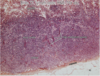
How does bone marrow appear histologically?

A
8
Q

What are the main supporting structures of lymph tissue?

A
  • Reticular fibers
  • **Except the thymus, which has keratin fibers
9
Q

Reticular Fibers

A
  • Type 3 collagen
  • Very thin compared to type 1 collagen
  • Supporting structure of most lymph tissues (except thymus, which uses keratin)
  • Special silver stain
10
Q

Summary features of thymus histologically

A
  • Diffuse organization
  • No nodules/follicles (b/c primary lymph organ)
    • But does have lobules defined by trabeculae
  • Fully encapsulated
    • See trabeculae
  • Has cortex and medulla
  • Hassel’s corpuscles is defining feature
  • **No afferent vessels to thymus, but there are efferent vessels
  • **Uses keratin instead of reticular fibers
11
Q

Histology of thymus

A
  • Cortex stains bluer than medulla
  • No nodules but there are lobules
12
Q

Other cells in the thymus aside from lymphocytes

A
  • Macrophages
  • Dendritic cells
  • Epithelioreticular cells
13
Q

epithelioreticular cells

A
  • Supporting cells of the thymus
  • Synthesize keratin for supporting structure
  • Involved in T-cell education
14
Q

Histology of epithelioreticular cells

A
  • They are found in the thymus
  • Recognize then by a nucleus that’s larger and euchromatic (lighter) than surrounding lymphocytes
15
Q

Thymic venule purpose

A
  • T-cells that finish differentiation into the thymus go into the general circulation (to travel to secondary lymphoid organs) via venules
  • Process is called intravasation
16
Q

Hassal’s corpuscles

A
  • Distinguishing feature of the thymus
  • Found in the medulla of the thymus, especially in the older thymus
17
Q

Young thymus vs adult thymus

A
  • Older thymus has more adipose tissue
  • T-cells leave thymus to populate other organs and the thymus shrinks as a person ages - this is called thymic involution
18
Q

Summary of MALT histology features

A
  • Nodules/follicles present (b/c secondary lymphoid organ where activation occurs)
  • Not encapsulated
    • No trabeculae
  • Located in lamina propria. Exists as a specialized type of connective tissue.
  • Can be either diffuse (in the gut) or nodular (in the esophagus)
19
Q

Primary vs. secondary nodules/follicles

A
  • Primary follicles do not have a lighter region inside of them. The lighter region is the germinal center, so primary follicles lack a germinal center.
  • Secondary follicles have a germinal center.
20
Q

Histology of primary follicle

A
  • No germinal center
  • Follicles only found in secondary lymphoid organs
21
Q

Histology of secondary follicle

A
  • Has a germinal center
  • Follicles only found in secondary lymphoid organs
22
Q

How to distinguish a B-cell from a plasma cell?

A
  • B-cell has nucleus that is very large
  • Plasma cells have eccentric nucleus, lots of endoplasmic reticulum, and a perinuclear hof (due to lots of golgi)
23
Q

Plasma cell histology

A
  • Eccentric nucleus
  • Perinuclear Hof (due to lots of golgi)
  • Can see tons of ER under electron microscope
24
Q

Intraepithelial lymphocytes

A
  • Specialized T-cells that can recognize free antigen without antigen presentation
  • They are found between epithelial cells in MALT
  • Considered part of innate immunity
25
Peyer's patches and appendix
* Both types of MALT * These are both GALT - gut associated lymphatic tissue
26
Tonsils
* Recognize secondary follicles * Half-encapsulated * Non-encapsulated side is the lumenal side * \*\*Crypt = distinguishing feature of tonsils
27
Histology of tonsilar crypts
* Crypts are distinguishing feature of tonsils * These are extensions of stratified squamous epithelium
28
Lymph Node purpose
* To filter (monitor) lymph fluids
29
How does lymph fluid get moved around the body if there is no pump in the lymph system?
* There are lots of one-way valves * Every time your skeletal muscle contracts, it moves lymph fluid
30
Organization of lymph node
* Secondary nodules/follicles * Fully encapsulated * Trebaculae exist * Cortex, deep cortex, medulla, lymphatic sinus * Unique to lymph node: high endothelial venule * \*\*Lymph nodes have both afferent and efferent vessels. (Contrast with thymus which has only efferent.) Lymph nodes are the only organs with afferent vessels.
31
High endothelial venule purpose
* Extravasation * Most of the lymphocytes coming to the lymph node arrive via the blood instead of afferent lymphatics * Lymphocytes move from the capillaries into lymph tissue via these high endothelial venules
32
Histology of high endothelial venules
* Low magnification recognize with unique appearance shown on left image * Bottom right image shows a typical venule, which is flatter. Top right image shows a high endothelial venule, whose cells appear more cuboidal. * Locate high endothelial venules in the deep cortex region
33
Medullary sinus
* The sinus of the lymph node * Lymph fluid drains into medullary sinus and then from here gets drained into efferent lymphatic vessels to leave the lymph node
34
Distribution of cells in the lymph node
* Outer layer: Cortex * Primary and secondary follicles seen here * B-cell area * Next layer: Deep cortex * Find high endothelial venules here * T-cell region * Inner layer: Medulla * Plasma cells (and B-cells) live here
35
Histology of lymph node at low magnification
* Recognize high endothelial venules
36
Purpose of the spleen
* Filter (monitor) blood * Destroys old red blood cells (in red pulp) * Has immune function (in white pulp)
37
Summary features of the spleen
* Has nodules/follicles * Fully encapsulated * Trabeculae present * Contains white pulp, red pulp, venus sinus * Unique features: central artery, PALS
38
Splenic circulation
* Blood enters via **splenic artery** * Blood gets taken deeper into spleen via **trabecular arteries** * When artery leaves trabecula it goes into white pulp area and is called a **central artery** * The central artery is surrounded by lymphocytes and we call this the peri-arterial lymphatic system **(PALS)** * PALS is a layer of lymphocytes covering the central artery and it's the main component of the white pulp * Blood from central arteries gets dumped into **marginal zone** (also white pulp) and then percolates into the **venus sinus** to return to blood circulation
39
White pulp
* Found in the spleen * PALS and marginal zone are in white pulp area * White pulp surrounds central artery
40
3 different sinuses and their functions
* Capillary sinusoid - in bone marrow - lymphocytes leave bone marrow and enter blood circulation * Lymphatic sinus - located in medulla of lymph node - collects lymphatic fluid that is carried away by efferent vessels * Venus sinus - located in spleen - carries blood from spleen back to general circulation
41
Histology of spleen
* White pulp stains blue * Central artery should be in the middle of the white pulp * Recognize follicles with germinal centers * Red pulp stains lighter than white pulp * Organized as cords and sinuses
42
Splenic venus sinus
* Located in red pulp * Collects blood from spleen and delivers it back to general circulation * Endothelial cells are unique b/c of ribbon-like appearance. This allows blood to come into venus sinus.
43
Where does T-cell development take place?
* Thymus
44
What cells are present in the thymus?
* Thymocytes * Dendritic cells * Epithelial cells
45
Cause of DiGeorge's Syndrome
* Thymus does not develop * Results in immunodeficiency
46
What is a double negative T-cell?
* The cell expresses neither CD4 or CD8 on its surface * This is the earliest stage of T-cell development
47
Which loci do RAG enzymes act on first in the T-cells?
* Beta, gamma, and delta * NOT alpha
48
Why does beta rearrangement usually occur instead of gamma or delta?
* There are 2 clusters of genes on the beta locus * If rearrangement fails at the first locus, rearrangement can still occur at the second cluster * Gamma and delta regions do not have 2 clusters
49
What is triggered by successful beta chain rearrangement (TCR)?
* A surrogate alpha chain pails with the beta chain --\> pre-TCR * Further beta rearrangement is stopped * Proliferative burst for successful beta-chain thymocytes --\> T cells express BOTH CD4 and CD8
50
What is a double positive T-cell?
* Expresses both CD4 and CD8 on its cell surface * Occurs after successful beta chain rearrangement --\> proliferative burst
51
Why does alpha rearrangement usually occur before gamma or delta?
* There are so many different V-alpha regions compared to the gamma and delta gene segments * Unproductive rearrangement at one V-alpha region --\> cell can try again with another alpha region. That's not true of gamma and delta segments
52
What happens after successful alpha chain rearrangement?
* Unlike with beta-chain rearrangement, the RAG enzymes maintain the ability to rearrange alpha chain again * This allows continued alpha-chain rearrangement through positive selection --\> allows T-cell a better chance at recognizing self MHC molecules
53
What is positive selection? When does it occur?
* Occurs after successful beta chain and alpha chain rearrangement * This is the process of selecting T-cells that recognize self-MHC for further development * Cortical epithelial cells play a role * After positive selection the thymocytes become either CD4 or CD8 T-cells
54
What cells in the thymus are responsible for educating T-cells during positive selection?
* Cortical epithelial cells * Display both MHC1 and MHC2 molecules on their surface
55
When do thymocytes begin expressing EITHER CD4 or CD8?
* After positive selection
56
When is TCR alpha rearrangement turned off?
* After positive selection
57
What is negative selection? When does it occur?
* The process of deleting T-cells that recognize self peptides * Occurs after positive selection
58
What is the major mechanism in T-cell development of preventing autoimmune disorders?
* Negative selection
59
What is the major mechanism in T-cell development that results in MHC restriction?
* Positive selection
60
What cells assist in negative selection?
* Dendritic cells
61
AIRE
* A transcription factor that occupies the promotor of MANY genes in the thymus * Drives really low levels of protein expression found in different tissues in the body * Major role in negative selection during T-cell development
62
APECED - autoimmune polyendocrinopathy-candiasis ectodermal dystrophy
* Autoimmune disorder resulting from mutations in AIRE transcription factor * T-cells cannot undergo proper negative selection
63
Function of T-regulatory cells
* These are generated during T-cell development * They prevent T-cells of the same clone from recognizing/attacking self peptides once they're all released into the body
64
What are the 2 mechanisms of central tolerance in T-cell development?
1. Negative selection 2. Production of T-regulatory cells
65
Bare lymphocyte syndrome revisited
* Dysfunction in TAP * Do not express MHC1 on cell surface * This means there are no MHC1 markers on thymic epithelial cells --\> body cannot generate CD8 T-cells (b/c they are educated using thymic epithelial cells)
66
Bone Marrow transplant implications of T-cell development
* Donor and recipient MHC markers must match * Donor bone marrow is transplanted * Dendritic cells in recipient come from donor bone marrow. They display donor MHC molecules. * T-cells in recipient come from donor bone marrow. But they are educated on thymic epithelial cells FROM THE RECIPIENT. * T-cells must be educated to recognize MHC markers that will allow them to recognize donor MHC markers on dendritic cells. * If MHC markers don't match, recipient cannot raise an immune response.
67
T-cell activation overview
68
What causes naive T-cells to leave the thymus and enter general circulation?
* Sphingosine-1-phosphate concentration gradient * (S1P is higher in the blood)
69
High endothelial venules
* Specialized endothelial cells in the lymph node through which T-cells enter lymph node
70
T-cell rolling
* T-cell is in blood circulation * When it gets to endothelial cells in lymph node, **L-selectin** on the T-cell interacts with residues on the endothelial cells. This causes it to slow down and is called "rolling.
71
Diapadesis of T-cell (from blood into lymph node)
* T-cell rolling causes T-cell to slow down * Binding of LFA-1 on T-cell to ICAM-1 on endothelial cell causes it to stop and then the T-cell can get through endothelial cells (diapadesis)
72
Two signals must occur for T-cell to be activated
1. Recognition of MHC-peptide 2. CD28 on T-cell must bind B7 on dendritic cell
73
How is B7 expression induced on dendritic cells?
* Dendritic toll-like receptors recognize pathogen --\> B7 expression on cell surface
74
What is special about dendritic cells?
* They express B7 on their cell surface (one of the few cells that does this) * They are central to T-cell activation * They can activate T-cells through both MCH1 and MHC2 pathways due to cross linking
75
What is cross presentation?
* The process that allows dendritic cells to express peptide in both MHC1 and MHC2 molecules * Dendritic cells can engulf a virus, which would NORMALLY lead to MHC2 presentation. But special interferon signaling allows dendritic cells to present viral peptides on MHC1 molecules without the dendritic cells themselves being infected first.
76
Naive T-cells receive both signals to become activated. What's next?
* Formation of the immunological synapse. * ITAMS on CD3 and TCR-zeta become phosphorylated --\> signaling cascade * NFAT, NF-kappaB, AP1 are all transcription factors that become activated * These result in **IL-2 transcription** * IL-2 binds to T-cell (autocrine mechanism) and result in **clonal expansion**
77
Deficiency in gamma portion of IL-2 receptor
* Causes a form of X-linked SCID * Results because gamma portion of IL-2 receptor is also used in IL-7 receptor, which is the cause of SCID
78
Mechanisms of T-cell negative regulation
1. T-cell anergy 2. T-cell apoptosis 3. CTL4 4. T-regulatory cells
79
T-cell anergy - what is it? what is its purpose?
* If TCR recognizes peptide-MHC but does NOT receive costimulatory signal (CD28 binding B7), the T-cell goes into a period of prolonged **HYPOresponsiveness** * This is a safeguard against hyperactiation of T-cells * T-cell only proliferates when B7 is present, which is only present on dendritic cells that have recognized a pathogen
80
What happens if T-cell does not receive costimulatory signal (CD28 binding B7)?
1. T-cell anergy 2. T-cell apoptosis (Fas and Fas ligand)
81
What is the mechanism of T-cell apoptosis when the costimulatory signal is absent?
* Fas receptor is always present on T-cell surface * When T-cell recognizes MHC-peptide but does NOT bind B7, Fas ligand expression is turned on in that T-cell * Fas ligand binds Fas receptor --\> apoptosis (autocrine signaling)
82
CTL4
* Important in negative regulation of T-cells * When T-cells proliferate in response to CD28 binding B7, there needs to be a way to turn them off. This is it. * CD28 binds B7 --\> clonal expansion AND simultaneous expression of CTL4 receptor * CTL4 receptor has a higher affinity for B7 than CD28 does. So it results in contraction of B-cell population after some time.
83
T regulatory cells
* Important in negative regulation of T-cells * Generated in the thymus during T-cell development * Express FoxP3 transcription factor * Recognize MHC2-peptide on antigen presenting cells and suppress the activation of CD4 cells
84
Autoimmune lymphoproliferative syndrome (ALPS)
* Results from mutations/defects in Fas or Fas ligand --\> T-cell populations cannot contract properly --\> enlarged lymph nodes
85
Immunodysregulation, polyendocrinopathy, enteropathy, X-linked (IPEX)
* Results from mutations in FoxP3 * Lack T-regulatory cells
86
CD4 T-cells
* Th1 * Th2 * Tfh * Th17
87
CD8 T-cells
* Cytotoxic T-cells
88
What is the function of Th1 cells?
* Activate macrophages for more effective killing
89
Which effector T-cells act at the site of infection?
* Cytotoxic T-cells * Th1 * Th17
90
Which effector T-cells remain in the lymph node?
* Th2 * Tfh
91
How do Th1 cells activate macrophages?
* CD40 ligand on Th1 binds CD40 receptor on macrophage * Th1 secretes IFN-gamma, which binds to macrophage * Both signals together activate macrophage
92
What happens if mycobacteria still survive even after macrophage activation?
* granuloma formation
93
What is the role of Th2 and Tfh?
* Help B-cells differentiate into plasma cells * B-cells need a signal from BCR AND help from one of these T-cells to become a plasma cell
94
How do Tfh and Th2 activate B-cells?
* Effector T-cells recognize peptide-MHC2 complex on B-cells * Effector T-cells have CD40 ligand, which binds CD40 receptor on B-cells * Effector T-cells release cytokine (IL-4 for Th4; IL-21 for Tfh), which bind to the B-cell. * Both signals - CD40 ligand and IL binding - cause B-cell clonal expansion and differentiation into plasma cells
95
What is the function of Th17?
* Recruit neutrophils to site of infection
96
What is the functon of cytotoxic T-cells?
* To kill virally infected cells * This is triggered by viral peptide recognition on MHC1 molecules * These are CD8 T-cells
97
How do cytotoxic T-cells kill their targets?
1. perforin and granzymes 2. Fas ligand on T-cell is induced; Binds Fas receptor on target cell 3. Secretes IFN-gamma --\> inhibits viral replication in cells in the vicinity, activates macrophages
98
Cytokine that triggers Th1 differentiation
* IL-12
99
Cytokine that triggers Th2 differentiation
* IL-4
100
Cytokine that triggers Tfh differentiation
* IL-6 and IL-21
101
Cytokine that triggers Th17 differentiation
* IL-6 * TGF-beta
102
Effector cytokine of Th1
IFN-gamma
103
Effector cytokine of Th2
IL-4
104
Effector cytokine of Tfh
IL-21
105
Effector cytokine of Th17
IL-17
106
What is the purpose of the primary immune response?
1. Clear the infection 2. Strengthens immune system temporarily against re-infection 3. Produce memory cells to provide long-term protection against the same pathogen
107
Overview of B-cell Activation
108
Two signals needed by a B-cell in order to activate
1. BCR signal - recognition of free antigen 2. Helper T-cell signals * CD40L on helper T-cell binds CD40 on B-cell * Cytokine (IL-4) from helper T-cell binds B-cell receptor
109
What is linked T-cell help?
* Mechanism by which T-cells help B-cells produce antibodies against non-protein antigens * T-cells can only recognize peptide fragments, but B-cells can recognize things like carbohydrates * B-cell recognizes carbohydrate antigen that is linked to the protein, chews it up, displays PROTEIN in MHC2 molecule * T-cell recognizes protein component but still helps B-cell proliferate and generate Abs for the carbohydrate component
110
Where does B-cell activation occur?
* Area between B-cell center (outer cortex) and T-cell area (inner cortex)
111
Where do B-cells go AFTER activation?
* Dark zone of the germinal center
112
What is Activated Induced Cytidine Deaminase (AID)?
* This enzyme gets expressed when B-cells are activated * It is the master regulator of somatic hypermutation and class-switching
113
Follicular dendritic cells
* Display the original antigen that the BCR recognized * These are in the light zone * The B-cell undergoes somatic hypermutation and then "samples" the original antigen from the surface of the FDC's. * Purpose is to ensure that somatic hypermutation does not result in B-cells that no longer recognize the original antigen and prevent B-cells that might actually recognize self
114
After B-cells undergo somatic hypermutation and Class switching, what happens?
* They can return to dark zone to proliferate more * They can exit germinal center as memory cells * They can exit the germinal center as plasma cells
115
What are the primary antibodies used/generated for vaccines?
* IgG (blood, skin, tissues) * IgA (mucosal surfaces) * IgM (blood, but less useful b/c IgM doesn't undergo any maturation)
116
Antibodies that neutralize pathogens
IgG IgA
117
Antibodies that do opsonization primarily
IgG
118
Antibodies that result in complement activation
IgM IgA
119
Antibody that binds virus-infected host cells and promotes lysis by NK cells
IgG
120
Subunit vaccines
* Generate antibodies against particular surface components on a virus * Ex: Hep B virus vaccine
121
Vaccines against bacterial toxins
* Generate antibodies that neutralize the bacterial toxins * Made by purifying the toxin and treating with formalin so it is inactive --\> toxoid * Ex: diptheria, tetanus
122
Vaccines against polysaccharides
* Directed against outer capsule of bacteria * Generate antibodies that promote complement fixation * a unique type of inactivated subunit vaccine composed of long chains of sugar molecules that make up the surface capsule of certain bacteria. Pure polysaccharide vaccines are available for three diseases: pneumococcal disease, meningococcal disease, and Salmonella Typhi.
123
Conjugate vaccines
* bacterial polysaccharide is conjugated to a carrier protein * Elicits linked T-cell help against polysaccharide antigens * ex: H.influenzae
124
Live vs. inactivated vaccines
* Inactivated = treated so they can no longer replicate * Live vaccines = treated so they are very poor at replication
125
Passive immunity
ex: IVIG infusions ex: used for antivenoms
126
Sabin vs. Salk vaccines
* Sabin = live, attenuated * Can replicate clunkily * Non-pathogenic * Humoral AND cell-mediated immunity (i.e. B-cells can work on it via humoral but cytotoxic T-cells work on it via cell-mediated) * Salk = inactive * Dead * Cannot replicate * Results in humoral response only (once the cell is infected, the B-cell cannot get to it) https://www.youtube.com/watch?v=Ovype5DUI04
127
Protein vs. polysaccharide vaccines
_[https://www.youtube.com/watch?v=-Qu2ROOfpLc](https://www.youtube.com/watch?v=-Qu2ROOfpLc)_
128
What are the Thelper cells in germinal centers
Tfh
129
How are memory T-cells produced?
130
Central memory T-cells vs. Effector memory T-cells
* Central memory T-cells express L-selectin and CCR7. L-selectin is an adhesive molecule; CCR7 is a chemokyne. These allow central T-cells to stay in lymphoid organs. * Effector memory T-cells do not express either of these molecules on their cell surface --\> they can leave lymphoid organs
131
what are the main cells of the acute inflammatory response?
neutrophils
132
what are the main cells of the chronic inflammatory response?
lymphocytes
133
Transudate vs. Exudate
* Transudate = low protein content * Exudate = high protein content
134
Common causes of transudates
* Heart failure * Renal disease * Associated with high proportion of non-cellular fluid
135
Common cause of exudates
* Inflammation
136
Types of exudates
* non-cellular * cellular * mixed
137
Example of non-cellular exudate
* fibrinous exudate
138
Types of cellular exudate
* Purulent * Suppurative * Cellulitis/phlegmonous
139
Key characteristic of purulent exudate
* No tissue destruction
140
Key characteristic of suppurative exudate
* Tissue destruction by liquefactive necrosis
141
Key characteristic of cellulitis
* Movement of inflammatory fluid * Requires clinical diagnosis, cannot diagnose on the slides
142
Septic shock
* Systemic response to acute inflammation
143
The triad that you see with septic shock
* Disseminated intravascular coagulation * Hypoglycemia * Hypotension/heart failure
144
Disseminated intravascular coagulation
* Sign of acute inflammation * Cytokines induce expression of a protein called tissue factor, which is involved in coagulation. This results in blood clots (thrombi) in your vessels and death of the tissue. * If you use up all of this tissue factor protein during coagulation response, you don't have enough leftover to clot effectively when you are actually bleeding. So a cut leads to bleeding excessively. * Paradoxical coagulation and bleeding is called "disseminated intravascular coagulation"
145
Granulomatous Inflammation
* A subtype of chronic inflammation * Main cells: macrophages
146
Hallmark of granulomatous inflammation
* macrophage mobs = granuloma * Giant cells = multinucleated fusion of macrophages * Lymphocytes surrounding the granuloma
147
Types of granulomatous inflammation
* Immune * ex: TB * Foreign body * ex: sutures
148
Outcomes of Inflammation
* Resolution * Injurious agent is removed * Healing by connective tissue replacement * Scarring * Chronic inflammation
149
What must be true for complete resolution of inflammation to occur?
* Injury must not cause permanent damage * Tissue must have regenerative capacity
150
What must be true for scar formation to occur?
* Tissue is not capable of regeneration * There is significant tissue destruction * There is abundant fibrinous exudate
151
What are the hallmarks of scarring?
* Removal of dead tissue by macrophages * Dead tissue replaced by granulation tissue * See proliferating fibroblasts * See blood vessels
152
**What process is going on if you see granulation tissue?**
* Healing * When there’s healing, we call it organization. Because the tissue will eventually organize into a scar. * Fibroblasts secrete collagen that will undergo restructuring to form a scar. * We call this granulation tissue **“organizing exudate”**
153
what happens with too much granulation tissue?
–exuberant granulation tissue
154
well-differentiated vs. poorly-differentiated
* Well-differentiated means the tumor resembles normal cells/tissue * Poorly-differentiated means it's hard to tell what tissue the tumor arose from
155
Characteristic of benign tumors
* Well-differentiated * circumscribed (clear borders)
156
Characteristic of malignant tumors
* METASTASIS * invasive
157
Dysplasia
* disordered growth * pre-cancerous
158
Hallmarks of dysplasia
* pleomorphism * nuclear abnormalities * High N-C ratio * Mitotic figures
159
Desmoplasia
* Stroma surrounds a malignant neoplasm * \*\*Characteristic of malignancy
160
What is the purpose of desmoplastic stroma?
* Host response: wall off the bad tissue * Cancer: Recruit blood vessels to help it grow
161
Barrett's esophagus
* Example of metaplasia (cells change from one type of epithelium to another in response to damage) * Example of the adeno-carcinoma sequence * Metaplasia turns to dysplasia turns to cancer
162
Adenoma
* Benign tumor of gland-forming epithelium
163
carcinoma
* Malignant tumor of the epithelium
164
sarcoma
malignant tumor of non-epithelium (mesenchyme)
165
adenocarcinoma
* malignant tumor of gland-forming epithelium
166
squamous cell carcinoma
* malignant tumor of squamous epithelium (as opposed to gland-forming epithelium)
167
polyp
tissue growing into any hollow organ.