Week 2: Lower Limbs and Glutes Flashcards
(27 cards)
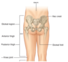
Where is the gluteal region and what is it’s significance?
The gluteal region is between the iliac crest and the bottom border of the buttock
Important nerves pass from the pelvis to the gluteal region and lower extremity
What are the main muscles (superficial and deep) of the gluteal region?
Superficial: gluteus maximus, gluteus medius, gluteus minimus
Deep: piriformis, which performs lateral rotation of the thigh

What are the functions of the superficial gluteal region muscles?
Gluteus maximus - extensor of thigh from the seated position
Gluteus medius and minimus - deep to gluteus maximus, abduction of hip, secure position of the pelvis when walking
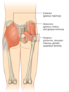
What is the main deep muscle of the gluteal region, and what does it do?
The piriformis is a part of the pelvic wall, and divides the greater sciatic foramen where important vessels pass above and below
It is also the lateral rotator of the thigh

What are the main nerves of the gluteal region?
The sciatic nerve, a VERY LARGE nerve that passes below the piriformis deep to the gluteus maximus, traveling to the posterior thigh
and
Pudendal nerve, which passes below the piriformis in the greater sciatic foramen and enters the perineum (innervates external genetalia elements)

What are the principal nerves of the lumbosacral plexus?
Sciatic
Pudendal
Femoral
Obturator
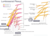
What are the functions of the nerves of the lumbosacral plexus?
Sciatic - innervates posterior leg, thigh and foot, feeds into tibial nerve (posterior leg/foot) and common fibular nerve (anterior/lateral leg/foot)
Pudendal - innervates the perineum and external genitalia (somatic)
Obturator - innervates the medial thigh
Femoral - innervates the anterior thigh

What are the three compartments of the thigh, and what are their common actions?
Anterior - extend knee
Medial - abduct thigh
Posterior - flex the knee
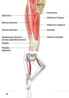
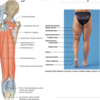
What are the main regions of the thigh, what muscles are within them, and what nerves are they innervated by?
The anterior compartment contains the sartorius, rectus femoris, and vastus lateralis, intermedius, and medialis (femoral nerve)
The medial compartment contains the gracilis, pectineus, and adductor group of the adductor longus, brevis, and magnus (obturator nerve)
The posterior compartment contains the hamstrings - biceps femoris, semitendinosus, and semimembranosus (sciatic nerve)

What are the muscles of the anterior compartment of the thigh and what do they do?
The sartorius flexes the knee and hip, allows for external rotation and abduction (“is there gum on my shoe?” muscle)
The quadriceps contain the rectus femoris, the vastus lateralis, intermedius and medialis. They all insert on the patellar tendon via the quadriceps femoris tendon.
Vastus group extends the knee
Rectus femoris extends knee AND flexes at the hip
Which two thigh muscles can act on the hip?
The rectus femoris and sartorius (both go “across” the thigh)

What muscles are in the medial compartment of the thigh and what is their function?
The gracilis and adductors (longus, brevis, and magnus) all function to adduct the femur/thigh and help stabilize you when standing. The adductor longus also participates in lateral rotation and flexion of the thigh. The pectineus muscle flexes and adducts the thigh.

What is the femoral triangle and what does it contain? How do we remember the organization of these elements from lateral to medial?
It is a bundle of nerves (femoral nerve), vessels (femoral artery and vein), and lymphatics
Lat - NAVeL - Med
Nerve Artery Vein Lymphatics
These structures are continuous inferiorly with the adductor canal, supply the lower limb, and allow passage of the femoral A/V down the medial side of the thigh and to the popliteal fossa

What are the major muscles of the hamstring and what is their collective action?
The hamstring contains the:
biceps femoris
semitendinosus
semimembranosus
They all work to flex the knee and extend the hip from the standing position.
What muscles insert on the medial side of the knee? What compartment do they come from? What is this structure ultimately called?
The gracilis (medial)
semitendinosus (posterior)
sartorius (anterior) muscles all insert medially
This is called the pes anserinus (literally the “goose’s foot”) and
What is the popliteal fossa and what does it contain?
It is the space on the posterior aspect of the knee, and contains the popliteal A/V (continuation of femoral A/V), tibial and common fibular nerves, as well as branches of the sciatic nerve

What are the three compartments of the leg and what is the general action of each?
Posterior (plantar flexion, flex toes, invert foot)
Lateral (eversion of foot)
Anterior (dorsiflexion, invert foot, extend toes)
What are the superficial and deep muscles of the posterior leg compartment? What are they innervated by?
Superficial: gastrocnemius, soleus
Deep: flexor hallucis longus, flexor digitorum longus, tibialis posterior
All of these are innervated by the tibial nerve
What are the muscles of the lateral leg compartment? What are they innervated by?
Fibularis longus and brevis, which are innervated by the common fibular nerve
What are the muscles of the anterior leg compartment? What are they innervated by?
The tibialis anterior, extensor hallucis longus, and extensor digitorum longus are all innervated by the common fibular nerve
What are the superficial muscles of the posterior leg compartment and where do they insert?
The gastrocnemius and soleus muscles enact plantarflexion (both) of the foot, and flex the knee (gastrocnem.)
They both insert via the calcaneal tendon onto the calcaneus bone (heel)

What are the deep muscles of the posterior compartment of the leg? What actions are they involved in?
The tibialis posterior is involved in inversion and plantarflexion (weak, acts on ankle)
and the flexor digitorum longus and hallucis longus allow for flexion of toes (tendons extend through plantar surface to the phalanges)

What are the muscles of the anterior compartment of the leg, and what actions are they involved in?
The tiabilis anterior attaches to the medial food and is involved in dorsiflexion and inversion of the foot
The extensor digitorum longus and hallucis longus are involved in dorsiflexion and extension of the toes

What are the muscles of the lateral leg compartment and what movement are they involved in?
The fibularis longus and brevis are involved in eversion of the ankle


