Week 3 - F - Small bowel - Obstruction/Ischaemia (acute/chronic)/Meckel's diverticulum/Tumours, Appendicitis (mass/abscess) Flashcards
(38 cards)
Which arteries does the small bowel blood supply come from?
Some bowel blood supply comes from the jejunal and ileal branches of the superior mesenteric artery (comes from aorta at L1)

What can wrong with the small bowel Many different things can go wrong - big problems include * Obstruction * Infarction * Bleeding
What are two main causes of small bowel obstruction?
Causes can be split into things that occur within the lumen, within the wall and more commonly - outside the wall
Outside the bowel wall - Intra-abdominal adhesions are the most common cause of small bowel obstruction, followed by hernias.
Rarer causes tend to occur within the bowel wall or within the lumen of the bowel itself What are examples of these causes of small bowel obstruction? (2)(3)
Small bowel obstruction caused within the lumen
* Gallstone ileus
* Food / Bezoar (a solid mass of indigestible material that accumulates in your digestive tract, sometimes causing a blockage)
Small bowel obstruction caused within the wall
* Tumour
* Crohn’s
* Radiation
What is the typical presentation in a patient with small bowel obstruction? What may be seen on examination?
Typical presentation
* Distension
* Vomiting
* Borborgymi - rumbling stomach
* Pain
* Faeculent vomiting
On examination may see scars meaning previous abdominal operation (predisposes to adhesions) or need to look for signs of small bowel hernia - both femoral and inguinal
How is the diagnosis of small bowel obstruction confirmed?
* Abdominal Xray (AXR)
* CT scan of abomen
Gastrogaffin studies may help with CT - radiographic dye is swallowed by the patient
How is AXR able to differentiate between small and large bowel obstuction?
In small bowel obstruction - AXR will show central gas shadows with plicae circularis (aka valvulae conniventes) that completely cross the lumen
In large bowel obstructin AXR will show peripheral gas shadows proximal t the blockage ie in caecum but not in rectum

What is the management of small bowel obstruction?
MANAGEMENT
Usually conservative - DRIP AND SUCK method
* Start IV fluids and correct any electrolyte disturbances (‘drip’)
* Make the patient nil-by-mouth (NBM) and insert a nasogastric tube to decompress the bowel (‘sucks out air and fluid to relive abdominal swelling’)
* Urinary catheter and fluid balance
* Analgesia as required with suitable anti-emetics
Adhesional small bowel obstruction resulting from previous surgery is treated conservatively in the first instance, with a success rate of around 80% Why do you not want to carry out surgery for adhesional small bowel obstruction? How long can drip and suck usually be continued up until? Does the drip and suck method treat hernias?
As far as prevention of adhesional small bowel obstruction is concerned, best approach is to avoid operation as any surgical procedure predisposes to adhesions
* Drip and suck method only treats adhesional small bowel obstruction -up to 72 hour is standard
* Hernias cannot be treated with drip and suck and usually require reduction - manually - to prevent stangulation and necrosis and if problematic surgical reduction
When should you surgically intervene in a patient with small bowel obstruction?
Intervene with emergency surgery if there is a strangulation of the bowel as this can cause ischemia and necrosis
Also intervene if there is small bowel perforation
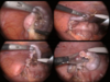
What is the surgical management of an obstructed small bowel - carried out if suspecting strnagulation or perforation? What type of incision? Which drugs are given?
Surgical management of small bowel obstruction is usually via a laparotomy using a midline incison
Antibiotics and antithromboembolic measures are also taken

Mesenteric ischaemia Mesenteric ischaemia can be classified as chronic or acute What are the main causes of both?
Acute mesenteric ischaemia usually occurs due
* to mesenteric artery occlusion (usually SMA) due to embolus or thrombosus
* mesenteric vein thrombosis
Chronic mesenteric ischaemia usually occurs due to (rare and difficult to diagnose)
* atherosclerotic disease in all three mesenteric arteries (coeliac, super mesenteric, inferior mesenteric)
Aside from ischaemia of the small bowel occurring due to * Mesenteric artery atherosclerosis * Thrombosis formation or embolism
What are non-occlusive perfusion insufficiency that can cause the ischaemia?
Non-occlusive perfusion insufficiency of the small bowel can occur due to
* Shock
* Strangulation obstructing venous return
* Drugs eg cocain
* Hyperviscosity
What is chronic mesenteric ischaemia often referred to as? What is its presentation?
Chronic mesenteric ischaemia is often referred to as angina of the gut Triad of
* Severe, colicky post-prandial pain (gut claudication)
* Decreased weight (eating hurts)
* Upper abdominal bruit (due to atherosclerotic vessels)
Also can occur with PR bleeding, malabsorption and nausea/vomiting
How does acute mesenteric ischaemia tend to present? Where do the emboli come from?
Acute mesenteric ischaemia tends to present as
* Acute severe abdominal pain - constant and central/around RIF
* No/minimal abdominal signs
* Rapid hypovolaemia (shock)
The emboli usually comes from atrial fibrillation, thrombus forms in Left atrium, breaks off and sticks in the narrow superior mesenteric artery
* AF with abdo pain always prompt thoughts of mesenteric ischaemia
Is chronic or acute mesenteric ischaemia more common?
Acute mesenteric ischaemia is more common however it can present as chronic
What is odd about the pain in acute mesenteric ischaemia? What is seen on blood tests in acute mesenteric ischaemia?
The pain in acute mesenteric ischaemia is out of proportion to the clinical findings
* Hb may be elevated
* WCC may be elevated
* Consistent metabolic acidosis - high lactate due to anaerobic bowel
What is used to try and diagnose mesenteric ischaemia? (acute or chronic)
CXR - may show evidence of a gasless abdomen
CT angiography is usually used to find evidence of ischaemia
Often the diagnosis is made at laparotomy if acute mesenteric ischaemia
What is the progression of acute mesenteric ischaemia as time goes on? Can be classified by the degree of infarction
Mucosal infarct - mucosal layer
Mural infarct - mucosal and submucosal layer affected
Transmural infarct - full thickness infarct of the gut

What are the complications of acute mesenteric ischaemia? Why is it important to carry out surgery in a patient with chronic mesenteric ischaemia?
Complications include
Fibrosis/stricture of the small bowel
Chronic ischaemia
Gangrene resulting from necrosed tissue
DEATH - poor prognosis
Important to carry out surgery if patient is diagnosed with chronic mesenteric ischaeima - angioplasty and stenting - this is to prevent ongoing risk of acute mesenteric ischaemia
What is the treatment for acute mesenteric ischaemia?
Resuscitate the patient if shock and provide antibitoics
* URGENT SURGERY
* Dead bowel must be resected
* Attempt to revascularise viable small bowel
What is meckel’s diverticulum?
Meckel’s diverticulum is the result of incomplete regression of the vitello-intestinal duct
The omphalomesenteric duct (omphaloenteric duct, vitelline duct or yolk stalk) normally connects the embryonic midgut to the yolk sac ventrally, providing nutrients to the midgut during embryonic development.
The vitelline duct narrows progressively and disappears between the 5th and 8th weeks gestation.
In Meckel’s diverticulum, the proximal part of vitelline duct fails to regress and involute, which remains as a remnant of variable length and location
What does meckel’s diverticulum contain?
Meckel’s diverticulum, formed due to failure of complete regression of the the vitello-intestinal duct contains ectopic ileal, gastric or pancreatic mucosa
What is the rule of 2s in Meckel’s diverticulum?
The tubular structure
Occurs in about 2% of the population
Only 2% of cases are symptomatic
Usually presents among children at the age of 2
Is 2 inches long
Is 2foot (60cm) from the ileocaecal valve

What is the presentation Meckel’s diverticulum?
Meckel’s diverticulum are diagnosed when complications manifest or incidentally in unrelated conditions such as laparotomy, laparoscopy or contrast study of the small intestine.
* Inflammation can cause symptoms that mimic appendicitis
* Can present as rectal bleeding
* Can present as intestinal obstruction