Week 1/2 - A(2) - Anatomy 3&4 - Liver, Spleen, Gallbladder, Pancreas and Small Intestines, arterial/venous supply/biliary tree Flashcards
(33 cards)
What are the foregut organs? What is the main artery that comes from the abdominal aorta to supply it and at what level? What does it trifurcate into?
Foregut organs - oeseophagus, stomach to mid duodenum (also liver, gallbladder, spleen, 1/2 of pancreas) The coeliac trunk (coeliac axis) supplies the organs of the foregut Coeliac trifurcates into the left gastric, spelnic and common hepatic artery

Different branches arise from the trifurcation of the coeliac trunk Can you name the branches arising from these arteries? * Left gastric - branches (2) * Splenic artery - branches (2) and arteries (2) * Common hepatic artery - arteries (2) Which goes to the fundus of the stomach? Which goes to greater curvature of the stomach?
Left gastric - gives gastric & oeseophageal branches Splenic - gives splenic branches & pancreatic branches, short gastric arteries (fundus) left gastro-omental artery (greater curvature) Common hepatic - proper hepatic & gastroduodenal artery

The two arteries that arise from the common hepatic artery have their own branches What are these?
Gastroduodenal artery - right gastro-omental artery and the superior pancreatico-duodendal artery Proper hepatic artery - right gastric, right and left hepatic artery Right hepatic artery gives the cystic artery

How would the route of the splenic artery be described? What are the anatomical relations of the spleen? Is it intra or retroperitoneal? What ribs protect it?
The splenic artery has a torturous route to the spleen The spleen has Diaphragm posteriorly Stomach anteriorly Splenic flexure inferiorly Left kidney medially Spleen is protected by ribs 9-11 - fracture could pierce it

What is the blood supply to the stomach? State which artery the branch arises from (no need to do this if arising from coeliac trunk) Which arteries anastamose?
Short gastric arteries (from splenic) - fundus Right gastric (from proper hepatic branch of common hepatic) and left gastric anastamose at the lesser curvature Right gastro-oemental (from gastroduodenal branch of common hepatic) and left gastro-omental (from splenic) anastamose at the greater curvature.

What is the blood supply to the liver - state what forms these vessels? Which ribs protect the liver? What are the four anatomical segements?
Hepatic artery (proper hepatic giving right and left) only supplies 20-25% Hepatic portal vein (union of spelnic vein with SMV) - supplies the rest Liver protected by ribs 7-11 4 anatomical segments * Right lobe * Left lobe * Caudate lobe (superior) * Quadrate lobe (inferior)

What are the anatomical relations of the liver? Do the hepatic veins or IVC have valves?
Diaphragm superiorly, anterior and posteriorly Anterior aspect of stomach medially Gallbladder posteriorly and inferiorly Hepatic flexure inferiorly Right kidney/adrenal gland, IVC and abdominal aorta posteriorly

What does each functional segment of the liver have to allow it function? What are the two clinically important areas of the peritoneal cavity related to the liver and which is the most inferior part of the peritoneal cavity when lying flat?
Each functional segment has its own blood supply (hepatic artery and portal vein), venous drainage and bile drainage Hepatorenal recess (between liver and posterior abdominal wall) - lowest point when lying flat (aka Morison’s pouch)* sub-phrenic recess (between liver and diaphragm)

Which arteries supply the foregut, midgut and hindgut? What vertebral level do the arteries arise? Which veins drain the foregut, midgut and hindgut? Where do the veins drain into?
Foregut - Coeliac trunk - T12. Splenic vein -> hepatic portal vein Midgut - Superior mesenteric artery- L1 Superior mesenteric vein-> hepatic portal vein Hindgut - Inferior mesenteric artery - L3. Inferior mesenteric vein -> splenic vein

Where does the hepatic portal vein drain and for what? Where does the blood from the liver drain?
Hepatic portal vein drains blood from the foregut, midgut and hndgut to the liver for first pass metabolism ‘Cleaned’ blood from the liver drains into sinusoids which drain into central veins which drain into hepatic veins - hepatic veins drain into the inferior vena cava which goes back to right atrium

What attaches the liver to the diaphragm? What attaches the liver to the anterior abdominal wall? * What lobes do the remnant of the umbilical vein and ductus venosus separate? * What are the remnants known as?
Coronary ligaments attach the liver to the diaphragm Falciform ligaments attach the liver to the anterior abdominal wall Ligamentum teres aka round ligament (umbilical vein remnant) separates the quadrate and left lobe Ligamentum venosum (ductus venosus remnant) separates the caudate and left lobe

What are the two parts of the gallbladder? Which part narrows to become the cystic duct and what is this a potential site for? What is the blood supply and where is pain felt?
Two parts are the body and neck of the gallbladder Neck narrows to become the cystic duct - potential site for gallastone impaction Blood supply is via the cystic artery (branch of right hepatic artery) Pain is felt in the right upper quadrant - can refer to right shoulder

Define jaundice?
Jaundice is a clinical sign describing yellow pigmentation of the skin, sclera, and mucous membranes due to raised plasma bilirubin, a bile pigment
What is bilirubin? Where is bilirubin formed? What is bilirubin used to form? Where does this product then travel?
Bilirubin is a normal by-product of red blood cells Bilirubin is formed in the spleen as this is where the breakdown of red blood cells mainly occurs The bilirubin is then used to form bile in the liver Bile travels through the biliary tree * a set of tubes connecting the liver to the 2nd part of the duodenum
What is the gallbladders role in bilirubin? What is bile important for? What does the pancreas also excrete into the 2nd part of the duodenum? What is this necessary for?
The gallbladder stores and concentrates the bile Bile is important for the normal absorption of fats from the small intestine The pancreas also excretes digestive enzymes into the 2nd part of the duodenum which is necessary for the digestion of food
Where is the portal triad found - both names? What does it consist of?
Portal triad is found in the free edge of the lesser omentum aka the hapatoduodenal ligament It consists of the Proper hepatic artery Hepatic portal vein Common bile duct

How is the common bile duct formed? What does this join with to form? Where does it drain?
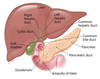
Common bile duct is formed from the cystic duct joining with the common hepatic duct The common bile duct joins with the pancreatic duct to form the Ampulla of vater which drains through the major duodenal papilla into the 2nd half of the duodenum
What is an anatomical sphincter? * What are the anatomical sphincters in the biliary tree? * What type of muscle are they?
An anatomical sphincter is a discrete area where muscle completely encircles the lumen of the tract. The three anatomical sphincters are formed from smooth muscle - Common bile duct sphincter Pancreatic duct sphincter Sphincter of oddi (at the major dudodenal papilla)

What is the purpose of the sphincter of oddi?
Purpose is to control the flow of bile and pancreatic juices into the dudoenum and prevent reflux of duodenal contents into the ducts
What does ERCP stand for? What does it do? How does it enter the biliary tree?
Endoscopic retrograde chlangiopancreatography Investigative and often treatment used to study the biliary tree & the pancreas Endoscope inserted though oral cavity to duodenum where a cannula is inserted into the major duodenal papilla- radio-opaque dye injected for visualization

Name two obstructions of the biliary tree that can cause post-hepatic jaundice? How does it lead to this?
Post-hepatic jaundice - eg due to gallstones or carcinoma of the head of the pancreas Cause bile to flow back into the liver leading to increased bilirubin levels
What are the 5 parts of the pancreas? What part does the duodenum form a C-shape around? What are the functions of the pancreas?
Pancreas - head, neck, body, tail and ucinate process The duodenum forms a C-shape around the head of the pancreas Exocrine pancreas secretes digestive enzymes into the pancreatic duct Endocrine pancreas is important in insulin + glucagon production

Exocrine pancreas secretes digestive enzymes into the pancreatic duct - what cells secrete this? Endocrine pancreas is important in insulin + glucagon production - what cells secrete this?
Exocrine pancreas - It is the acinar cells that secrete the pancreatic digestive enzymes into the pancreatic duct Endocrine pancreas - it is the islets of Langerhans that secrete insulin and glucagon into the blood stream

One of the reasons for pain arising from the pancreas is secondary to inflammation - pancratitis What are the different causes of pancreatitis? (GET SMASHED) Where is pain felt and where can it radiate to?
Pancreatitis Gallstones, Ethanol (alcohol), Trauma, Steroids, Mumps, Autoimmune, Scorpion bites, Hyperlipidaemia / hypercalcaemia, ERCP, Drugs Pain is usually felt epigastric and/or umbilical, can radiate to patients back


