10/16- Appendix and Peritoneum, Pathology Flashcards

1
Q

>95% of biliary tract disease is attributable to ____

A

>95% of biliary tract disease is attributable to cholelithiasis

  • > 20M Americans in the US have gallstones
  • Cholecystectomy done 600,000/yr (one of most common abdominal operations)
2
Q

Describe the anatomy of the gallbladder

A
3
Q

Describe the histology of the gallbladder (layers)

A
  • Mucosa
  • No discrete muscularis mucosae/submucosa
  • Fibromuscularis layer
  • Subserosal fat with vessels
  • Serosa
  • Except no serosa on hepatic bed
4
Q

Characteristics of gallstones

  • Prevalence in developed countries
  • More silent or symptomatic
  • Percentage undergoing surgery
  • Risks/associations
  • Common stone composition
A
  • Prevalence in developed countries: 10-20% of adults
  • More silent (>80%)
  • Percentage undergoing surgery: > 50%
  • Gallbladder cancer in long-standing cholecystitis with gallstones
  • Cholesteral stones are common (80%)
  • > 50% crystalline cholesterol monohydrate
5
Q

What is seen here?

A

Gallstones (?)

6
Q

What are the different types/presentations of cholecystitis?

A

Acute

  • Acalculous (without stones)
  • Calculous (with stones)

Chronic

  • Almost always in the setting of a gallstone
7
Q

What is seen here?

A

Acute cholecystitis

8
Q

What is seen here?

A

Acute cholecystitis

9
Q

What is seen here?

A

Chronic cholecystitis

10
Q

Characteristics of gallbladder carcinoma

  • Common or Rare
  • Age of onset
  • Gender
  • Most common type
  • Associated factors
A

Intestinal metaplasia -> dysplasia

  • Relatively uncommon
  • most > 60 yo (average 72 yrs)
  • 75% women
  • Mostly adenocarcinoma (90%)
  • Associated factors: gallstones (2/3)
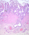
11
Q

What is seen here?

A

Gallbladder carcinoma

12
Q

What is seen here?

A

Gallbladder carcinoma

13
Q

What is seen here?

A

Gallbladder carcinoma

14
Q

What pathology can occur with the appendix?

A
  • Acute and chronic inflammation
  • Tumors
15
Q

Describe anatomy of the appendix- what is it?

  • Length
  • Diameter
  • Layers
A
  • Long, narrow and worm-shaped tube
  • Normal true diverticulum of the cecum
  • 1-9” long; 3” in diameter on average
  • Mucosa, submucosa, muscularis propria(?) and serosa
  • Mucosa: abundant lymphoid tissue especially in young people
16
Q

Layers of appendix?

A
  • Mucosa with crypts of Lieberkuhn
  • Submucosa
  • Circular muscle
  • Longitudinal muscle
  • Serosa (visceral peritoneum
  • Mesoappendix
17
Q

What is seen here?

A

Appendix

  • Mucosa
  • Submucosa
  • Muscularis mucosa
  • Serosa
18
Q

What is the function of the appendix?

A
  • Uncertain
  • Mucosal immunity
  • B cell lymphocyts from appendix migrate and populate distant sites of gastrointestinal lamina propria and evolve into functional IgA secreting plasma cells
19
Q

Characteristics of acute appendicitis:

  • Age
  • Lifetime risk
  • Gender
  • DDx includes
A
  • Most in adolescents and young adults
  • Lifetime risk = 7%
  • More in males
  • Confused with other intra-abdominal or pelvic pathologies
20
Q

How does acute appendicitis present?

A
  • Periumbilical pain, RLQ pain
  • N/V, low-grade fever
  • Mildly elevated WBC
  • Classic physical finding: McBurney’s sign
  • On spinoumbilical line

Diagnosis of acute appendicitis in young children and elderly is problematic (atypical clinical presentations)

21
Q

What are complications of acute appendicitis?

A
  • Perforation
  • Pyelophlebitis
  • Portal venous thrombosis
  • Liver abscess
  • Bacteremia
22
Q

What is seen here?

A

Removed appendix (acute appendicitis?)

23
Q

Describe the pathology of acute appendicitis

  • Early
  • Diagnosis
  • Severe
A

Early phase:

  • Congestion of subserosal vessels with
  • Modest perivascular neutrophilic infiltrate of all layers of the wall

Diagnosis:

  • Neutrophilic infiltration of the muscularis propria

More severe cases:

  • Prominent neutrophilic exudate generates a serosal fibrinopurulent reaction
24
Q

What is seen here?

A

Appendicitis

  • Focal ulceration of mucosa
  • Collection of neutrophils
25
Q

What is seen here?

A

Neutrophils in muscularis mucosa

26
Q

Describe the pathology of chronic appendicitis

A

(or is this more acute pathology??)

  • Focal abscesses within the wall (acute suppurative appendicitis)
  • Large areas of hemorrhagic ulceration and gangrenous necrosis that extends to the serosa (acute gangrenous appendicitis)
  • Rupture and suppurative peritonitis
27
Q

What is seen here?

A

Appendicitis

  • Neutrophils in muscularis mucosa
28
Q

Characteristics of acute appendicitis

  • Associated with what
  • Pathogenesis
A
  • Associated with obstruction in 50-80% of cases, usually due to a fecalith
  • Obstruction causes increased intraluminal pressure, collapse of draining veins, ischemia, mucosal injury and ulceration, bacterial overgrowth -> more edema, more ischemia
29
Q

What are the indications for removing appendix in suspected appendicitis?

A

Surgeon needs 10% risk to remove

30
Q

What is the most common well-differentiated neuroendocrine tumor?

A

Carcinoid tumor

31
Q

What is the most common tumor of the appendix?

A

Carcinoid tumor

32
Q

Characteristics of carcinoid tumor

  • Gender predominance
  • Age predominance
  • Symptoms or silent
  • Location
A
  • # 1 well-differentiated NE tumor
  • # 1 tumor of appendix
  • More in females (2x)
  • Peak in young adult

Usually incidental finding

  • >70% in tip of appendix
  • 20% in mid-portion of appendix
  • Remainder at base

Carcinoid syndrome: very rare and associated with metastatic disease

33
Q

Describe gross features of appendix carcinoid tumor

A
  • Round or oval
  • Gray to yellow cut surface
  • Usually under 1 cm in diameter and at the tip
34
Q

Describe microscopic features of appendix carcinoid tumor

A
  • Insular pattern of variably sized nests, often with some peripheral nuclear palisading
  • Tumor cells: typically polyhedral, with round or oval nuclei, speckled chromatin, and inconspicuous nucleoli
  • Eosinophilic or amphophilic cytoplasm
  • Rare mitoses, no necrosis
  • Stroma: delicate and vascular in small lesions but can be dense and fibrous in large ones
35
Q

What is seen here?

A

Appendix carcinoid tumor

36
Q

What is seen here?

A

Appendix carcinoid tumor

  • Insular pattern of tumor cells
37
Q

What is seen here?

A

Appendix carcinoid tumor

  • Cells with nuclei that have salt and pepper chromatin pattern
  • Neuroendocrine granules
38
Q

What is the prognosis for appendix carcinoid tumor?

A

Prognosis good

  • Metastasis rare and usually to regional lymph nodes
  • Metastasis in lesions >2cm
39
Q

What is the treatment for appendix carcinoid tumor?

A

Carcinoid in appendectomy

  • Right hemicolectomy in:
  • Tumors > 2 cm
  • Regional lymph node metastases
  • High mitotic count (more than two mitoses per square millimeter) and
  • Vascular invasion
  • Tumor at the surgical margin: local re-excision
40
Q

Describe features of adenoma and adenocarcinoma of the appendix

  • Presentation (broadly)
  • Pathogenesis
A
  • Obstruction and enlargement
  • Mimics acute appendicitis
  • Mucocele, a dilated appendix filled with mucin
  • Mucinous cystadenoma and Mucinous cystadenocarcinoma
41
Q

Describe the peritoneum- what is it?

  • Layers
A
  • Moist, slippery serous membrane (or serosa) that lines the peritoneal cavity

Two layers

1. Parietal peritoneum lines abdominal and pelvic walls and the undersurface of the diaphragm

2. Visceral peritoneum covers the intraperitoneal parts of the digestive system and the suspensory folds, such as mesenteries and omentum

42
Q

What parts of the GIT are covered by visceral peritoneum? Parietal only?

A

Two layers

  1. Parietal peritoneum lines abdominal and pelvic walls and the undersurface of the diaphragm
  2. Visceral peritoneum covers the intraperitoneal parts of the digestive system and the suspensory folds, such as mesenteries and omentum
    - A serosa, which constitutes the visceral peritoneum, covers the stomach and intestines; suspensory folds also support these parts of the digestive tract
    - In contrast, parts of the duodenum and colon are retroperitoneal and are covered only anteriorly by parietal peritoneum
43
Q

What is seen here?

A

Reactive mesothelial cells in ascitic fluid

44
Q

What is seen here?

A

Window between mesothelial cells

45
Q

Wat is pointed out here?

A

Microvilli

46
Q

What is pointed out here?

A

Mesothelial flat cells at rest

47
Q

What is seen here?

A
  • Flat at rest
  • Becoming round and columnar when irritated
48
Q

What is seen here?

A
  • Mesothelial cells become round and columnar when irritated
  • Here, they have proliferated and present as multiple layers of cells
  • Still not “malignant” (just dysplastic?)
49
Q

Describe the composition of the peritoneum’s serosa

  • Cell types/layers
A
  • One layer of mesothelial cells, which face the peritoneal cavity, an underlying basal lamina, and a deeper layer of loose connective tissue
  • Mesothelial cells have intercellular junctions and microvilli
  • Produce a thin film of serous fluid: a slippery surface over which abdominal viscera can glide freely
50
Q

What is peritonitis?

  • Clinical features
  • What causes it
A
  • Localized or diffuse inflammation of the peritoneum

Clinical features:

  • Severe abdominal pain and distention, nausea, vomiting, and diarrhea

Causes:

  • Usually due to entry of bacteria into the peritoneal cavity via an internal perforation of the digestive tract or an external penetrating wound
  • Infecting bacteria are most commonly Escherichia coli and Enterococcus faecalis
  • Gastric (peptic) ulcer
  • Appendicitis
  • Diverticulitis
  • Cholecystitis
  • Gangrenous obstruction of the small intestine

A medical emergency: can be life-threatening if untreated!

51
Q

What is seen here?

A

Peritonitis

  • Irritated mesothelial cells may be plump
  • … or mesothelial cells gone and replaced with neutrophils
52
Q

What is seen here?

A

Peritonitis

  • Layers of neutrophils
  • If irritation is sudden, reactive mesothelial cells and layers of neutrophils are seen
53
Q

What is seen here?

A

Peritonitis

  • Layers of neutrophils
  • Reactive mesothelial cells
54
Q

What is a peritoneal cyst?

  • What causes it
  • Origin
A
  • Cysts may develop within the abdominal cavity and are frequently attached to the peritoneum
  • Can be quite large: palpable abdominal masses

Diverse origin:

  • “Blind” lymphatic channels
  • Walled-off infections
  • Sequela of pancreatitis (pseudocyst)
55
Q

What is seen here?

A

Peritoneal cyst

56
Q

What is seen here?

A

Peritoneal cyst??

57
Q

Describe primary peritoneal carcinoma

  • Average age
  • Presentation
  • Genetic?
A
  • Mean age: 50–65 years
  • Abdominal pain and ascites
  • Has a familial basis
  • Germline BRCA mutation
58
Q

Describe peritoneal carcinoma

  • Gross features
  • Location
  • Microscopically
A
  • Generally bulky and widespread with extensive peritoneal carcinomatosis
  • Almost always involves the omentum
  • Occasionally, smaller tumors as discrete nodules
  • Microscopically, the tumor primary in the peritoneum is morphologically identical to serous adenocarcinoma arising in the ovary
59
Q

What is seen here?

A

Peritoneal carcinoma

60
Q

What is seen here?

A

Peritoneal carcinoma

61
Q

What is seen here?

A

Peritoneal carcinoma

62
Q

How should primary peritoneal carcinoma be managed/treated?

A
  • Management: similar to advanced ovarian cancer
  • Surgical exploration, cytoreductive surgery, and cytotoxic chemotherapy
  • No specific feature distinguish these tumors from advanced-stage ovarian serous tumors
  • Should only be diagnosed following exclusion of ovarian, fallopian tube and endometrial primaries