16 - Disorders of the Shoulder Flashcards
In general, how will a shoulder dislocation present?
- Deformed shoulder
- Swelling or bruising
- Movement of shoulder restrictred

What is the most common type of shoulder dislocation and the subcategories of this type?
- Anterior (90%)
- Subcoracoid (60%): head of humerus anterior to glenoid fossa, inferior to coracoid
- Subglenoid (30%): head of humerus antero-inferior to glenoid fossa

What will the position of the arm be when there is an anterior dislocation of the shoulder?
- External rotation
- Slight abduction

What is the mechanism of injury for an anterior dislocation?
- Arm in abduction and external rotation (hand behind head) and something pushing posteriorly
- Direct blow to posterior shoulder

How would an x-ray of an anterior dislocation of the shoulder look?
This has large Hill-Sachs lesion

What are some common complications of shoulder dislocations?
- Bankart Lesion (with or w/o fracture)
- Hill-Sachs lesion

What is a Bankart Lesion?
- Force of humerus popping out of socket can tear glenoid labrum (mainly anterior part as loose)
- Can cause bit of bone to be torn off too

Why would a Bankart Lesion mean someone is more likely to dislocate their shoulder again?
There are nerve endings in the labrum that could be disrupted and the shoulder has less proprioreception so doesn’t know when shoulder in dangerous position
What is a Hill-Sachs lesion?
- Due to tone of infraspinatus and teres minor, humeral head can get jammed on anterior lip of glenoid fossa causing a dent fracture
- 50% under 40 with anterior shoulder dislocation and 80% with recurrent dislocations have these
- Increased risk of secondary arthiritis

Why do posterior dislocations occur?
THINK VIOLENT MUSCLE CONTRACTION

- Seizure
- Electrocution
- Lightning strike
- Blow to anterior shoulder
- Arm flexed across body and pushed posteriorly
How do posterior dislocations present?
- Interal rotation (subscapularis)
- Adducted
- Flattening/squaring of shoulder with prominent coracoid process
- Arm cannot be externally rotated in anatomical position

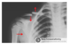
How would an xray of a posterior dislocation look?
- Can be missed on AP
- Widened glenohumeral space
- Light bulb sign

How else can you diagnose a posterior dislocation by x-ray?
Scapular y view

What are complications of posterior dislocations?

Why do inferior dislocations occur and what are the complications?
Forceful traction of arm like grabbing tree as falling

What are the complications of any shoulder dislocation?
- Recurrent dislocations: 60% risk overall that decreases with age. Due to damage to stabilising tissue. Risk of OA. Lose elasticity as get older
- Axillary artery damage: elderly as less elastic b.v. Haematoma, absent pulse and cool limb
- Nerve damage: mainly axillary as wraps around neck of humerus but can damage cords of brachial plexus or other branches like musculocutaneous
- Fractures: 1/4 and more common in traumatic injury, first time dislocation or over 40. Mainly humeral head, greater tubercle, clavicle or acromion
- Rotator Cuff tears: more common in elderly and inferior dislocations. Integrity of muscles must be checked after reduction

What are the symptoms of an axillary nerve damage, how does it mainly occur and how is it treated?
- Shoulder dislocation
- Regimental badge area
- Often resolve once shoulder is reduced and make full recovery

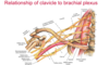
What structures does the clavicle protect?
- Apex of lung
- Subclavian vessels
- Trunks and divisions of brachial plexus
Where does the clavicle most commonly fracture and what is the mechanism of injury?
- Middle third
- Fall onto affected shoulder or outstretched hand
How does a displaces middle clavicle fracture present and why?
Medial: subscapularis and pec major

Who are clavicle fractures most common in and how are they treated?
- Peak in children and young adults
- Conservative (sling) unless….

What are some complications that can arise from a clavicle fracture?
- Non-union
- Malunion
- Pneumothorax
- Suprascapular nerve (C5,C6) damage by medial elevation
- Supraclavicular nerve (C3, C4) so paraesthesia in upper chest
- Subclavian vein and artery injury

Which rotator cuff is most commonly torn and what functions does it compromise?
- Supraspinatus tendon as passes beneath coraco-acromial arch, tearing at site of insertion on humerus
- Mainly tendon not muscles that tear
- Compromises stability of glenohumoral, external and interal rotation

What are the different mechanisms of shoulder cuff tears?
- Acute: e.g shoulder dislocation or trauma
- Chronic (Degenerative Microtrauma Model): extended use and age-related degeneration. With age blood supply to tendon decreases so cannot repair microtrauma.

What are risk factors for rotator cuff muscle tears?
Tear more likely in dominant arm but once torn one, more likely to tear in opposite shoulder too

What is the clinical presentation of a supraspinatus (rotator cuff muscle tear)?
- Can be asymptomatic
- Most commonly anterolateral shoulder pain radiating down arm, may be during activity above horizontal

Why in a supraspinatus tear do patients often not complain of weakness of abduction of shoulder?
- Found on clinical examination as supraspinatus only responsible for first 15 degrees of abduction then deltoid takes over so patient may tilt body to get first 15 degrees
- It is the pain that makes them seek medical attention

How can you diagnose and treat a supraspinatus tear?
- History, examination, MRI and ultrasound
- May be conservative (analgesia and rest) or operative

What is impingement syndrome?
- Supraspinatus tendon impinges on coraco-acromial arch leading to irritation and inflammation
- Causes: thickening of coracoacromial ligament, inflammation of supraspinatus tendon, OA causing subacromial osteophytes
- Not diagnosis, it is clinical sign

What are the symptoms of impingement syndrome?
- Pain, weakess and reduced motion when shoulder flexed or abducted as narrows space
- Pain worsened by overhead movement and bad at night
- Grinding or popping sensation when moving shoulder
- Dull long pain (not sharp) if gradual causing difficulty sleeping
- Painful arc

What is calcific supraspinatus tendinopathy and the symptoms?
- Hydroxyapatite (crystal CaPO4) deposits in supraspinatus tendon
- Can occur in any tendon but mainly this one

What is the pathology of calcific supraspinatus tedinopathy?
- Multifactorial
- Theory 1: regional hypoxia, tenocytes converted to chondrocytes, endochondrial ossification
- Theory 2: Metaplasia of MSC into osteogenic cells, ectopic bone formation

How does calcific supraspinatus tendinopathy appear on radiographically and how is it treated?
- White deposits in resting crystalline phase
- Phagocytes reabsorped and it looks like toothpaste, very painful, cloudy on x-ray
- Treat: rest and analgesia, surgery if persists

What is adhesive capsulitis?
- Frozen shoulder
- Glenohumoral joint inflamed and stiff
- Pain is constant, worst at night and exacerbated by movement and cold weather
- Restricted movement
- May be autoimmune, triggered by trauma

What are risk factors for adhesive capsulitis?
- Parkinson;s
- Polymyalgia rheumatic
- Can be alongside other shoulder problems

Why do patients with frozen shoulder often get depression?
Severe pain and sleep deprivation for prolonged period interfering with work and daily activities of living

How do you treat adhesive capsulitis?
- Physiotherapy
- Analgesia
- Anti-inflammatory medication
- Manipulation under anaesthesia to restore range of motion by breaking adhesive scar tissue, intense physio straight after to maintain movemet

What is the recovery rate for frozen shoulder?
- Most recover in time and get back 90% of shoulder motion
- Once it is resolved, opposite shoulder becomes affected in 6-17% of patients in five years suggesting autoimmune

What joint does OA of the shoulder mainly affect and who does it mainly affect?
- Acromioclavicular joint over glenohumeral joint
- Affects mainly over the age of 50

What is treatment for OA of the shoulder?
- Activity modification
- Analgesia
- NSAID
- Nutritional supplement
- Arthroscopy to remove damaged cartilage
- Hemiarthroplasty or total shoulder replacement
