19 - Hand and Wrist Flashcards
Label the following diagram

Some Lovers Try Positions That They Cannot Handle

Label the following diagram of the posterior forearm.


Label the following diagram of the wrist.


Label the following diagram of the muscles of the hand.


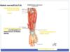
What are the muscles of the posterior forearm, how are they organised and what are they innervated by?
- Split into superficial and deep separated by fascia
- Innervated by radial nerve
- Rules of 3
- Superficial: brachioradialis, ECRL, ECRB, ED, EDM, ECU, Anconeus
- Deep: Supinator, APL, EPB, EPL, EI

What muscles originate from the common extensor origin?
- ECRB
- ED
- ECU
- EDM
- Supinator
At lateral epicondyle

What is the origin, insertion, action and innervation of the superficial muscles of the posterior forearm?
- All innervated by the radial nerve
- ECRB and Supinator: innervated by deep brranch of radial nerve as origin distal to the branching of radial nerve into superficial and deep
- ED, EDM and ECU: posterior interosseous branch of radial nerve

How do you test the function of the extensor digitorun?
Forearm pronated and extend fingers against resistance
Why can’t the middle or ring finger be fully extended if the other fingers are flexed? Why can the IP joints still fully flex but the MCP joints can’t?
- Juncturae Tendinum: fibrous bands on the dorsum of the hand linking the extensor digitorum tendons
- Index and little finger can fully extend as they have a second extensor each
- Extension of IP achieved by lumbricals not ED

When does the radial nerve become the posterior interosseous branch of the radial nerve?
When the deep branch of the radial nerve passes between two heads of supinator and enters posterior forearm

What are the origins, insertions, innervations and actions of the deep muscles of the posterior forearm?
All innervated by radial nerve
- Deep branch of radial nerve: supinator
- Posterior interosseous: APL, EPL, EPB, EI

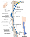
What are the sensory and motor functions of the radial nerve?
Sensory: skin of posterior arm, lower lateral arm, posterior forearm, dorsal surface of radial hand, dorsal surface of lateral three and a half digits
Motor: triceps brachii and extensors of forearm

What is the anatomical course of the radial nerve?
- Arises in axilla posterior to the axillary artery
- In base of triangular interval it gives off branch to long and lateral head of triceps and posterior cutaneous nerve of arm
- Enters radial spiral groove and passes laterally across humerus to give off branch to medial triceps and two more cutaneous branches (inferior lateral cutaneous nerve of arm and posterior cutaneous nerve of forearm)
- Emerges laterally from radial groove and gives branch to brachioradialis, ECRL and sometimes brachialis
- Pierces lateral fascia and travels anterior to lateral epicondyle of humerus through cubital fossa where it divides into deep (motor) and superficial (sensory) branch
- Deep branch innervates ECRB and exits cubital fossa posterior to two heads of supinator which it innervates
- Becomes posterior interosseous nerve after supinator and innervates rest of the posterior muscles
What does the wrist joint consist of, what type of joint is it and what movements are possible?
- Distal radius, triangular fibrocartilage complex, scaphoid and lunatte NOT ulnar
- Ellipsoid
- Flexion, extension, adduction, abduction, circumduction

What ligaments stabilise the wrist joint?
- Dorsal and palmar radiocarpal ligaments (ensure hand follows radius during pronation and supination)
- Ulnar and radial collateral ligaements

What muscles cause each movement at the wrist and what is the innervation of the wrist?
Flexion: FCU, FCR, PL (long flexors like FDS assist)
Extension: ECU, ECRB, ECRL (long extensors assist)
Adduction: FCU, ECU
Abduction: ECRB, ECRL, FCR
Using hilton’s law the wrist nerve supply is median, ulnar and radial

How are the carpal bones arranged?
Into proximal and distal rows, SLTP in proximal

What is the significance of the hook of Hamate?
- On palmar surface
- Forms ulnar border of carpal tunnel
- Forms radial border of Guyon’s canal
- Flexor retinaculum and FCU attach here

Label the carpals on the x-ray.


What is the blood supply like to the scaphoid and why is this relevant?
- Dorsal carpal branch of radial artery that enters dorsal distal scaphoid and supplies scaphoid by retrograde
- High rate of non-union or AV necrosis if fractured

What carpal does each of the metacarpals articulate with?
1: trapezium
2: trapezoid
3: capitate
4 and 5: hamate

What movements can occur at the fingers and thumb?
- Circumduction
- Radial abduction in coronal plane
- Palmar abduction in sagittal plane

What are the movements of the fingers?

What are the different groups of muscles in the hand?
- Intrinsic: thenar, hypothenar, adductor and central compartment
- Extrinsic: ED, FDS, FDP

What are the origin, insertions, actions and innervations of the thenar muscles?
All innervated by median nerve apart from deep head of flexor pollicis brevis which is supplied by ulnar nerve

What are the origin, insertions, actions and innervations of the adductor compartment of the hand?
- Supplied by ulnar nerve
- Palmar and radially adducts, not just one like abductor pollicis brevis

What are the origin, insertions, actions and innervations of the hypothenar muscles?
All innervated by the ulnar nerve

What are the origin, insertions, actions and innervations of the central muscle compartment of the hand?
- Radial two lumbricals: Median nerve
- Ulnar two lumbricals, palmar/dorsal interossei and palmaris brevis: Ulnar nerve

What is the role of the lumbricals?
- To connect the FDP tendon and ED tendon by going from FDP tendon to extensor expansion of ED.
- Extensor expansion: widening of extensor tendon over dorsium of MCPJ

Compare and contrast the palmar and dorsal interossei.
- Both innervated by ulnar nerve
- Dorsal are bipennate and have 4
- Palmar are unipennate and have 3
IAN GLOVE TRICK

Why is there no dorsal interossei for the little finger and no palmar interossei for the thumb?
- Little: has abductor digiti minimi
- Thumb: adductor pollicis
- Middle finger: reason there is only 3 palmar interossei is because middle moved side to side by second and third dorsal interossei

How do the extrinsic muscles of the hand insert onto the hand?
- ED: at wrist plits into four tendons each tendon splits into central and lateral slip that converge and insert on dorsal base of distal phalanx
- FDP: at wrist splits into four and passes through carpal tunnel to insert on base of distal phalanx
- FSP: at wrist splits into four and passes through carpal tunnel to insert on base of middle phalanx. FDS tendon divides into two slips at proximal phalanx and FDP pases through gap created

What happens if you rupture the central slip of the ED tendon?
Boutonniere Deformity

What are the borders of the carpal tunnel and what passes through it?
Deep: carpal bones
Superficial: flexor retinaculum (transverse carpal ligament)
- FPL, FDS X 4, FDP X 4, Median Nerve
FDS middle and ring finger tendons pass superficial to the others when passing through the tunnel

What are the attachments of the flexor retinaculum?
Lateral: scaphoid and trapezium
Medial: hook of hamate and pisiform

What is Guyon’s canal?
- Canal that allows passage of ulnar nerve and artery
- Superficial to flexor retinaculum so ulnar nerve not involved in carpal tunnel. Can be palpated just radial to the pisiform bone
- Roof: palmar carpal ligament

What are the borders of the anatomical snuff box and what are the contents?
- Triangle depression on radial dorsal hand at carpal bones when thumb radially abducted
- Radial:* Tendons of APL and EPB
- Ulnar:* tendon of EPL
- Proximal:* styloid of radius
- Floor:* scaphoid and trapezium
- Roof:* skin

What is the arterial supply to the hand?
- Deep and superficial palmar arches
- Ulnar: crosses anterior to flexor retinaculum and lies radial to pisiform and ulnar. Divides into two branches in hand to anastomose and form arches. Contributes mainly to superficial arch which gives off common palmar digital arteries
- Radial: enters betweeen tendons of brachioradialis and FCR and crosses snuffbox before entering palm between two heads of adductor pollicis. Contributes mainly to deep arch and therefore supply to thumb and radial side of index finger

What muscles of the upper limb does the ulnar nerve supply?
- Some anterior forearm
- All intrinsic muscles bar LOAF which are done by median nerve

Why are the dorsal fingertips supplied by the same nerve that innervates their palmar side?
During embryological development the fingernails begin on the volar aspect of hand and they are dragged to dorsal so take their nerve supply with them

What is the sensory supply to the hand and where do the nerves originate from?
- Palmar cutaneous branch of median nerve: proximal to carpal tunnel
- Palmar cutaneous branch of ulnar nerve: proximal to Guyon’s canal
- Palmar digital branches: arise in the palm from median and superficial branch of ulnar nerve
- Dorsal cutaneous branch of ulnar: branches from main nerve in forearm

How do you test peripheral nerves of the hand in practice?
There is a lot of overlap like dermatomes so need to check regions with consistant supply
- Radial: dorsum of first webspace
- Ulnar: ulnar border of hand
- Median: palmar surface of tip of index

How can you test nerve supply to the hand of an unconscious patient?
- Put hand in water and see if wrinkles
- Tenodesis effect
- Sticky, sweaty hand

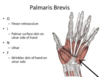
Why might a patient come in with a twitchy hand?
Their palmaris brevis may be in spasm as it is not under voluntary control
How can you test blood supply to the hand?

What is the posterior interosseous branch of the radial nerve?
The name of the deep branch of the radial nerve once it has passed the two heads of supinator and entered the posterior arm