12 - Ophthalmology Conditions 2 Flashcards

1
Q

What are some causes of optic neuritis?

A
  • MS
  • Infection e.g syphillis
  • Drugs e.g ethambutol, methanol
  • Diabetes
  • Vitamin deficiency
2
Q

What are some risk factors for developing optic neuritis?

A
  • Age 20-40
  • Female
  • Caucasian
  • FHx of MS
3
Q

How does optic neuritis present?

A
  • Unilateral loss of vision over hours or days
  • Dyschromatopsia/Red desaturation
  • Eye movements painful
  • Flashing lights
  • RAPD
4
Q

What is the prognosis with optic neuritis?

A
  • Usually resolves in 2-6 weeks
  • 50-80% go on to develop MS in next 15 years
5
Q

What is the treatment for optic neuritis?

A
  • High dose methylprednisolone IV for 72hrs

then

  • PO Prednisolone for 11 days
6
Q

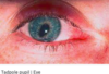
What is the difference between orbital and periorbital cellulitis?

(IMAGE IMPORTANT)

A

Orbital: Infection of the tissues posterior to the septum and it is life threatening so an emergency. Usually from paranasal sinus, dental or occular infection

Periorbital: Infection of the tissues anterior to the septum. Usually from sinus infections or facial skin lesions

7
Q

What is the presentation of periorbital cellulitis and how can you rule out orbital cellulitis?

A
  • Swollen red hot eyelids

- Features that rule out orbital: absence of painful eye movements, absence of visual impairment, absence of diplopia

  • If in doubt treat as orbital cellulitis as this is life threatening
8
Q

What is the treatment of periorbital cellulitis?

A

- PO co-amoxiclav

  • Admit if child or high risk of progression to orbital cellulitis
  • Safety net to return if any red flags or not responding after 48 hours
9
Q

How does orbital cellulitis present?

A
  • Swollen red eye
  • Pain on eye movement and reduced eye movements
  • Diplopia
  • Proptosis
  • RAPD
  • Fever
  • Reduced visual acuity
10
Q

What are the complications with orbital cellulitis that make it an emergency?

A

- Subperiosteal and orbital abscess

- Visual loss due to optic neuritis and CRAO

- Meningitis

- Brain abscess

- Cavernous sinus thrombosis

11
Q

When should you CT in orbital cellulitis and what are some red flags that an orbital cellulitis needs urgent action?

A

Eyelid oedema and erythema OR failure to respond to 48hr abx PLUS ONE RED FLAG:

  1. Proptosis
  2. Chemosis (swelling ov conjunctiva)
  3. Ophthalmoplegia
  4. Relative afferent pupillary defect (RAPD)
  5. Systemically unwell 6. Painful eye movement
  6. Altered visual acuity
12
Q

How is orbital cellulitis managed?

A

- Admit for promt CT (if indicated as radiation exposure to young), ENT and Optho assessment

- IV Abx (Ceftriaxone +/- Metronidazole if sinus infection)

- Surgical drainage of any abscesses to prevent meningeal involvement and cavernous sinus thrombosis

13
Q

How can you tell the difference between episcleritis and scleritis?

A

Episclera lies superficially so the vessels will move when probed with a cotton bud and blanch when 10% phenylephrine is put on them

This won’t happen with sclerla vessels as they are deeper

14
Q

How does episcleritis present?

A
  • Typically not painful but can be mild pain
  • Segmental redness (rather than diffuse) in lateral sclera
  • Foreign body sensation
  • Dilated episcleral vessels
  • Watering of eye
  • No discharge
15
Q

What types of patients does episcleritis usually present in?

A

MOSTLY IDIOPATHIC

  • Young middle aged women
  • Inflammatory disorders e.g IBD, RA
16
Q

How is episcleritis managed?

A
  • Self limiting in 1-4 weeks
  • Symptomatic relief with simple analgesia, cold compresses, artificial tears
  • Safety net
17
Q

What is scleritis and why is it more serious than episcleritis?

A
  • Generalised inflammation of the sclera with oedema of the conjunctiva
    scleral thinning, and vasculitic changes

- URGENT REFERRAL as necrotising scleritis can lead to perforation of the sclera and is sight threatening

18
Q

What are the causes of scleritis?

A

Usually not infectious, it is autoimmune related

19
Q

How does scleritis present?

A
  • Diffuse red eye
  • Severe dull pain made worse on occular movements
  • Headache
  • Photophobia
  • Reduced visual acuity
20
Q

How is scleritis managed?

A

URGENT OPHTHO REFERRAL

Anterior/Non-necrotising:

  • Oral high-dose prednisolone
  • Oral NSAIDs

Posterior or Necrotising:

  • Immunosuppression for underlying autoimmune condition e.g clophosphamide or rituximab
  • Course of methylprednisolone
  • If globe perforation need surgery
21
Q

How can you detect a cataracts with a pen torch?

A

COME BACK TO

22
Q

What are indications of cataract surgery and what can you expect to change with your vision from it?

A

Indications: Symptoms are troubling, lifestyle is restricted, or if unable to read a number-plate at 20 metres (and they need to drive)

Changes:

  • Improve colour vision
  • Dazzle/glare often remains after
  • Improved visual acuity as put refractive lens in, may need distance glasses
  • Visual acuity may not return to normal as may be underlying co-existing AMD
23
Q

What are Drusen in AMD?

A
  • Soft Drusen lift RPE from Bruch’s membrane. This can cause VEGF release so encourages neovascularisation and progression from dry to wet AMD
24
Q

How does neovascularisation in wet AMD cause vision loss?

A

- Disciform scar in macula that leads to a blind spot (scotoma)

25
Q

What is Charles Bonnet synrome?

A

Most people with AMD may experience this

Vivid hallucinations in response to central vision loss

Reassure them it is normal

26
Q

What is the pathophysiology of retinal detachment and why is it an emergency?

A

Retins separates from the choroid, usually due to a tear allow vitreous humour to get under retina.

Sight threatening as outer retina gets its blood supply from choroid

Rhegmatogenous retinal detachment—tear in retina causes fluid to pass
from vitreous space to subretinal space between sensory retina and
the retinal pigment epithelium. Caused by trauma

Exudative retinal detachment - Retina detaches without
a tear, eg hypertension, vasculitis, macular degenerative conditions, tumour

Tractional retinal detachment—pulling on the retina, eg proliferative reti- napathy, myopic eyes

27
Q

What are some of the risk factors for retinal detachment?

A
  • Posterior vitreous detachment
  • Diabetic retinopathy
  • Trauma to the eye
  • Retinal malignancy
  • Older age
  • Family history
  • Myopia
  • Cataract surgery (vitreous haemorraghe)
28
Q

If a retinal tear is found before retinal detachment, what can you do to prevent this from developing into retinal detachment?

A

Cryotherapy or Laser Retinopexy

Creates adhesions between the retina and the choroid

29
Q

How does a retinal detachment present?

A

4 F’s!!!

- Flashes

- Floaters

- Field loss (usually peripheral)

- Fall in acuity (painless vision loss like a curtain or shadow over vision)

30
Q

How does retinal detachment look on fundoscopy?

A
  • Grey opalescent retina ballooning forward
  • May see tear
  • Crinkling of retinal tissue
31
Q

How is retinal detachment managed?

A

- URGENT REFERRAL AND MANAGEMENT

- Cryotherapy or laser coagulation to seal any tear

- REST! Lie flat if superior tear, lie head 30 degrees up in inferior tear

- Surgery to reattach retina: vitrectomy, scleral bulking, pneumatic retinopexy

32
Q

What is posterior vitreous detachment and what is it caused by?

A

Vitreous body keeps retina pressed against the choroid. It is made of collagen and water

As we age it becomes less firm so the posterior vitreous may no longer sit on the retina

Normal part of ageing

33
Q

How does posterior vitreous detachment present?

A
  • Often asymptomatic
  • Painless spots of vision loss
  • Floaters
  • Flashing lights

No decline in vision or loss of visual fields

34
Q

How is posterior vitreous detachment managed?

A
  • Refer for fundoscopy as can predispose to retinal tears and detachment
  • If the above is ruled out no treatment neccessary as brain will adjust
35
Q

What is a vitreous haemorraghe and what is it caused by?

A

Bleeding into the vitreous body

  • Retinal neovascularisation (DM, CRVO)
  • Retinal tears
  • Retinal detachment
  • Trauma
36
Q

How does vitreous haemorraghe present and how is it managed?

A

Presentation

  • Patient will see black floaters or ring like floaters with clear centre
    Patients typically present with an acute or subacute onset of:
  • painless visual loss or haze (commonest)
  • red hue in the vision
  • floaters or shadows/dark spots in the vision

Mx

  • If small may undergo spontaneous reabsorption
  • If dense then do vitrectomy to remove blood
  • Give anti-VEGF if neovascularisation or photocoagulation for diabetic retinopathy

Signs:
- decreased visual acuity: variable depending on the location, size and degree of vitreous haemorrhage
- visual field defect if severe haemorrhage

37
Q

What is this and what could have caused it?

A

Subconjunctival Haemorraghe

One of the vessels in the conjunctiva has popped and blood pooled between sclera and choroid. It is painless

Causes: heavy coughing, weight lifting, straining when constipated, trauma

38
Q

What are some conditions that predispose people to subconjunctival haemorraghes?

A
  • Hypertension
  • Bleeding disorders (e.g thrombocytopenia)
  • Whooping cough
  • Medications (warfarin, NOACs, antiplatelets)
  • Non-accidental injury

CHECK INR AND BP

39
Q

How is a subconjunctival haemorraghe managed?

A

- Resolve spontaneously in 2 weeks

  • Can give lubricating eye drops if foreign body sensation
  • Consider underlying disorders e.g HTN
40
Q

What is retinitis pigmentosa?

A

Inherited condition of the retina (X-Linked, Auto Dom or Auto Rec) usually affecting males

Degeneration of the rod photoreceptors followed by degeneration of the cones

Can be associated with other systemic diseases e.g Usher Syndrome, Refsum syndrome

41
Q

How does retinitis pigmentosa present and what does it look like on fundoscopy?

A

Presentation

  • Starts with night blindness as rods affected
  • Then progresses to peripheral vision loss then central vision loss
  • Eventually causes complete blindness

Fundoscopy

- Bone spicule pigmentation especially in mid-peripheries

  • Can have pale optic disc
  • Narrowing of arterioles
42
Q

How is retinitis pigmentosa managed?

A

Nothing to slow the progress of the disease just manage it

43
Q

What is corneal abrasion and what are some of the causes of this?

A

Damage to the epithelium of the cornea

  • Contact lenses (consider pseudomonas infection)
  • Foreign bodies
  • Finger nails
  • Eyelashes
  • Entropion (inward turning eyelid)
44
Q

How does a corneal abrasion present?

A
  • History of contact lenses or foreign body
  • Painful red eye
  • Foreign body sensation
  • Watering eye
  • Blurring vision
  • Photophobia
45
Q

How is a corneal abrasion diagnosed?

A

- Fluorescein stain and blue light slit lamp exam: will collect in the area of abrasion

- Evert eye lid to look for foreign body

46
Q

How is a corneal abrasion managed?

A
  • Will usually heal over 2-3 days, bring back in a week to check
  • Simple analgesia
  • Lubricating eye drops
  • Chloramphenicol drops if infection
47
Q

What is a corneal ulcer and what are some of the causes of this?

A
  • White opaque structure on the cornea.
  • Fx: Can be very painful, red eye, photophobia, blurred vision
  • A corneal ulcer describes a defect in the cornea, typically secondary to an infective cause.
  • RFs: contact lense use, Vit A deficiency

- Causes: infection (keratitis: bacterial viral, fungal), trauma, contact lens wear, vasculitis from RA

48
Q

How is a corneal ulcer managed?

A
  • Refer to specialist urgently for urgent diagnostic smear/Gram stain and scrape
  • Remove contacts
  • Prescribe aciclovir if opthalmic shingles or herpes keratitis
  • Alternate chloramphenicol drops (for Gram +ve) with ofloxacin drops (for Gram –ve) until cultures known
  • Once recovery started can add steroid drops
49
Q

Why do you need to be careful when using steroid eye drops?

A
  • Risk of glaucoma
  • Risk of cataract formation
  • Risk of corneal melt if ulcer
  • Can prolong viral conjunctivitis
50
Q

What is thyroid eye disease and what is the main risk factor?

A

In people with Grave’s disease and doesn’t correlate with whether euthryoid, hyperthyroid or hypothyroid

Smoking is biggest risk factor

Avoid radioiodine treatment as can worsen it. Causes retrooribtal inflammation and lymphocyte infiltration

51
Q

What are some of the signs and symptoms of thyroid eye disease?

A

Symptoms

  • Eye discomfort
  • Grittiness
  • Diplopia
  • Photophobia
  • Decreased visual acuity
  • RAPD

Signs

  • Exopthalmos
  • Proptosis (look from above)
  • Corneal ulceration
  • Papilloedema
  • Loss of volour vision
  • Opthalmoplegia especially on upward gaze
52
Q

Why should you seek urgent advice if a patient with thyroid eye disease has an RAPD?

A

Could mean optic nerve compression so needs urgent decompression

53
Q

How is thyroid eye disease diagnosed?

A
  • Clinical
  • Do CT or MRI of orbits and will see enlarged eye muscles
54
Q

How is thyroid eye disease managed?

A

- Treat hyper/hypothyroidism

- Stop smoking

- Avoid radioiodine

- Symptomatic: artificial tears, sunglasses, avoid dust, elevate bed when sleeping to prevent periorbital oedema, eyelid surgery

- Diplopia: Fresnl prism stuck to glasses

- Opthalmoplegia or Oedema: high dose steroids and oral radiotherapy

- Sigh threatening: surgical decompression

55
Q

What nerves cause pupil dilation and pupil constriction?

A
56
Q

What are some disorders that cause an abnormal pupil shape?

A

Anterior uveitis: scarring posterior synechiae

Rubeosis iridis: neovascularisation in iris due to diabetic retinopathy

Coloboma: congenital

Tadpole pupil: temporary, associated with migraines

Acute angle closure glaucoma: oval pupil

Trauma to sphincter muscles: from cararact surgery

57
Q

What are some causes of mydriasis (dilated pupil)?

A

- Third nerve palsy

- Holmes-Adie syndrome: usually women with tonic pupil, absent knee/ankle jerks and impaired sweating

- Raised intracranial pressure: uncal herniation

- Congenital

- Trauma

- Stimulants such as cocaine

- Anticholinergics

58
Q

What are some causes of miosis (constricted pupil)?

A
  • Horners syndrome
  • Cluster headaches
  • Argyll-Robertson pupil (in neurosyphilis)
  • Opiates
  • Nicotine
  • Pilocarpine
59
Q

How does a third nerve palsy present?

A

Occulomotor

- Ptosis

- Dilated non-reactive pupil

- Divergent strabismus: “down and out”

60
Q

What are some causes of a third nerve palsy?

A

Can be idiopathic

Pupil Sparing (microvascular cause as no compression of parasympathetic fibres)

Diabetes

Hypertension

Ischaemia

Full Palsy

Idiopathic

Tumour

Trauma

Cavernous sinus thrombosis

Posterior communicating artery aneurysm

Raised intracranial pressure

61
Q

What are some causes of Horner’s syndrome? (disruption of sympathetic fibres)

A

4 Ss, 4 Ts, 4Cs + Congenital (Heterochromia)

Can tell location by the pattern of anhidrosis

Central lesions cause anhidrosis of the arm and trunk as well as the face. Pre-ganglionic lesions cause anhidrosis of the face. Post-ganglionic lesions do not cause anhidrosis

62
Q

How is Horner’s syndrome diagnosed?

A
  • Cocaine eye drops (stop noradrenaline reuptake)
  • Adrenaline eye drops (dilates pupil)
63
Q

What is Holmes-Adie Pupil?

A
  • Damage to post-ganglionic PS fibres due to virus
  • Unilateral dilated pupil that is slow to react to light with slow dilation of the pupil following constriction
  • Over time pupil will get smaller

Holmes Adie Syndrome: Holmes Adie pupil with absent ankle and knee reflexes

64
Q

What is a Argyll-Robertson Pupil?

A
  • Found in neurosyphillis and diabetes. (Prostitute Pupil - Accomodates but does not react)

- Constricted pupil that accommodates when focusing on a near object but does not react to light.

  • They are often irregularly shaped
65
Q

Why should you be concerned if somebody presents with a unilateral third nerve palsy?

A

Posterior Communicating Artery Aneurysm until proven otherwise

Need to send for CT angiography as high risk of rupture

Will involve pupil but doesn’t necessarily have down and out

66
Q

What are some causes of a RAPD?

A

Do swinging light test

67
Q

How does internuclear opthalmoplegia present?

(IMPORTANT IMAGE)

A

- Weakness of aDduction of the affected eye

􏰀 AND

- Horizontal jerk nystagmus of aBducting other eye

Due to lesion in MLF that connects the 3rd and 6th cranial nerves. MS

68
Q

What is hypertensive retinopathy and what are some symptoms a patient might experience?

A

Damage to the blood vessels in the retina due to chronic hypertension or malignant hypertension

  • Asymptomatic until late stage
  • Reduced vision
  • Eye swelling
  • Bursting of a blood vessel
  • Double vision accompanied by headaches
69
Q

What are some signs of hypertensive retinopathy?

A

- Silver/Copper wiring: walls of the arterioles become thickened and sclerosed causing increased reflection of the light

- Arteriovenous nipping: arterioles cause compression of the veins where they cross due to sclerosis and hardening of the arterioles

- Cotton wool spots: ischaemia and infarction in the retina causing damage to nerve fibres.

- Hard exudates: damaged vessels leaking lipids into the retina

- Retinal haemorrhages: damaged vessels rupturing and releasing blood into the retina.

- Papilloedema: ischaemia to the optic nerve resulting in optic nerve swelling (oedema) and blurring of the disc margins

70
Q

How is hypertensive retinopathy graded?

A

Keith-Wagener Classification

Stage 1: Mild narrowing of the arterioles

Stage 2: Focal constriction of blood vessels and AV nicking

Stage 3: Cotton-wool patches, exudates and haemorrhages

Stage 4: Papilloedema

71
Q

What are the complications with hypertensive retinopathy?

A
  • At risk of developing retinal pathology
  • Increased CVD and stroke risk
72
Q

How is hypertensive retinopathy managed?

A
  • Controlling the blood pressure e.g antihypertensives, less salt
  • Reduce risk factors such as smoking and blood lipid levels
73
Q

Why does increased ICP cause issues with the eyes?

A

Optic nerve is surrounded by meninges and CSF. Increase in CSF pressure can compress the optic nerve and the vessels that supply it. The vein gets occluded first so slow venous return so oedema of the retina

74
Q

When a patient has visual field loss, what questions do you need to ask yourself to work out the location of the lesion?

A
75
Q

What visual field defects would you get with the following lesions?

(Important concept card)

A

Optic nerve: total blindness of that eye. Direct pupillary reflex is absent, indirect remains intact. Lesions tend to occur in the horizontal meridian when in the optic nerve. When the lesions are behind the optic chiasma, the boundaries are in the vertical meridian. Acuity tends to be affected .

Optic chiasma: Bitemporal hemianopia. Fibres coming from the nasal halves of both retinas are involved. There is normal direct, consensual light reflex and accommodation reflex

Optic tracts A lesion on the optic tract causes a contralateral homonymous hemianopia. There is a normal direct light reflex, consensual light reflex and accommodation reflex.

Optic radiation: contralateral homonymous hemianopia. Lesions over the lateral portions of the lateral geniculate body which received impulses from the inferior rental quadrants (superior visual field). Lesions in the nerve fibres passing more directly posterior to the cortex cause an inferior quadrantic hemianopia.

Visual cortex Destruction of the the primary visual cortex produces contralateral homonymous hemianopia. Macula is often spared because in some people there is an anastomosis between the posterior and middle cerebral arteries.

76
Q

What tests can you do to figure out somebodys visual fields?

A

- Finger confrontation

- Hat-pin test (Red for central vision, White for peripheral vision)

- Amsler Grid

77
Q

If somebody has a homonymous hemianopia with macular sparing, what is the location of the lesion?

A
  • Occipital Lobe Visual Cortex
  • Means they have likely had a PCA stroke

- Both MCA and PCA supply the macula so if PCA knocked out MCA can compensate. All other parts of visual cortex only supplied by MCA