19 - ENT History, Examination and Presentations Flashcards

1
Q

What are some ENT emergencies?

A
2
Q

How would you perform an examination of the ear in an OSCE?

https://geekymedics.com/hearing-ear-examination-osce-guide/

A

1. Introduction: wash hands, introduce, 3 point ID, gain consent, any pain?

2. General Inspection: hearing aids, walking aids (vestibulocochlear)

3. Gross Hearing Assessment: any changes? whisper word at 60cm whilst rubbing other tragus

4. Weber’s and Rinne’s: midline forehead and mastoid process

5. Otoscopy:

- Any pain? Inspect pinna, mastoid and pre-auricular area. Palpate the tragus and regional lymph nodes

  • Pull pinna up and backwards and put in otoscope holding in left hand for left ear and vice versa. Hold like a pencil
  • Inspect auditory canal e.g oedema, ear wax, erythema
  • Inspect tympanic membrane e.g colour, shape, light reflex (absent in otitis media), perforation, scarring
  • Inspect other ear

6. Complete Exam

  • Dispose of speculum in clinical waste
  • Thank patient
  • Extra exams e.g CN exam, tympanometry, audiometry
3
Q

Where is the cone of light viewed?

A

In a healthy normal ear drum should be able to see the cone of light

5 o clock in right ear

7 o clock in left ear

4
Q

How do you interpret the results of a Weber’s and Rinne’s test?

A
  • Rinne positive means AC>BC (normal)
  • Mastoid –> Temporal Bone –> Cochlear
5
Q

What does tragal tenderness point to?

A

Otitis externa

6
Q

What should you be able to visualise on the TM with otoscopy?

A
  • Lateral process of malleus
  • Umbo
  • Cone of light
  • Pars tensa and pars flaccida
7
Q

What tuning fork do you need to use in a Weber’s and Rinne’s test?

A

512Hz

Small fork for small body part

8
Q

What does a pure tone audioram measure and how does the assessment take place?

A
  • Assesses any patient aged >4 hearing threshold at different frequencies
  • Performed in soundproof booth
  • Air conduction tested by headphons
  • Bone conduction tesred by bone conductor placed over mastoid process. Looks at sensorineural hearing. If any hearing discrepancy need to play sound in opposite ear to distract it whilst testing bone conduction as sound can travel from one mastoid to both cochlears
9
Q

What is consider normal on an audiogram?

A

Anything above 20dB

10
Q

What are some causes of conductive hearing loss and how will this present on an audiogram?

A

Anything to do with external or middle ear e.g wax, otitis media with effusion

Audiogram will have normal bone conduction but redcucd air conduction thresholds

11
Q

What causes sensorineural hearing loss and how will it appear on audiogram?

A

Problem between cochlear and auditory cortex of brain e.g presbyacusis, acoustic neuroma

Any unilateral sensorineural hearing loss needs MRI to look for Acoustic Neuroma (Vestibular Schwanoma)

Audiogram: reduced bone and air conduction, no air bone gap

12
Q

What is a tympanogram used for and how do you do this test?

A

Measures the compliance of the tympanic membrane and can provide information about the middle ear and eustachian tube

Insert probe into external ear canal into patient of any age. Probe changes pressure in the ear canal

Compliance vs Pressure

COMPLIANCE PEAKS WHEN PRESSURE IN THE CANAL EQUALS THAT OF THE MIDDLE EAR

13
Q

What are the three different tympanogram tracings and what do they show?

A

Type A

  • Normal result
  • Peak centred at 0daPa on x-axis

Type B

  • Flat tracing
  • Suggests middle ear effusion or perforation
  • Look at canal volume on side of tympanogram, if middle ear effusion volume will be normal (around 1), if perforation volume will be much larger as measuring middle ear and outer ear

Type C

  • Peak of tracing has negative pressure
  • Suggests ET (eustachian tube) dysfunction
14
Q

How do you perform an examination of the nose in an OSCE? (Anterior Rhinoscopy)

https://geekymedics.com/nasal-examination-osce-guide/

A

Equipment needed: Light source (headlight, pen torch or otoscope) and Nasal Speculum (or Otoscope with large speculum)

1. Introduction

2. External Inspection: from front, side and back looking for deformities or lesions

3. Nasal Cavity Inspection: look externally by lifting tip of nose and using light source then use nasal speculum. Look at vestibule, inferior turbinates and septum

4. Nasal Cartilage and Bone Palpation: palpate bone, cartilage, infraorbital ridge and eye movements

5. Nasal Airflow: metal surface or occlude one nostril

6. Thank patient and further exam: olfactory assessment, regional lymph node exam, oral cavity exam (as palate is the floor of the nose), flexible nasendoscopy

15
Q

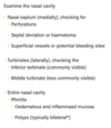
What sorts of things in the nasal cavity are you look for on anterior rhinoscopy?

A
16
Q

Why is it important that we also do an exam of the posterior nose?

A
  • Middle meatus (between middle and inferior turbinates) is where the sinuses ventilate
  • Postnasal space (nasopharynx) contains the ET orifices & the pharyngeal recess, and may contain adenoids or naso-pharyngeal cancer
17
Q

How do you do a oral cavity exam in an OSCE?

https://geekymedics.com/oral-cavity-examination-osce-guide/

A

Equipment: Pen/Headtorch, Tongue Depressors

1. Introduction: inc remove dentures if wearing any

2. General Inspection: any parotid or submandibular swelling

3. Closer Inspection: open mouth, lips, teeth and gums, tongue, buccal mucosa and parotid duct, depress tongue and look at palate and uvula, tonsils and pharyngeal arches, floor of mouth by lifting tongue

4. Bimanual Palpation of the Mouth: if examiner and patient allow

5. Thank patient and further exams: examine neck, ears, TMJ, flexible nasoendoscopy to look at oropharynx, FNA of any lumps

18
Q

How do you do an examination of a neck lump in an OSCE?

https://geekymedics.com/neck-lump-examination-osce-guide/

A

1. Introduction: inc expose clavicles, ask if patient can point out lump and ask about pain

2. General Inspection: any scars, cachexia, hoarse voice

3. Inspect lump: Site, Size, Shape, Consistency, Fluctuance, Temperature, Overlying skin changes, Pulsatility, Transilluminates, Bruit, Tongue protrusion, Swallow

4. Lymph nodes palpation: including para-tracheal

5. Thyroid Exam

6. Submandibular Exam

7. Thank and summary: do thyroid status exam and TFTs, examine oral cavity, US, FNA, ?urgent referral to ENT

19
Q

What are some differentials for neck lumps in the following areas:

  • Midline
  • Anterior Triangle
  • Posterior Triangle
A

Midline:

  • Lymph node
  • Lipoma
  • Dermoid cyst
  • Epidermoid cyst
  • Enlarged thyroid gland
  • Thyroid nodule
  • Thyroglossal cysts
  • Laryngocele
20
Q

How do you do a quick thyroid and submandibular exam in a neck lump exam?

A

Submandibular: bimanual palpation

Thyroid: lobes and isthmus

21
Q

What are rigid and flexible nasendoscopies used for?

A

To view the back of the nose, ET tube, pharynx and the larynx. Can also aid treatment of epistaxis, foreign body etc

Rigid endoscope has a better image quality and is better for treatment procedures as one hand for scope and one hand for treatment. However cannot see the larynx

Flexible endoscope cannot see structures as clearly but easier to do and can see the larynx

22
Q

If a patient comes in to have an endoscopy how do you explain to them what the procedure involves and what complications could occur due to the procedure?

A
  • Whilst sitting upright a small flexible tube with a light and camera will be placed into your nose to view the back of your nose and throat. We will ask you to make some noises and pull some faces to see how your throat moves
  • Before starting we will use local anaesthetic to numb the area so it won’t be painful but may be a strange sensation. The anaesthetic can make you feel like your throat is swelling and like you cannot swallow but this is normal

- Only takes a few minutes

  • After anaesthetic you can’t eat or drink for an hour because there is a risk you could choke or burn your mouth because you can’t feel it

- Complications: temporary dizziness, nosebleed, coughing, laryngospasm

23
Q

How would you triage the following ENT scenarios?

  • Unstable epistaxis
  • Stable epistaxis
  • OM/OE
  • Stridor
  • Periorbital cellulitis
  • Foreign body exc batter
  • Quisy
  • Bell’s Palsy
  • Post tonsillectomy bleed
  • Laryngectomy/Tracheostomy issues
  • Acute vestibulopathy
  • Acute sensorineural hearing loss
A

URGENT: Post tonsillectomy bleed, Laryngectomy issues, Unstable epistaxis, Stridor (airway compromise)

SOON (30 MINS): Periorbital cellulitis, Foreign body, Stable epistaxis, Quinsy

ADVICE: OE/OM, Bell’s Palsy, Acute sensorineural hearing loss, Acute vestibulopathy

24
Q

What are some cardinal ear, nose and throat symptoms?

A
25
Q

What are the borders of the anterior and posterior triangle of the neck?

A
26
Q

What are some important questions to ask a patient when they present with a neck lump?

A

- Duration of onset

- Any recent change in size

- Associated symptoms (especially red flag)

- Relevant PMHx: smoking status, alcohol intake, and known radiation exposure.

27
Q

What are some red flag symptoms with a neck lump that raise the suspicion of a head and neck malignancy?

A
  • Hard and fixed lump
  • Associated otalgia, dysphagia, stridor, or hoarse voice
  • Epistaxis or unilateral nasal congestion
  • Unexplained weight loss, night sweats, or fever or rigors
  • Cranial nerve palsies

In children: presence of a supraclavicular mass, lumps larger than 2cm, and a previous history of malignancy.

28
Q

What are some differentials for a neck lump (surgical sieve)?

A

Infective: reactive lymphadenopathy, sialadenitis (e.g parotitis)

Malignancy: lymphoma, head and neck malignancy, metastatic spread, lipoma (benign)

Vascular: carotid body tumour

Inflammatory: sarcoidosis

Autoimmune: Grave’s

Congenital: Cystic hygroma, Thyroglossal cyst, Branchial cyst, Dermoid cyst

29
Q

What are some differential diagnoses for a neck lump based on the location?

A

Midline: Dermoid cyst, Thyroglossal cyst, Thyroid mass, Chondroma

Anterior triangle: Lymphadenopathy, Lymphoma (ask B symptoms), Branchial cyst, Carotid body tumour, Laryngocele, Parotid tumour

Posterior triangle (Behind SCM): Cystic hygroma, Pharyngeal pouch, Cervical rib, Lymphadenopathy

30
Q

What investigations should you do when a patient presents with a neck lump?

A

Imaging

- 1st line: US +/- FNA if suspicious features

  • CT or MRI
  • Nuclear medicine: PET for metastases or toxic thyroid nodules

Bloods

  • See image
31
Q

What is important to examine when a patient comes in with a neck lump?

A

General examination

  • Ear, nose and throat infections (e.g.reactive lymph nodes)
  • Weight loss (e.g. malignancy or hyperthyroidism)
  • Skin pallor and bruising (e.g. leukaemia)
  • Focal chest sounds (e.g lung cancer)
  • Clubbing (e.g lung cancer)
  • Hepatosplenomegaly (e.g.leukaemia)
32
Q

What are the NICE guidelines for a 2 week referral for a neck lump?

A
  • An unexplained neck lump in someone aged 45 or above
  • A persistent unexplained neck lump at any age

If there is a lump growing in size and urgent US is needed in 48 hours if aged <25, or 2 weeks if aged >25

33
Q

What are some causes of lymphadenopathy in the neck?

A
  • Reactive e.g tonsillitis
  • Infected lymph nodes e.g TB, HIV or EBV
  • Inflammatory conditions e.g SLE or sarcoidosis
  • Malignancy e.g lymphoma, leukaemia or metastasis
34
Q

What are some features of lymphadenopathy that suggest a malignancy?

A
  • Unexplained (e.g., not associated with an infection)
  • Persistently enlarged (>3cm)
  • Abnormal shape (normally oval shaped where the length is more than double the width)
  • Hard or “rubbery”
  • Non-tender
  • Tethered
  • Night sweats, weight loss, fatigue or fevers
35
Q

What is a cystic hygroma and how does it present?

A
  • Benign fluid-filled sac caused by a malformation of the lymphatic system
  • Usually in posterior triangle of neck or axilla
  • Usually diagnosed before 2 as soft painless fluctuant masses** that **transilluminate
  • Can be associate with Turners Syndrome
  • Can grow large enough to cause dysphagia and airway obstruction
36
Q

How are cystic hydromas treated?

A

Only treat if symptomatic

  • Surgical excision
  • Lymphatic sclerotherapy
37
Q

What are carotid body tumours and how do they present?

A

- Benign neuroendocrine tumour (paraganglionoma) that arise from the paraganglion cells of the carotid body

  • Pulsatile painless neck lump,with bruit on auscultation
  • Can be moved sided to side but not up and down

- Slow growing, but can become large enough to compress cranial nerves so CN palsies (e.g can cause Horners)

38
Q

How are carotid body tumours treated?

A

On imaging can see separatingof the internal and external carotid arteries (Lyre sign).

  • Monitoring with serial imaging
  • Surgical excision with ENT and vascular surgeons
  • Radiotherapy if unresectable
39
Q

What is a thyroglossal cyst and how do they present?

A
  • Congenital fluid filled sac that usually present in childhood
  • Painless midline mass that moves up and down on tongue protrusion
  • If infected can become large and painful
40
Q

How are thyroglossal cysts treated?

A

Sistrunk Procedure:

  • Remove central body of the hyoid bone
  • Remove whole thyroglossal tract
  • If don’t remove centre of hyoid high chance of recurrence

Rarely patients may develop thyroglossal duct cyst carcinoma from ectopic thyroid tissue in the cyst (papillary carcinoma)

41
Q

What is a branchial cyst and how does it present?

A

Congenital neck lump. Branchial clefts form ridges known as branchial arches, involved in the formation of a number of structures in the head and neck. Incomplete obliteration of these clefts will result in the formation of branchial cysts.

Palpable mass anterior to SCM

If infected can become large and cause dysphagia, dysphonia, and difficulty breathing

42
Q

How are branchial cysts treated?

A

Ix: Need to do ultrasound-guided FNA as there is a differential of a cystic metastasis from a H+N SCC

Mx:

- Surgical excision

- US guided sclerotherapy

43
Q

What is stridor and stertor?

A

Stridor: High pitched noise heard on inspiration from partial obstruction of the larynx or large airways, EMERGENCY

Stertor: Inspiratory snoring noise from obstruction of the pharynx

Anything from the bronchi onwards will cause a wheeze

44
Q

Why is stridor more worrying in children?

A

Pouiselle’s Law

  • Children have smaller airways
  • Exponential increase in airflow resistance with decrrease in radius
    e. g 1mm oedema in a neonate’s 4mm airway increases resistance 16–fold
45
Q

What are some causes of stridor? (airway compromise)

A

- Congenital: Laryngomalacia, web/stenosis, vascular rings

  • *- Inflammation:** Laryngitis, epiglottitis, croup, anaphylaxis
  • *- Tumours:** Haemangiomas or papillomas

- Trauma: Thermal/chemical

46
Q

How does the timing of stridor help to figure out where an obstruction is?

A

- Inspiratory stridor suggests a laryngeal obstruction

- Expiratory stridor suggests tracheobronchial obstruction

- Biphasic stridor suggests a subglottic or glottic anomaly

47
Q

Stridor itself is a red flag. What are some other red flags associated with stridor that could point to an impending airway obstruction?

A

- Volume of the stridor decreases: patient is becoming tired

  • Swallowing difficulty/drooling
  • Torticollis
  • Trismus (lockjaw)
  • Cyanosis
  • Use of accessory muscles of respiration
  • Poor response to initial management
48
Q

What investigations are done for stridor?

A

Acute: Diagnosis is clinical, avoid any airway exam due to risk of airway closure from Croup or Epiglottitis

Chronic: Fibreoptic nasal endoscopy, CT for masses, Bronchoscopy if want to see past vocal cords (supraglottic)

49
Q

How is acute stridor managed? (airway compromise)

IMPORTANT CARD

A
  • Start high-flow oxygen or Heliox as less dense

- Alert suitable senior specialists (ENT and Anaesthetics) and move to HDU setting

  • IV hydrocortisone 200mg

- Nebulized adrenaline (1mL of 1:1000 with 1mL saline)

  • Try to suction secretions or clear any foreign body from airway if obvious or visible
  • Take bloods, including an ABG or cultures if indicated
  • In emergency situations, be prepared to perform or assist with an emergency cricothyroidotomy or intubation
50
Q

What is epiglottitis and how does it present?

A

- EMERGENCY as can respiratory arrest from airway obstruction

  • Progressive inflammation of the epiglottis due to HiB (also S.Pneumoniae, HSV, P.Aeruginosa) but declining incidence due to vaccination

- Presentation: sore throat, fever, dyspnoea, absence of a cough, drooling, painful swallowing, tripodding

51
Q

How is a suspected case of acute epiglottitis investigated and managed?

A
  • Do not do anything to distress patient. Clinical diagnosis
  • Summon ENT or anaesthetist and move to HDU or ITU setting

- High flow oxygen

- Nebulised adrenaline and IV dexamethasone

- Broad spectrum IV abx (Cefotaxime)

- Analgesia

- IV fluids

  • Once initial management started then do Examination Under Anaesthesia (EUA) and intubation in theatre
  • Chemoprophylaxis
52
Q

What is the chemoprophylaxis for epiglottitis?

A

For anyone that is a contact and is unvaccinated give Rifampicin

53
Q

What is Croup and what causes it?

A

Inflammation of the larynx, trachea and bronchus

95% are viral causes e.g parainfluenza, influenza, RSV, rhinovirus

54
Q

How does Croup present?

A

- Barking cough and dyspnea that were preceded by a URTI

- Stridor

- Usually worse at night at gets wose over 48 hours before improve

Usually affects 6 months to 2 years

55
Q

How is the severity of croup decided?

A

Grade 1: Expiratory Stridor

Grade 2: Pulsus Paradoxus

Grade 3: Cyanosis or Reduced Cognition

56
Q

How is croup managed?

A

- Single dose of oral dexamethasone (0.15mg/kg)

- Paracetamol and Ibuprofen for fever and pain

  • Explain to patient it is self-limiting over 48 hrs so only admit if moderate and not settling or severe. Come back if severe signs
  • Explain not to give humidified air
  • Fluids if dehydration
  • Supplemental oxygen if needed

- Neb adrenaline and ICS for inpatients

57
Q

When should you admit a patient with Croup?

A
  • Known structural upper airway obstruction
  • History of severe croup
  • Immunocompromised
  • Uncertain diagnosis
  • An unwell child with inadequate oral intake
  • Less than 6 months old
58
Q

What is laryngomalacia and how does it present?

A

Congenitaly abnormality of the larynx. Tissues above the vocal cords are soft so collapse on inspiration causing stridor and breathing/feeding difficulties

Stridor noticable in certain sleeping positions or when excited/upset

59
Q

How is laryngomalacia managed?

A

- Reassure most improve by 2 years and only have complications when laryngeal infection

- Ranitidine for reflux

  • If very symptomatic can have aryepiglottoplasty surgery
60
Q

How does vocal cord palsy present? (use oxford and hoarse voice from teach me surgery)

A
61
Q

What does a hoarse voice mean and what investigations should you do when somebody presents with a hoarse voice?

A

Weak or altered voice

Investigations

- Flexible Nasoendoscopy (FNE): to view larynx and the true and false vocal cords

- Microlaryngobronchoscopy (MLB): can be done under GA to view bronchi too

- Stroboscopy: done in special clinc to assess vocal cord movements

- Ask about: GORD, dysphagia, smoking, stress, singing/shouting

62
Q

Why can a hoarse voice be a red flag?

A

If >3 weeks could be laryngeal carcinoma! Especially if smoker

63
Q

What are some differentials for a hoarse voice?

A
  • Laryngeal cancer
  • Vocal Cord Nodules
  • Muscle tension dysphonia
  • Vocal cord polyps
  • Laryngitis (Viral or Reflux)
  • Laryngeal papilloma
  • Reinke’s oedema
  • Acute epiglottitis
  • Recurrent laryngeal nerve palsy
64
Q

What are vocal cord nodules and how are they managed?

A
  • Secondary to chronic phonotrauma causing hoarse voice
  • Benign nodules
  • Usually bilateral at the junction between the anterior and middle folds of the vocal cords

Mx: Speech and Language Therapy (SALT) or surgical excision

65
Q

What is muscle tension dysphobia and how is it managed?

A

Hoarse voice worsening towards the end of the day or following prolonged use. Voice tires easily. Due to abnormal laryngeal muscle tension

Associated with stress

Mx: Diagnose with stroboscopy, Reassurance, SALT therapy

66
Q

What are vocal cord polyps and laryngeal papillomas and how are they managed?

A

Vocal Cord Polyps

  • Unilateral so need to be surgical excised to exlude malignancy

Laryngeal Papilloma

  • Cause hoarse voice, can grow and cause airway obstruction. Due to HPV
  • Need excision and debulking if growing
67
Q

Reflux and Viral laryngitis can both cause a hoarse voice. How are they both managed?

A

Reflux Laryngitis

  • Acid reflux irritates larynx
  • OGD to diagnose
  • PPIs +/- H.Pylori eradication

Viral Laryngitis

  • Supportive
  • Can give Penicillin V if not cleared after a week as may be secondary bacterial infeciton
68
Q

What is Reinke’s Oedema and how is it treated?

A
  • Deep gruff voice. Due to chronic cord irritation from smoking ± chronic voice abusse. Causes fusiform enlargement (oedema) of the cords
  • Usually in females, smokers, elderly

- Mx: smoking cessation, voice therapy, laser therapy

69
Q

What are some symptoms of vocal cord paralysis and what nerve is involved in this paralysis?

A

Due to recurrent laryngeal nerve palsy (from vagus nerve) as this innervates muscle that abduct/adduct vocal cords

  • Weak ‘breathy’ voice
  • Weak cough
  • Repeated coughing/aspiration
  • Exertional dyspnoea (a narrow glottis reduces air flow)
70
Q

What are some of the causes of vocal cord paralysis?

A

Cancers (larynx, thyroid, oesophagus, lung)

Iatrogenic (after parathyroidectomy, oesophageal surgery)

MS

Stroke

Aortic aneurysm

71
Q

What are some investigations you should do into vocal cord paralysis?

A

ALWAYS KEEP IN MIND MALIGNANCY

  • Neck and Cranial Nerve exam
  • FNE

- 1st investigation: CXR

- Subsequent investigations: CT from skull base to hilum, US thyroid, OGD

72
Q

What is some advice you can give to a patient on vocal hygiene?

A
73
Q

What is the path of the facial nerve and what are the branches of this nerve?

A

Two roots come from the pons, into the internal acoustic meatus (inner ear) then the facial canal. After facial canal goes out through stylomastoid foramen and gives off first extracranial branches. Then goes into parotid and gives off TZBMC

Intracranial branches: Greater petrosal nerve, Nerve to stapedius, Chorda tympani

Extracranial branches: posterior auricular nerve, nerve to digastrics, and nerve stylohyoid, in parotid gives off TZBMC

74
Q

What are some risk factors for Bell’s Palsy?

A

- Concurrent viral infection: HSV-1, CMV, EBV

  • Pregnancy
  • Diabetes
75
Q

What are some causes of facial nerve palsy?

A

- Intracranial: Brainstem tumours, strokes; polio, MS, cerebellopontine angle lesions (acoustic neuroma, meningitis)

- Intratemporal: Otitis media, Ramsay Hunt syndrome, cholesteatoma.

- Infratemporal: Parotid tumours; trauma leading to a complete palsy is an indication for urgent CT

- Others: Bell’s Palsy, Diabetes

BELL’S PALSY IS DIAGNOSIS OF EXCLUSION

76
Q

What are the clinical features of Bell’s Palsy?

A

Painless unilateral LMN lesion. ACUTE ONSET

  • Hyperacusis
  • Inability to close eye
  • Metallic taste
  • Reduced lacrimation (greater petrosal nerve)
  • No forehead wrinkling
77
Q

How can you tell the difference between a facial nerve palsy from Bell’s and Stroke?

A

Bell’s is LMN lesion so paralyses whole nerve. Stroke is UMN lesion so forehead sparing

78
Q

How do you grade the severity of a facial nerve palsy?

A

House-Brackmann Classification

Can see if treatment is working by rescoring them

79
Q

What investigations can you do for a patient with a facial nerve palsy?

A
  • Usually clinical

Can do viral serology but won’t change management

- Look in ear for choleasteatoma and Ramsey Hunt

  • Examine parotid for lumps
80
Q

How is Bell’s Palsy managed?

A

- Conservative: Reassurance will likely resolve fully, Eye care (eye drops hourly, patch at night - MOST IMPORTANT)

- Medical: If <72h since onset give PO Prednisolone +/- Aciclovir for 10 days

- Surgical: Refer to ENT if bilateral, recurrent or persistent over 1 month. Can refer for botox injections for synkinesis or to Ophthalmology for closure of eye

81
Q

What are some complications of Bell’s Palsy?

A

Majority will make full recover. Risks for incomplete recovery are:

  • Complete palsy
  • No signs of recovery within 3 weeks
  • Age >60yrs
  • Associated pain
  • Ramsay Hunt syndrome
  • Associated hypertension, diabetes mellitus, or pregnancy
82
Q

What is Ramsey Hunt syndrome and how does it present?

A

Herpes Zoster Oticus (Reactivation in geniculate nucleus so facial nerve palsy)

  • Initially moderate to severe ear pain
  • After a few days there is a facial nerve palsy, ipsilateral vertigo, hyperacusis, and tinnitus. Also develop vesicles on ear, anterior 2/3 tongue and soft palate
83
Q

How is Ramsey Hunt syndrome diagnosed and managed?

A
  • Clinical diagnosis but needs prompt diagnosis as facial nerve palsy can often be permanent if left untreated

- Management: Prednisolone + Aciclovir as early as possible

- Complications: tinnitus, vestibular dysfunction

84
Q

What are some of the important causes of:

  • Nasal Blockage
  • Epistaxis
  • Rhinitis
A

Nasal Blockage: sinusitis, septum deviation, foreign body, nasal polyps, influenza, enlarged adenoids, allergies

Epistaxis: dry air, nose picking, bleeding disorders, anticoagulants, cocaine use, foreign body, tumours, trauma

Rhinitis: allergies, aspirin, weather changes, environmental irritants

85
Q

What are some of the important causes of:

  • Deafness
  • Referred Otalgia
  • Pain in the throat
A

Deafness: Acoustic neuroma, Ear wax, TM perforation, Otosclerosis, Presbyacusis, Noise damage

Otalgia: C-Spine issues, TMJ dysfunction, Tongue cancer, Laryngeal or Oesophageal Cancer

Pain in throat: Lymphoma, Thyroid pathology, Tonsillitis, Cancer, Acute epiglottis, Croup

86
Q

What are some important causes of:

  • Dysphagia
  • Neck swelling
  • Facial pain
A

Dysphagia: Cancers, CN palsies, MS, Stroke, MND, Thyroid disease

Neck Swelling: Reactive lymphadenopathy, lymphoma, metastases, primary cancer, branchial cyst, thyroid pathology,thyroglossal duct cyst

Facial Pain: Sinusitis, GCA, TMJ dysfunction, Post-Herpetic Neuralgia, Headaches

87
Q

What are some important causes of:

  • Headaches
  • Hoarseness
A

Headaches: SOL, Meningitis, raised ICP, Benign Intracranial HTN, Migraine, SAH, Head trauma, Encephalitis

Hoarseness: Cancers, Singers nodules, Reinke’s oedema

88
Q

According to the UHL guidelines, what are the best antibiotics for the following:

  • Acute mastoiditis
  • Acute otitis media
  • Otitis externa
  • Otitis externa with cellulitis/extension
  • Malignant otitis externa
A

- Acute mastoiditis: IV co-amoxiclav

- Acute otitis media: PO amoxicillin

- Otitis externa: Topical acetic acid spray for 2 days

- Otitis externa with cellulitis/extension: PO flucloxacillin

- Malignant otitis externa: IV piperacillin-tazobactam

89
Q

According to the UHL guidelines, what are the best antibiotics for the following:

  • Acute sinusitis uncomplicated
  • Acute sinusitis complicated (abscess or cranial involvement)
  • Chronic sinusitis
  • Acute epiglottitis
A

- Acute sinusitis uncomplicated: PO phenoxymethylpenicillin

- Acute sinusitis complicated: IV ceftriaxone and IV metronidazole

- Chronic sinusitis: PO co-amoxiclav

- Acute epiglottitis: IV ceftriaxone

90
Q

According to the UHL guidelines, what are the best antibiotics for the following:

  • Ludwig’s Angina
  • Neck abscess
  • Peritonsillar abscess
  • Sore throat/Pharyngitis/Tonsillitis
  • Facial cellulitis
A

- Ludwig’s Angina: IV benzylpenicillin and IV metronidazole

- Neck abscess: IV co-amoxiclav

- Peritonsillar abscess: IV benzylpenicillin and IV metronidazole

- Sore throat/Pharyngitis/Tonsillitis: PO phenoxymethylpenicillin

- Facial cellulitis: IV flucloxacillin

91
Q

Why should people not use cotton buds?

A
  • Can impact wax
  • Trauma to ear canal
  • Traumal to TM
92
Q

What investigations do you need to do with a TM perforation?

A
  • Otoscopy
  • Pure tone audiometry
93
Q

What are the complications of mastoiditis?

A
94
Q

What do you expect to see on a Dix-Hallpike test with BPPV?

(IMPORTANT CARD)

A

When the affected ear is undermost:

  • Latency
  • Torsional geotropic nystagmus
  • Fatigue
  • Habituation on repeating the test
95
Q

How does cocaine damage the nasal septum?

A

It causes loss of blood supply to the cartilage of the septum

96
Q

What blood vessels cause a posterior nose bleed?

A

Anterior ethmoidal and sphenopalatine

97
Q

What are the anatomical boundaries of the submandibular gland?

A
98
Q
A
99
Q

What is the anatomical location of the larynx?

A
100
Q

What is the management of an airway emergency?

A
101
Q

What are some causes of dizziness?

A