15. Embryonic Development (TT) Flashcards
(219 cards)
What part of which germ layer do the urinary and genital systems originate from?
Intermediate mesoderm

Compare excretion in humans and aquatic vertebrates, and how this relates to the structure of the urinary system.
Aquatic vertebrates:
- Ammonia is used mostly, since it is simple to produce
- However, it is toxic, so lots of water is required to excrete it, and therefore it is only really used by aquatic animals
Humans:
- Urea is used
- The kidney is more important in terrestrial vertebrates because it allows excretion while conserving animals
Summarise the two important functions of the kidney.
- Removal of waste
- While conserving water
What are the three stages of kidney development in mammals?
[EXTRA]
There is a progression of 3 stages:
- Pronephros
- Mesonephros
- Metanephros
These form in the craniocaudal sequence.

What happens to each of pronephros, mesonephros and metanephros in humans?
- Pronephros and mesonephros degenerate during embryogenesis
- Metanephros forms the definitive kidney (IMPORTANT)
The definitive kidney forms from…
The metanephros of intermediate mesoderm.
What is the cloaca?
- It is the point at the caudal end of the embryo through which the urinary, intestinal and genital systems exit.
- Humans have a cloaca during development, but it later partitioned into a separate rectum and bladder.
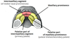
What are the mesonephric ducts in kidney development?
- Two epithelial tubes that run craniocaudally along the entire embryo
- There are formed by mesenchymal to epithelial transitions in the intermediate mesoderm
- They drain the mesonephros in the embryo into the cloaca, and they give rise to the ureteric buds in the formation of the metanephros
Describe the formation of the pronephros, mesonephros and mesonephric ducts. [EXTRA]
Pronephros and mesonephric ducts:
- Near the start of the 4th week, mesenchymal to epithelial transitions in the intermediate mesoderm lead to the formation of two epithelial tubes, known as mesonephric ducts. They begin in cervical region, but inductive mechanisms cause them to extend in the caudal direction.
- Simultaneous to this formation, the pronephros forms also form in the cervical region of the intermediate mesoderm, medial to the mesonephric ducts.
- The pronephros takes the form of a series of hollow epithelial buds, but it is not functional and within two days of its formation, it begins to degenerate.
Mesonephros
- As the mesonephric ducts develop caudally, they induce the intermediate mesoderm caudal to the pronephros to form into mesonephric buds, which begins around day 25.
- Within each bud, mesonephric tubules form, forming a total of around 40 tubule pairs, although earlier tubules regress as more caudal ones develop, and by the end of the 5th week there are only 20 pairs (in the lumbar region).
- Each of these tubules is similar in structure and function to the nephrons in the definitive adult kidney. The most cranial mesonephric tubules fuse on their lateral side to the mesonephric ducts, which have extended towards the cloaca and fused with its ventral side (around day 26), draining the tubules.
- The tubules are functional and produce urine between approximately the 6th and 10th weeks, although in females they regress. In males, the mesonephric duct forms the genital duct in the adult.

When does the metanephros start to develop?
Around the end of week 4 (day 28).
What are the two portions of the definitive kidney? What part of the intermediate mesoderm is each derived from?
- Excretory portion (nephron) -> Metanephric mesenchyme
- Collecting portion -> Ureteric bud
Describe the formation of the metanephros.
- The metanephros begins to develop around the very end of week 4 with the formation of the ureteric buds at the caudal section of each mesonephric duct. This occurs due to induction from the metanephric mesenchyme, which is intermediate mesoderm in the sacral region.
- The ureteric buds each enter the metanephric mesenchyme occurs by day 32.
- The epithelial-mesenchymal interaction between the ureteric bud (epithelial) and metanephric mesenchyme induces the ureteric bud to begin branching (branching morphogenesis). Bifurcation continues until around the 32nd week.
- There is also a reciprocal induction, which is the way in which the ureteric buds induce the mesenchyme to form glomerular units.
- The branching forms the ureter, pelvis, major and minor calyces, and collecting tubules (see other flashcard for this)

What two tissues does the metanephros form from?
Ureteric bud and surrounding mesenchyme
Describe how these structures are formed by the branching of the ureteric bud:
- Pelvis
- Major and minor calyces
- Ureter
- Collecting ducts
- Ureter -> This is the part of the ureteric bud before any branching occurs
- Pelvis -> Formed by the first bifurcation
- Major calyces -> After 4 generations of branching by simple bifurcation, the branches formed coalesce to form the major calyces
- Minor calyces -> Further branches coalesce to form the minor calyces
- Collecting ducts -> Form at the tips of the minor calyces by further branching

Is the process of branching of the ureteric bud continuous?
No, there are periods of branching and periods of coalescing (which are required to form the calyces).
Describe how nephrons develop in the metanephros.
- First appear around 10 weeks, in the distal region of the metanephros
- The ampullae at the tip of each collecting duct interact with adjacent mesenchyme and induce nephron formation.
- Nephric vesicles form ‘comma’ and then ‘S’ shaped tubules. These ultimately form a Bowman’s capsule, proximal convoluted tubules and loops of Henle.
- Ampullae extend deeper into cortex and continue to branch, inducing the formation of more nephrons .

What is Wt1 and what is its importance?
- Wilm’s tumour suppresor gene
- Expressed in the early metanephric mesenchyme for induction of other tissues
- It is required for the initial formation of the ureteric bud from the mesonephric duct
What is Wilm’s tumour?
- A form of kidney cancer that is common in children
- It is frequently caused by alterations to the Wt1 gene
- Results in a palpable mass, pain in the abdomen, poor appetite and fever.
- It is highly responsive to treatment.
Aside from Wt1, what are two factors that are important in inductive processes in kidney development? What does each do?
- Ret (expressed in the ureteric bud) -> Important in branching morphogenesis + Transduces extracellular signals to inside the cell by phosphorylating tyrosine in downstream targets
- Gdnf (expressed in the metanephric mesenchyme) -> Important in branching morphogenesis + It is the ligand for Ret receptor
They have strikingly complementary expression in the two tissues.
When in development do the kidneys ascend and how does this happen?
- Between the 6th and 9th weeks, the metanephric kidney ascends to the lumbar region from its original caudal position
- Caused primarily by differential growth -> The kidneys stay in place while the embryo lengthens
- It remains retroperitoneal (behind the peritoneum)
- It changes its blood supply to progressively higher segmental arteries (branches from the aorta) as it ascends
What happens when a kidney fails to ascend in development?
[EXTRA]
It can result in a pelvic kidney.

What is horseshoe kidney?
[EXTRA]
When the kidneys fuse before they ascend, so they are trapped under the inferior mesenteric artery.

What does the bladder form from?
- Lower end of allantois
- Caudal ends of mesonephri ducts
- Urogenital sinus
What is the allantois?
It is an outgrowth from the hindgut.