7. Musculoskeletal Anatomy Flashcards

(561 cards)

1
Q

What is radiology?

A

The medical discipline that uses medical imaging to diagnose and treat diseases within the bodies of humans.

2
Q

What are the different imaging technologies in current clinical use?

A
  • X-rays
  • Computer tomography (CT)
  • Magnetic resonance imaging (MRI)
  • Radiographic contrast agents -> Barium radiology + Angiography
  • Ultrasound
3
Q

What are some radiology safety measures?

A
  • Dose should be kept as low as reasonably achievable (ALARA)
  • Radiation techniques should be avoided in pregnancy
  • Non-radiation techniques should be used where possible and appropriate
4
Q

In radiology, what is the dose measured in?

A

mSv

5
Q

Describe the principle on which radiography works.

A
  • High energy EM waves (usually x-rays) are absorbed by different materials to varying extents.
  • This means that the detector or film receives different intensities of the waves depening on what they pass through.
  • The creation of 2D images using this technique is called projection radiography.
6
Q

What are some advantages and disadvantages of plain x-rays?

A

ADV:

  • Quick
  • Cheap
  • Great detail
  • Widely availabke

DIS:

  • 2D image only
7
Q

How are x-rays produced?

A

High energy electrons striking a tungsten target within a vacuum tube.

8
Q

Is it acceptable to call the image produced by an x-ray an x-ray?

A

No, it is better to call it a radiograph, image or film.

9
Q

What colour will the following appear on a x-ray/CT image:

  • Air
  • Fat
  • Bone
  • Metal
  • Calcium
  • Organs, Muscles, Soft tissues
A
  • Air -> Black
  • Fat -> Black
  • Bone -> White
  • Metal -> White
  • Calcium -> White
  • Organs, Muscles, Soft tissues -> Shades of grey
10
Q

In radiography, how are the different views of the image named?

A

It is named according to the direction of the x-rays, such as the anterposterior, posteroanterior, oblique and lateral views.

11
Q

What are some applications of x-rays?

A
  • Bone fractures
  • Basic anatomy checks -> e.g. Dextrocardia of the heart, lung collapse, soft tissues
  • Detecting abnormalities, such as tumours
12
Q

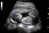
Describe the principle on which ultrasound imaging works.

A
  • High frequency ultrasound is passed into the tissue
  • At every boundary where there is a change in density, part of the signal is reflected, which is picked up by the detector at the surface and used to produce an image
  • The greater the difference in density, the more of the signal is reflected at that boundary.
13
Q

What is the frequency of ultrasound typically used in imaging?

A

2-15 MHz

14
Q

What are some advantages and disadvantages of ultrasound imaging?

A

ADV:

  • High quality information about soft tissue
  • No ionising radiation
  • Inexpensive
  • Flow information

DIS:

  • Gas and bone block US beam
  • Obesity degrades image quality
  • Operator dependent
  • Pixel brightness is not quantitive
15
Q

Check how to interpret an ultrasound scan!!!

A

Not sure if it detects densities - more likely just differences between them.

16
Q

What are some applications of ultrasound imaging?

A
  • Obstetrics (pre-natal scan)
  • Pediatric brain
  • Testicle and prostate
  • Female pelvis
  • Abdomen (liver, kidneys, pancreas, gall bladder) -> Variable appearance can show infection, cancers, etc.
  • Vascular disease
  • Rotator cuff of the shoulder
17
Q

What is a Doppler ultrasound?

A
  • A form of ultrasound that employs the Doppler effect to generate imaging of the movement of tissues and body fluids (usually blood), and their relative velocity to the probe.
  • A colour scale can be used to represent the direction of blood flow.
18
Q

What is another name often used for ultrasound imaging?

A

Ultrasonography

19
Q

Describe the principles of nuclear imaging.

(Note: This is not core material)

A

Nuclear imaging involves the use of a radioactive tracer within the body that emits usually gamma radiation. This can be detected by a detector and used to understand internal body structure.

20
Q

What is the most common radionuclide used in nuclear medicine imaging studies?

(Note: This is not core material)

A

99mTc -> Half-life of 6 hours and a gamma emitter.

21
Q

What are some of the scan types used in nuclear medicine imaging?

A
  • 2D scintigraphy -> Bone, renal
  • 3D -> PET
22
Q

What is a PET CT scan?

A

A nuclear medicine imaging technique that uses both PET and CT scanning simultaneously. The images are superimposed.

23
Q

What does CT stand for?

A

Computer tomography

24
Q

Describe the principle on which CT works.

A
  • An x-ray tube rotates around a patient, on the other side of which are multiple rows of detectors. This is used to assemble a cross-section of the patient.
  • A CT can take multiple slices per rotation (up to 64).
  • In spiral CT, the patient may move past the x-ray tube so that a multi-layer image is formed.
  • The detector works by converting the x-rays to a flow of electrons, which is digitised to Hounsfield units (HU).
25
What are the colours on a CT scan homologous to?
The colours on an x-ray, since a CT is essentially just a series of x-ray images.
26
Describe the Hounsfield scale for CT scans.
The scale assigns the following values: * +1000HU = Bone * 0HU = Water * -1000HU = Air
27
What are some advantages and disadvantages of multislice CT?
ADV: * Static or movie images * Noninvasive * Rapid filming gives few motion artifacts * Good spatial resolution DIS: * Expensive
28
What are some applications for CT?
* Trauma * Intercranial haemorrhage * Abdominal injury (especially to organs) * Fractures * Spinal alignment * Diagnosis and staging of cancers * Detection of foreign bodies in joints
29
What are radiographic contrast agents and how do they work?
* Chemicals that absorb radiation in a CT or x-ray scan, so that the tissue of interest can be ssen more clearly * They work by changing the density of the tissue, changing its opacity to x-rays
30
Are radiographic contrast agents the same as radionuclides?
No, radiographic contrast agents do not emit radiation (like radionuclides used in nuclear medicine imaging), but change the opacity of the tissues of interest.
31
Name some radiographic contrast agents and their uses.
* Barium sulphate -\> Gastrointestinal imaging * Iodinated contrasts -\> Vascular snd lymphatic imaging, Contrast CTs, Cavities * Air -\> Used alongside other contrast to provide double contrast, since the air is less opaque than the contrast tissue
32
What is an angiograph and how is it done?
* Imaging technique used to study blood vessels. * Radiographic contrast (iodine) is injected into the blood vessels, which are then imaged using an x-ray based technique.
33
What is an arthrogram and how is it done? (Note: This is not core information)
* An x-ray image or CT scan of a joint after a contrast has been injected into it * The soft tissue is made clearer, so that injuries can be diagnosed more easily
34
What does MRI stand for?
Magnetic Resonance Imaging
35
Describe the principle on which MRI works, including the parts of the machine.
The machine is a tunnel consisting of: * High strength magnets -\> Apply a magnetic field to the patient, polarising the sample * Shim coils -\> Correct the magnetic field * Gradient coils -\> Localise the region to be scanned * Radiofrequency coils -\> Excite the sample and detect the resulting NMR signal The time for each proton to stop resonating determines the type of tissue. The de-excitation of protons occurs in two phases (T1 and T2), each of which releases energy that can be detected.
36
What contrast agents are used in MRI?
Magnetically active agents, such as gadolinium.
37
What are some advantages and disadvantages of MRI?
ADV: * Static or movie images * Multiple plane images * Good contrast * No known health hazards * Good for soft tissue injuries DIS: * More expensive than CT * Long scans are not pleasant for those with claustrophobia
38
Whereas CT and x-ray imaging look at tissue density, what does MRI look at?
Proton energy
39
What colour will the following appear in an MRI scan: * Air * Fat * Bone * Bone Marrow * Organs, Muscles, Soft tissues * Gadolinium * Water
* Air -\> Black * Fat -\> White (or grey in T2) * Bone -\> Dark * Bone Marrow -\> White (or grey in T2) * Organs, Muscles, Soft tissues -\> Shades of grey * Gadolinium -\> White * Water -\> Dark (but WHITE in T2)
40
Describe how a T1 and T2 MRI scan can be differentiated.
Although fat will appear darker in T2, water will appear much brighter, so that, for example, fluids will be much lighter. Overall, T2 scans frequently seen lighter in general.
41
What are some safety precautions taken with MRI?
Aside from no metal being allowed in the room, these are some contraindications: * Pacemakers * Inner ear implants * Cerebral aneurysms clipped by ferromagentic clips * Metallic foreign bodies in and around the eyes * Pregnancy (especially in the 1st trimester)
42
What are some applications of MRI?
* Neuroimaging -\> Detection of tumours (better than CT because it separates grey and white matter * Cardiac * Musculoskeletal -\> Spinal, joints and tumours * Liver and gastrointestinal * Angiography
43
Remember to practise differentiating between an MRI and CT scan, which can look similar.
Do it. ## Footnote This website is very very useful: https://www.embodi3d.com/blogs/entry/373-how-to-easily-tell-the-difference-between-mri-and-ct-scan/
44
CT
45
MRI
46
Ultrasound
47
X-ray
48
What type of tissue is bone?
Connective tissue
49
Give an example of a disease which causes the conversion of one organ systemm to another.
Fibrodysplasia ossificans progressiva (FOP) is a very rare condition where muscle tissue and connective tissue such as tendons and ligaments are gradually replaced by bone (ossified), forming bone outside the skeleton (extra-skeletal or heterotopic bone) that constrains movement.
50
What is the average number of bones in the human skeleton?
206
51
What can alter the number of rib or digits in a human skeleton?
Homeotic mutations
52
What is sesamoid bone? Give an example.
A bone embedded in tendon (or muscle). An example is the patella.
53
Give some examples of variable human skeletal anatomy and how this is useful.
The human skeleton varies by age and geographic variation. Humans are moderately **sexually dimorphic** so on average female bones are smaller than those of males. This has an importance in forensic medicine.
54
Describe the divisions of the skeleton.
* Axial skeleton -\> Head, Neck, Trunk * Appendicular skeleton -\> Upper and lower extremities (including the pectoral and pelvis girdle)
55
What are the pectoral and pelvic girdles?
The skeletal frameworks which provides attachment for the upper and lower limbs respectively. They consist of: * Pectoral girdle = Scapulas + Clavicles * Pelvic girdle = Hip bones, Sacrum + Coccyx
56
Describe some of the functions of the human skeleton.
* Providing a rigid framework * Protection of soft tissues * Facilitation of movement * Resistance to forces (e.g. gravity) * Surface for muscular attachment * Maintenance of blood calcium * Red marrow is important in blood cell formation
57
Describe the basic structure of bone as a tissue.
It is connective tissues so it contains cells embedded within a composite ECM containing: * **Organic matrix** -\> Mostly collagen, which provides tensile strength and some flexibility * **Inorganic material** -\> Hydroxyapatite (mostly calcium phosphate) that gives hardness and rigidity (the crystals impregnate the collagen matrix)
58
What are the two main materials in bone? Describe how the properties of both can be deduced experimentally.
* Collagen -\> Heat can be used to disrupt the peptide bonds, which makes the bone brittle -\> This shows that collagen provides elasticity and tensile strength * Hydroxyapatite -\> Acid can be used to remove the Ca2+ ions, which makes the bone curl up and be soft -\> This shows that hydroxyapatite provides hardness and rigidity
59
60
Give an example of a bone disease caused by abnormal collagen.
Osteogenesis imperfecta ("brittle bone disease")
61
What are the different morphological types of bone?
* Flat bones * Long bones * Short bones * Irregular bones * Sesamoid bones
62
Where are flat bones found?
* Cranial vault * Thoracic cage * Scapula
63
Where are long bones found?
* Limbs * Hands * Feet
64
Where are short bones found?
* Wrist * Ankle (Tend to be cuboidal)
65
Where are irregular bones found?
* Facial skeleton * Pelvis * Vertebral column
66
What is the function of sesamoid bones?
Act as pulleys and protect the tendons from excessive wear.
67
Describe the histological divisions of bone.
* Bone is divided into woven (immature) and lammellar (mature) * Woven (or immature) bone is the first bone to develop when the skeleton forms, and it is also found in bone repair and tumours * Lammellar (or mature) bone is what makes up all normal adult bones, and is divided into compact and spongy/trabecular bone * Compact bone -\> Dense bone found around the external surfaces of the bone, including the shaft * Trabecular bone -\> Lightweight bone found in the ends of long bones, short bones and in the bodies of vertebrae
68
What are the different names for roughened areas for the attachments of muscle tendons or ligaments?
* Tuberosities * Trochanters * Tubercles * Ridge * Line * Epicondyle
69
What is the purpose of grooves on bones?
They are usually where blood vessels, nerves and tendons lay close to the bone.
70
What are holes in bone called? What is the purpose of these?
Foramina - these are usually the site of passage of nerves or blood vessels through the bone.
71
What is the name for a concave depression in a bone?
Fossa
72
What are condyles and epicondyles?
* Condyles -\> Smooth round surfaces at the ends of bones, usually acting as articulating surfaces * Epicondyles -\> Rough projections on the condyle that act as attachment
73
What two types of bone are adult bones composed of? Describe the location and appearance of each.
1. Compact (a.k.a cortical) bone -\> Dense form of bone with a Haversian canal structure. Found on the external surfaces of the bone, including the shaft. 2. Trabecular (a.k.a. spongy or cancellous) bone -\> Lightweight form of bone with honeycomb structure. Found in the ends of long bones, short bones and in the bodies of vertebrae.
74
Describe the histological structure and parts of adult bone.
* Compact (a.k.a cortical) bone -\> Dense form of bone with a Haversian canal structure. Found on the outside of the bone, including the shaft. * Trabecular (a.k.a. spongy or cancellous) bone -\> Lightweight form of bone with honeycomb structure. Found in the ends of long bones, short bones and in the bodies of vertebrae. * Red bone marrow may be found within trabecular bone. * Medullary cavity (a.k.a. marrow cavity) is in the centre of long bone shafts -\> Contains yellow marrow, which contains fat cells * The periosteum is a layer of dense irregular tissue that covers the entire outside of bones (except the joint surfaces) * The endosteum is like the periosteum, except it lines the inside of the medullary cavity
75
Where is red bone marrow found in bones and what is its function?
* In trabecular bone. * It contains stem cells that ultimately give rise to RBCs, WBCs and platelets.
76
What is the name for the production of blood cells and platelets in the bone marrow?
Haemotopoiesis
77
Where is yellow bone marrow found, what is it made of and how does it change over time?
* In the medullary cavity of long bones * It is made of fat cells * Over time, red bone marrow is converted to yellow bone marrow
78
What are the different areas of a long bone?
* Diaphysis -\> Shaft, which develops from the primary ossification centre * Epiphyses -\> Ends,which develop from secondary ossification centres * Metaphyses -\> Expanded areas of the bone shafts, to which the epiphyses fuse in bone growth
79
Describe the structure of compact bone.
* Consists of units called osteons (or Haversian systems). * Osteons are cylindrical structures that contain a mineral matrix and living osteocytes. * Each osteon consists of lamellae, which are layers of compact matrix that surround a central canal called the Haversian canal. The Haversian canal (osteonic canal) contains the bone’s blood vessels and nerve fibers. * The lamellae are joined by caniculi (microscopic canals). * Osteons in compact bone tissue are aligned in the same direction along lines of stress and help the bone resist bending or fracturing. Therefore, compact bone tissue is prominent in areas of bone at which stresses are applied in only a few directions.
80
Describe the structure of spongy bone.
* Consists of trabeculae, which are lamellae that are arranged as rods or plates. * Red bone marrow is found between the trabuculae. * Blood vessels within this tissue deliver nutrients to osteocytes and remove waste.
81
Describe how bone is adapted for strength and force transmission.
* Compact bone around the outside * Trabecular bone is present in parts of the bone where forces may be exerted in various directions * Trabeculae are also largely arranged in the direction of stress and tension lines
82
Define ossification.
The laying down of bone material by osteoblasts (i.e. bone formation).
83
What are the two types of ossification and where does each occur?
Intramembranous ossfication (rapid): * Bone is deposited directly onto an embryonic connective tissue membrane. * This occurs in parts of the crania, mandible and most of the clavicle. Endochondral ossification (slow): * Hyaline cartilage model of the bone is formed, which is gradually replaced by bone. * This occurs in the rest of the skeleton.
84
Draw a diagram showing where each type of ossification occurs.
85
What are the embryonic derivatives of bone and what does each form?
* Neural crest cells -\> Form the skull, mandible and clavicle (by intramembranous ossification) * Somites (sclerotome) -\> Rest of the _axial_ skeleton (by endochondral ossification) * Lateral plate mesoderm -\> Long bones (by endochondral ossification)
86
How can you remember which type of ossification occurs where in the body?
* Intramembranous ossification is required to form strong bone where it is necessary for protection from early-on, such as in the skull * Endochondral ossification is required for growth over time
87
Describe the process of intramembranous ossification.
* Neural crest derived mesenchymal cells differentiate into osteoblasts and group into ossification centers * Osteoblasts become entrapped by the osteoid they secrete, transforming them to osteocytes * Trabecular bone forms * Mesenchymal cells on the surface form a membrane called the periosteum * Cells on the inner surface of the periosteum differentiate into osteoblasts and secrete osteoid parallel to that of the existing matrix, thus forming layers of cortical bone * Blood vessels in the centre form the red marrow
88
Describe the process of endochondral ossification.
* Mesoderm-derived mesenchymal cells differentiate into chondrocytes and form the cartilage model for bone * Chondrocytes near the center of the cartilage model undergo hypertrophy and alter the contents of the matrix they secrete, enabling mineralization * Chondrocytes undergo apoptosis due to decreased nutrient availability * Blood vessels invade and bring osteogenic cells * Primary ossification center forms in the diaphyseal region of the bone -\> This occurs where the major blood vessel enters * Secondary ossification centers develop in the epiphyseal region after birth
89
What are the two types of growth in a long bone and how does each occur?
* Appositional growth -\> Thickens the bone -\> Bone is reabsorbed at the endosteal surface and added at the periosteal surface * Interstitial growth -\> Makes the bone longer -\> At the epiphyseal plate, cartilage grows towards the extremity of the bone and is replaced by bone
90
When do the primary and secondary ossification centres form?
* Primary -\> Before birth * Secondary -\> After birth (EXCEPT the distal femur and proximal tibia i.e. near the knee)
91
92
What is the name for the site of lengthening of long bones?
Epiphyseal plate (a.k.a. growth plate)
93
What is the metaphysis?
The section of the bone that is next to the epiphyseal plate, on the side of the diaphysis. It marks the growth of the bone.
94
How many ossification centres do bones have?
* Long bones -\> Two secondary, one primary * Short bones -\> Form from just primary osssification (and may have many primary ossification centres)
95
When does the lengthening of the bone stop and what happens when this goes wrong?
* When the epiphyseal plate stops dividing and the epiphysis fuses with the metaphysis * Premature ossification leads to premature interruption of limb extension
96
Can epiphyseal plates be seen on an x-ray?
They are seen as empty spaces that look sort of like a big fracture, but aren't!
97
Name the divisions of the upper limb.
* Arm * Forearm * Wrist * Hand
98
What bone is in the arm and what does it articulate with?
* Humerus * Articulates with: Scapula, Radius, Ulna
99
What is it worth noting about the upper and lower limb in the anatomical position?
They are rotated differently so that the big toe and thumb are on opposite sides.
100
State some functional differences between the upper and lower limb.
* Glenohumeral joint is capable of a wider range of movement than the hip joint due to joint depth * Forearm bones are capable of pronation and supination, which cannot happen in the leg * Thumb allows opposition, which cannot happen in the foot
101
What are the bones in the wrist and hand what do they articulate with?
* Carpals, Metacarpals, Phalanges (proximal, intermediate, distal) * Carpals articulate with the radius at the wrist
102
What is the proper name for the shoulder joint?
Glenohumeral joint
103
What are all of the bones of the upper limb?
(Shoulder = Scapula + Clavicle) * Humerus * Radius * Ulna * Carpals * Metacarpals * Phalanges (x3)
104
What is the pectoral girdle composed of?
* Clavicle * Scapula
105
What does the clavicle articulate with and what are the names of these joints?
* Medially -\> With manubrium of the sternum (sternoclavicular joint) * Laterally -\> With acromion of scapula (acromioclavicular joint a.k.a. AC joint)
106
What are the main landmarks and attachments of the clavicle?
Landmarks: * Acromial facet (laterally) * Sternal facet (medially) * Conoid tubercle (for coracoclavicular ligament) Muscle attachments: * Trapezius (insertion) * Deltoid (origin) * Pectoralis major (origin) * Subclavius muscle (insertion) * Sternocleidomastoid (origin) Ligment attachments: * Acromioclavicular ligament (laterally) * Coracoclavicular ligament (laterally) * Sternoclavicular ligament (medially) * Costoclavicular ligament (medially)
107
What is the clinical relevance of the clavicle?
The clavicle is a very frequent site of fracture, with fractures most commonly occuring in the middle third of the bone.
108
Is this the anterior or posterior view of the scapula?
Posterior
109
What does the scapula articulate with and what are the names of these joints?
* Humerus -\> Glenohumeral joint * Clavicle -\> Acromioclavicular joint
110
What are the main landmarks and attachments of the scapula?
Landmarks: * Subscapular fossa -\> Subscapularis origin * Coracoid process -\> * *Muscles* - Pectoralis minor (attachment), coracobrachialis (origin) and short head of biceps brachii (origin) * *Ligaments* - Coracoclavicular, coracoacromial, coracohumeral * Glenoid fossa -\> Site of glenohumeral joint (glenohumeral ligaments) * Supraglenoid tubercle -\> Long head of biceps brachii * Infraglenoid tubercle -\> Long head of triceps brachii * Spine -\> Deltoid, Trapezius * Acromion (at end of spine) -\> Site of AC joint (AC ligaments), Coracoacromial ligament * Infraspinous fossa -\> Infraspinatus muscle * Supraspinous fossa -\> Supraspinatus muscle Other muscle attachments: * Latissimus dorsi * Serratus anterior * Teres minor * Teres major * Levator scapulae * Rhomboideus major * Rhomboideus minor Other ligment attachments: * Suprascapular ligament
111
What are the major palpable landmarks of the shoulder? Where are they found?
* Acromion (palpable above shoulder) * Coracoid process (palpable below lateral clavicle)
112
Label this.
113
What is some clinical relevance related to the appearance of the scapula?
* The scapula may be winged when pushing with the arm * This usually indicates damage to the long thoracic nerve, which innervates the serratus anterior muscle * The serratus anterior muscle is responsible for holding the scapula close to the ribcage
114
What does the humerus articulate with?
* Scapula (glenohumeral joint) * Head of radius + Trochlear notch of ulna (elbow joint)
115
What are the main landmarks and attachments of the humerus?
Landmarks: * Head -\> Glenohumeral joint * Greater tuberosity (lateral) -\> Supraspinatus, Infraspinatus, Teres minor * Lesser tuberosity (anterior) -\> Subscapularis * Bicipital groove (a.k.a. intertubercular groove) -\> Tendon of long head of biceps passes through this, Latissimus dorsi, Pectoralis major and Teres major attaches here * Deltoid tuberosity (lateral side of shaft) -\> Deltoid * Radial groove (diagnal on posterior surface) -\> Radial nerve and profunda brachii artery run through this * Medial and lateral supracondylar ridge -\> Lateral is rough and provides attachment for forearm extensor muscles * Medial and lateral epicondyles -\> Medial is larger and features a groove where the ulnar nerve passes through * Trochlea (more medial) -\> Articulates with trochlear notch of ulna * Capitulum (more lateral) -\> Articulates with head of radius * Radial and coronoid fossa -\> Receive forearm bones during flexion * Olecranon fossa -\> Receives olecranon of ulna during extension Other muscle attachments along shaft: * Anteriorly: Coracobrachialis, deltoid, brachialis, brachioradialis. * Posteriorly: Medial and lateral heads of triceps Ligaments: * Medial collateral * Lateral collateral * Glenohumeral ligaments
116
Describe the position of the greater and lesser tubercles of the humerus. What attaches to each?
Greater tubercle: * Lateral, with both posterior and anterior surfaces * Supraspinatus attaches to superior facet * Infraspinatus attaches to middle facet * Teres minor attaches to inferior facet Lesser tubercle: * More medial, with anterior surface only * Subscapularis attaches here
117
How can you remember the attachments of at the lips of the bicipital groove (intertubercular groove)?
"A lady between two majors" The latissimus dorsi muscle attaches between the pectoralis major and teres major.
118
What are the two necks of the humerus? What is the significance of each?
* Anatomical neck -\> Just below the head * Surgical neck -\> Further down the humerus -\> It is a frequent point of fracture
119
Why might a knock to the elbow elicit a tingling feeling?
The ulnar nerve passes posterior to the medial epicondyle of the humerus, where it is very superficial and can easily be stimulated.
120
What is often seen with a fracture at: * Surgical neck of the humerus * Shaft of the humerus
* Surgical neck of humerus -\> Damage to the axillary nerve * Shaft of humerus -\> Radial nerve
121
Label this:
122
Label this:
123
What does the radius articulate with?
Proximally: * With capitulum of humerus (elbow joint) * With radial notch of ulna (proximal radioulnar joint) Distally: * With head of ulna (distal radioulnar joint) * With scaphoid and lunate (radiocarpal / wrist joint)
124
What does the ulna articulate with?
Proximally: * Trochlea of humerus (elbow joint) * Head of radius (proximal radioulnar joint) Distally: * Ulnar notch of radius (distal radioulnar joint)
125
What are the main landmarks and attachments of the radius?
Landmarks: * Head -\> Site of elbow joint (with capitulum of humerus) * Radial tuberosity -\> Biceps brachii * Shaft * Styloid process (lateral) -\> Radial collateral ligament of the wrist + Site of radiocarpal joint * Ulnar notch -\> Site of distal radioulnar joint (with head of ulna) Other muscles: * Pronator teres * Pronator quardatus * Supinator teres * Finger and thumb flexors and extensors * Brachioradialis Other ligaments: * Radial collateral ligament (at elbow) * Radial collateral ligament (at wrist) * Radiocarpal ligaments Note the intraosseous membrane between the radius and ulna.
126
What are the main landmarks and attachments of the ulna?
Landmarks: * Trochlear notch - Site of elbow joint (with trochlea of humerus) * Olecranon - Triceps brachii * Coronoid process - Brachialis + Ulnar collateral ligament of elbow * Ulnar tuberosity - Brachialis * Radial notch - Site of proximal radioulnar joint + Annular ligament * Styloid process - Ulnar collateral ligament of wrist * Head - Site of distal radioulnar joint (with ulnar notch of radius) Other muscles: * Supinator * Anconeus * Finger and thumb flexors and extensors Other ligaments: * Radiocarpal ligaments * Ulnar collateral ligament (wrist)
127
Does the head of ulna articulate with the bones of the wrist?
No, it is prevented from doing so by a cartilaginous articular disc.
128
Give some clinical relevance of the radius.
* Colles' fracture * Most common type of radial fracture * Caused by fall on outstretched hands * Fracture at distal end, so that the wrist area is dislocated posteriorly -\> Gives dinner fork appearance * Smith's fracture * Opposite of Colles' fracture * Caused by fall on back of hands
129
Label this image.
130
What must you remember exists between the radius and ulna?
Interosseous membrane
131
Which way does the palm face in pronation and supination of the forearm?
* Pronation -\> Down * Supination -\> Up
132
During pronation and supination of the forearm, which bone rotates?
Radius
133
Describe all of the types of bones in the hand, starting from the tip of a finger.
* Distal phalanges * Middle phalanges * Proximal phalanges * Metacarpals * Carpals
134
Describe all of the joint in the hand, starting from the tip of a finger.
* Distal interphalangeal joint (DIP) * Proximal interphalangeal joint (PIP) * Metacarpophalangeal joint * Carpometacarpal joints
135
Describe the bones in the thumb.
There is one metacarpal (as with the other fingers), but only two phalanges (there are 3 in the other fingers).
136
Label this.
137
How many carpals are there?
8
138
Name all of the carpals, starting from the side of the thumb.
Distal: Trapezium, Trapezoid, Capitate, Hamate Proximal: Scaphoid, Lunate, Triquetrum, Pisiform
139
Give a good mnemonic for remembering the carpals.
Some lovers try positions that they can't handle.
140
Out of the triquetrum and pisiform, which is on the palmar side of the hand?
Pisiform
141
Describe how a scaphoid fracture might occur, symptoms and why it is dangerous.
* Occurs when falling onto outstretched hand * Characterised by pain in the anatomical snuffbox * In many people the blood supply to the scaphoid is only in the distal to proximal direction, so a fracture can cause avascular necrosis of the proximal portion of the scaphoid
142
What stops the ulna articulating with the carpals?
An articular disc.
143
What is the wrist joint?
Articulation between the lower end of the radius and the articular disc covering the distal ulna, with the scaphoid, lunate and triquetrum.
144
Describe how the fingers are numbered.
From 1 to 5, with the thumb being number 1.
145
What are the divisions of the lower limb? What are the bones in each?
* Thigh -\> Femur * Leg -\> Tibia, patella and fibula * Ankle * Foot -\> Tarsals, metatarsals and phalanges
146
What are some adaptations of the lower limb that make it different to the upper limb?
* Hip joint is deeper than shoulder joint, which allows for greater stability + Rigid pelvic girdle * Bicondylar angle of the femur allows for the centre of gravity to be directly above the knee, so that walking can happen * Knee joint reinforced by internal ligaments * Leg bones do not rotate around each other like the forearm bones do
147
What is another name for the hip bone?
Os coxae or innominate bone
148
What bones make up the hip bone? What is the position of each?
* Ilium -\> Top * Ischium -\> Bottom, Back * Pubis -\> Bottom, Front
149
Describe the structure of the pelvis.
* Made up of two hip bones and the sacrum * Each hip bone consists of the ilium, ischium and pubis, while the sacrum fits between the ilium of each hip bone * The coccyx may be considered part of the pelvis too
150
Where are the three bones of the hip bone all united?
Acetabulum
151
Note that for the lower limbs I have not gone into as much detail about landmarks, ligaments and muscles as I have with the upper limb.
Ok.
152
What is the function of the pelvis?
It protects the lower abdominal and pelvic organs and forms points of articulation with the lower limbs.
153
Compare the pelvis and pelvic girdle.
The pelvis also includes the sacrum, while the pelvic girdle does not.
154
What does the pelvis girdle (two hip bones) articulate with?
* Sacrum (sacroiliac joint) * Head of femur (hip joint One hip bone also articulates with the other at the pubis symphysis.
155
Describe the development of the hip bone.
The three bones all have their own primary ossification centre and then eventually fuse together around the ages of 16 to 18.
156
What are the main landmarks of the hip bone?
Ilium: * Iliac crest (palpable) * Anterior superior iliac spine (ASIS) (palpable) * Posterior superior iliac spine (PSIS) (palpable) * Anterior inferior iliac spine (AIIS) * Greater sciatic notch Ischium: * Ischial spine * Ischial tuberosity (palpable) Pubis: * Pubic tubercle (palpable) All 3: * Acetabulum -\> Articulates with head of femur
157
Name some important palpable landmarks of the hip bone.
* Iliac crest * Anterior superior iliac spine (ASIS) * Posterior superior iliac spine (PSIS) * Ischial tuberosity * Pubis tubercle
158
What is some clinical significance of the iliac crest?
A line drawn between the highest point on each iliac crest crosses at the level of the spine of the L4 vertebrae, which is useful when finding the spot to insert a lumbar puncture needle.
159
What is the significance of the ischial tuberosity?
The ischial tuberosities are what you sit on.
160
What is the acetabulum?
It is the point on the hip bone where the femur articulates.
161
What does the femur articulate with?
* Acetabulum of hip bone (hip joint) * Tibia and patella (knee joint)
162
What are the main landmarks of the femur?
* Head -\> Articulates with acetabulum * Neck * Greater trochanter (lateral and slightly posterior) -\> Gluteus medius and minimus * Lesser trochanter (medial and posterior, below neck) -\> Powerful hip flexor iliopsoas * Linea aspera (posterior) -\> Adductors * Lateral and medial epicondyles -\> Articulate with tibia
163
What is the danger of a fracture of the neck of the femur?
If the fracture is inside the joint capsule, blood supply to the femoral head might be disrupted.
164
Name all of the articulations at the knee.
* 2 x Femur with the tibia * 1 x Femur with the patella
165
Label this.
166
Is the fibula part of the knee joint?
No, it is sort of off to the side.
167
Name the surfaces that articulate between the femur and tibia.
The lateral and medial femoral condyles articulate with the lateral and medial tibial condyles. There is a raised intercondylar eminence on the tibia.
168
What type of bone is the patella?
Sesamoid
169
Give an interesting piece of information about the sesamoid bones near the knee.
A small fraction of people have a very small sesamoid bone that exists in the calf muscle tendon. It is called the fabella (small bean).
170
What is a name for the top of the tibia?
The tibial plateau
171
What does the tibia articulate with?
* Femur (tibiofemoral / knee joint) * Fibula (x2) (proximal and distal tibiofibular joints) * Talus (ankle joint) Note that there is also an interosseous membrane between the tibia and fibula.
172
What are the main landmarks of the tibia?
* Tibial plateau -\> Articulates with femur * Intercondylar eminence -\> ACL and PCL, Menisci of tibia * Medial and lateral tibial condyles * Tibial tuberosity (anterior) -\> Quadriceps * Tibial shaft * Medial malleolus -\> Articulates with talus
173
What does the fibula articulate with?
* Tibia (x2) (proximal and distal tibiofibular joints) * Talus (ankle joint)
174
What are the main landmarks of the fibula?
* Head of fibula -\> Articulates with tibia * Fibula shaft * Lateral malleolus -\> Articulates with talus
175
Compare the main functions of the tibia and fibula.
* Tibia -\> Weight bearing * Fibula -\> Muscle attachments
176
Label this.
177
Describe the articulations at the ankle joint.
The medial malleolus of the tibia and the lateral malleolus of the fibula "grip" the talus (a tarsal) bone.
178
Describe the bones in the foot.
* 7 tarsals -\> Talus, Calcaneus, Cuboid, Navicular, Cuneiforms (x3) * 5 metatarsals * 14 phalanges (only 2 in the hallux) Therefore the structure is homologous to the hand.
179
Name all of the tarsals.
* Calcaneus (forms the mass of the heel, very large) * Talus (forms ankle joint) * Cuboid * Navicular * Cuneiforms x 3 (Medial, intermediate, lateral)
180
Give the clinical relevance of the tubersity on the 5th metatarsal.
It is prone to an aversion fracture when the foot is everted excessively.
181
What is the technical name for the big toe?
Hallux
182
Do inversion and eversion of the foot happen at the ankle joint?
No, it is a hinge joint. Inversion and eversion happen at the joints between the tarsals.
183
Label this.
184
185
What are the three bones in the shoulder girdle?
Clavicle, scapula and humerus
186
Label this.
187
What are some features of the clavicle?
* First bone to ossify * Among the last the fuse * Most commonly fractured bone * Only bony attachment of the upper extremity is via the sternoclavicular joint
188
\* What component affects the direction of a break in the clavicle?
* Ligament action pulls the fragments in specific directions (medial fragment drawn upwards) * These breaks aren’t overly benign as close to lung and brachial plexus - has been shown that fixing them surgically is a better treatment than just leaving them to recover
189
What type of joint is the sternoclavicular joint?
Synovial - with articular disc
190
Which ligament tears in a medial clavicle dislocation?
The ligament between the sternum and the clavicle No operating on this kind of dislocation, just try and reduce it
191
What is the AC joint?
The acromioclavicular joint - between the clavicle and the acromium process
192
How many ligaments are there that attach to the coracoid process and what are they?
There are 3: * Coracoacromial ligament, between acromium and coracoid processes (both on scapula) * Coracoclavicular ligament -\> Trapezoid and conoid ligaments, attach the clavicle to the coracoid process
193
How can you remember the order of the coracoclavicular ligaments?
Medial to lateral: "CT" scan to diagnose (conoid then trapezium)
194
What is an acromioclavicular separation?
This is where the coracoclavicular ligaments tear, but the AC joint is only partially damaged
195
What happens in an AC dislocation?
Both the AC and coracoclavicular ligaments tear, resulting in full dislocation of the distal end of the clavicle
196
What type of joint is the AC joint?
Synovial, but with little movement
197
\* What is a good indication for the side to which a scapula belongs?
The direction in which the coracoid process is pointing - it will always be pointing towards the arm and is on the front/anterior side of the bone
198
\* How are humeral head fractures treated?
Conservative management is disproportionately successful (length/alignment/rotation), use a collar and cuff. Alternatively, surgical intervention can be used.
199
Where is and what are some characteristics of the glenohumeral joint?
The joint between the humeral head and the glenoid fossa/cavity Shallow joint, stability improved by the glenoid labrum and the ligaments Manipulated by the rotator cuff muscles amongst others Synovial joint, ball and socket
200
What are the three ligaments in the glenohumeral joint?
* SGHL: superior glenoid-humeral ligament * MGHL: medial glenoid-humeral ligament * IGHL: inferior glenoid-humeral ligament
201
How do the shoulder and hip joints compare? (In one sentence)
They are opposites - hip has a more restricted range of movement but is more stable, shoulder has a larger range of movement but is less stable. Shoulder is likened to a golfball on a golf tee
202
Which ribs articulate with the scapula?
Ribs 2-7 Major contributors to shoulder motion - bursas are present
203
What is a bursa?
Small fluid-filled sac contained within synovial joints - act as a cushion
204
How many muscles attach the upper limb to the axial skeleton?
17 Ones we need to know are: * Deltoid * Rotator cuff muscles (x4) * Biceps/attachment of the biceps tendon
205
What is innervated by the axillary nerve?
Deltoid and teres minor (amongst others)
206
What happens if the axillary nerve is damaged?
Shoulder is weakened, ability to abduct is severely lessened
207
What are the four rotator cuff muscles?
Subscapularis, infraspinatus, supraspinatus, teres minor
208
What innervates supraspinatus and infraspinatus?
The suprascapular nerve
209
What innervates teres minor?
The axillary nerve
210
What innervates subscapularis?
The subscapular nerve
211
What acronym can be used to remember the four rotator cuff muscles/tendons?
SITS: * Supraspinatus * Infraspinatus * Teres minor * Subscapularis
212
Where do the rotator cuff muscles attach?
* Supraspinatus, infraspinatus and teres minor attach to the greater tuberosity of the humerus * Subscapularis attaches to the lesser tuberosity of the humerus (only one) The other ends of the muscles attach to the scapula.
213
What is the function of the rotator cuff muscles?
* Move shoulder joint * Provides stability (stops dislocation) * Keeps deltoid from pulling humeral head up
214
Why is the biceps muscle called the biceps?
Bi - two Ceps - cephalus, means ‘head’ The biceps has two ‘heads’, the long head and the short head - long head goes over the humeral head, short goes anteriorly, both attach to the scapula.
215
Where are the origins and insertions of the biceps muscle?
Origins: * Long head: Supraglenoid tuberosity (of scapula) * Short head: Coracoid process Insertions: * Distal biceps tendon: Radius (at radial tuberosity)
216
What are the origins and insertions of the brachialis muscle?
Origins: * Halfway up the humerus Insertions: * Coronoid process and tuberosity of the ulna.
217
\* What is the failsafe mechanism of the biceps?
There are two heads, so if one ruptured the muscle is still functional as the other remains attached - contraction still results in movement of the bone. Results in ‘popeye’ muscle
218
\* What are some of the problems that can occur within the shoulder?
* Adhesive capsulitis (frozen shoulder) * Impingement syndrome * Rotator cuff tears * Dislocation * Arthritis
219
\* How can age affect outcome of shoulder dislocations?
* Younger patients have a higher dislocation rate * Older patients have a higher rate of rotator cuff tears
220
\* How should you manage a dislocated shoulder?
* Best to reduce the dislocation at time of accident, then short period of immobilisation (though external rotation splint or simple sling) * Can be managed surgically, usually after recurrent dislocations (labrum can tear and will need to be repaired).
221
What is the most commonly dislocated joint?
The shoulder - inherently unstable (shallow joint)
222
\* What is adhesive capsulitis?
Aka frozen shoulder, inflammation and thickening of the shoulder capsule eventually results in hugely reduced range of movement (especially external rotation). Associated with diabetes, but often idiopathic (arises spontaneously with unknown cause). Not self limiting (so won’t resolve itself without treatment), just stops hurting over time, doesn’t really improve.
223
\* How can you treat adhesive capsulitis?
* Used to be conservative treatment/‘wait and see’ * Now can have glenohumeral injections * More and more commonly manipulated under anaesthesia - only in stiff phase, and these is a slight risk of humeral fracture * Generally resolved with time (1-2 years), residual stiffness is expected. But can only get it once in each shoulder!
224
What is shoulder impingement?
This is where the rotator cuff tendons within the coracoacromial arch are impinged - trapped or limited mobility of tendons, or inflamed bursa within the shoulder. This affects nearby nerves and causes pain. Association with ‘hooked’ acromion (specific shape, morphologically hooked around joint therefore increases risk of impingement)
225
What are the risk factors for shoulder impingement?
* Can occur at any age * Classically involves anterior 1/3 of acromion * Incidence does occur with age
226
What is the treatment for shoulder impingement?
* Usually conservative - subacromial injections and physio * 2/3 resolve (but only 1/2 in over 60s) * Can also have surgical decompression
227
What happens if a rotator cuff tendon is torn?
The patient will be unable to elevate their arm fully as only deltoid is able to work on the humerus - other abductors are compromised. Incidence increases with age, and management is hotly debated.
228
What is another word for arthroscopic surgery?
Keyhole surgery
229
What is current treatment for damaged rotator cuff muscles?
Therapy to strengthen the remaining cuff, traditional open or mini-open repair (both arthroscopic)
230
How common is glenohumeral arthritis?
Least common of the major joints
231
What is glenohumeral arthritis?
This is joint inflammation of the shoulder, where the space between the glenoid fossa and the humeral head is greatly reduced, causing pain. Can be secondary to trauma, chronic rotator cuff deficiency and necrosis of the humeral head (this can occur when a fracture cuts off blood supply)
232
How can a massive rotator cuff tear cause glenohumeral arthritis?
The rotator cuff muscles help to hold the humerus down/oppose the action of the deltoid muscle - when the rotator cuff tears, the deltoid can draw up the humerus, reducing joint space and therefore causing arthritis
233
What are the treatments for glenohumeral arthritis?
Conservative: injections Operative: replacement
234
What position is the humeral head in relation to the transepicondylar axis of the distal humerus?
Humeral head is retroverted (tilted backwards) relative to the transepicondylar axis - joint is most unstable anteriorly, to by turning shoulder back by ~30 degrees improves stability
235
Where does pectoralis major insert?
To the bicipital groove and the deltoid tuberosity on the humerus (lateral to the intertubercular groove)
236
Where does latissimus dorsi insert?
Floor of the intertubercular groove
237
Where does teres major insert?
The medial lip of the intertubercular groove
238
How can the order of the insertions of pectoralis major, latissimus dorsi and teres major be remembered?
‘A lady between two majors’, pectoralis is the most lateral, latissimus is in the middle, teres major the most medial
239
What attaches to the greater tuberosity of the humerus?
Supraspinatus, infraspinatus, teres minor (C5-C6)
240
What attaches to the lesser tuberosity humerus?
* Subscapularis - allows for internal/medial rotation and adduction * Upper (C5) and lower (C6) subscapular nerve ???
241
Patient with a subscapularis rupture would have what findings?
* Positive Gerber lift off test (arrange hand so that it is behind the back, back of hand touching back of the person. Ask the patient to try and lift their hand from their back - if positive, patient should be unable to do this with either inability or pain as a limiting factor). * Weak internal rotation * Increased passive external rotation
242
What is the triangular space in the shoulder?
The space formed between the teres minor (superiorly), teres major (inferiorly) and long head of triceps (laterally). The scapula circumflex vessels pass through here (axillary, subscapular and scapular circumflex arteries).
243
What is the quadrangular space in the shoulder?
The space formed by the teres minor (superiorly), teres major (inferiorly), long head of triceps (medially) and neck of humerus (laterally). The axillary nerve and the posterior circumflex humeral artery + vein pass through here EVERYTHING THROUGH THIS GAP EITHER HAS 4 SYLLABLES OR 4 WORDS
244
What provides the blood supply to the humeral head?
Anterior circumflex humeral artery Posterior circumflex humeral artery
245
On the upper limb, where is the relative position of the neurovascular bundle?
Medial
246
What is flexion?
Movement that decreases the angle between two body parts
247
What is extension?
Movement that increases the angle between two body parts
248
What is dorsiflexion?
The action of moving the foot/toes upwards/towards the shin (remember this through the idea of creating a dorsal fin)
249
What is plantarflexion?
The action of moving the foot/toes downwards/towards the ground if the foot was suspended (remember this through the idea of 'planting' your foot on the ground)
250
To what body part are dorsi- and plantarflexion specific?
The ankle joint/foot
251
What is abduction?
The action of moving a body part away from the midline/laterally (remember this through the idea of abduction as stealing a human)
252
What is adduction?
The action of moving a body part towards the midline/medially (remember this through a-d-duction, 'adding')
253
What is eversion?
Movement of the foot so that the sole is facing outwards/has been moved away from the midline
254
What is inversion?
Movement of the foot so that the sole is facing inwards/has been moved inwards - this is what happens when you twist your ankle
255
Why are inversion and eversion useful?
Important for walking across uneven ground
256
What is lateral flexion?
This is the bending of the neck or body away from the midline, i.e. to the left or right
257
What is medial rotation?
The rotating of a joint so that it faces more internally
258
What is lateral rotation?
The rotating of a joint so that it faces more externally
259
What is rotation?
Movements made about the longitudinal axis and in the transverse plane
260
What is pronation?
The act of rotating the forearm so that the palm is facing down (remember: the whip sequence)
261
What is supination?
The act of rotating the forearm so that the palm is facing upwards (remember: lying down as if supine)
262
What is circumduction?
The movement of a limb or extremity so that the distal end completes a circle whereas the proximal end remains in one place (performed best at ball and socket joint)
263
What are the different movements of the scapula?
* Elevation (moving upwards) * Depression (moving downwards) * Retraction (moving inwards) * Protraction (moving outwards) * Medial rotation * Lateral rotation
264
What are the movements of the pollux/thumb?
* Abduction (movement away from the hand along sagittal plane) * Adduction (movement towards the hand along sagittal plane) * Extension (movement away from the hand along the coronal plane) * Flexion (movement towards and across the hand along the coronal plane) * Opposition (UNIQUE, ability to touch little finger with thumb) * Reposition (UNIQUE, ability to return hand to normal shape after opposition)
265
What are the three types of joint and all their subtypes?
* Fibrous * Suture (flat bones) * Syndesmosis (long bones) * Cartilaginous * Synchondrosis (primary) * Symphysis (secondary) * Synovial * Uniaxial * Biaxial * Multiaxial
266
What are the movements of the fingers?
* Abduction - splaying the fingers * Adduction - closing the fingers
267
What are the different types of fibrous joint, where are they made of and where are they found?
* Suture * Join together flat bones * Made of dense connective tissue * Syndesmosis * Between long bones * Made of dense connective tissue
268
What are the different types of joint that are lined with hyaline cartilage?
* Plane joint (small gliding movement e.g. between cuneiform bones of foot) * Saddle joint (e.g. interphalangeal) * Hinge joint (e.g. knee) * Pivot joint (e.g. wrist) * Ball and socket joint (e.g. shoulder, hip) * Ellipsoid/condylar joint (e.g. where the radius and ulna meet the wrist)
269
What is the function of a fibrous joint?
Hold bones very tightly together, allowing very little movement.
270
What are the different types of cartilaginous joint, where are they found and what are they made of?
* Primary cartilaginous (synchondroses) * In developing bone, seen in epiphyseal plates * Made of hyaline cartilage * Secondary cartilaginous (symphyses) * Midline joints/intervertebral discs (annulus fibrosus) * Made of hyaline cartilage AND fibrocartilage composite
271
Describe the general structure of a secondary fibrocartilaginous joint.
It is a composite fibrocartilaginous and hyaline joint: * Articulating bones covered with hyaline cartilage * Thick, fairly-compressible pad of fibrocartilage between them
272
What defines a synovial joint?
The presence of synovial fluid within the joint capsule.
273
What tends to strengthen a hinge joint?
Collateral ligaments
274
What are the ligaments involved in the pectoral girdle?
* Coracoacromial ligaments * Acromioclavicular ligaments (AC) * Coracoclavicular ligaments * Glenohumeral ligaments
275
\* Where does osteoarthritis usually occur? What are the risk factors?
In weight bearing joints Risk factors: - Age - Bodyweight
276
\* What causes osteoarthritis?
Destruction of articular cartilage (and/or bone) at a joint, presents as a severely reduced joint space in imaging and pain/reduced mobility of the joint
277
\* Where does rheumatoid arthritis usually occur?
In smaller joints, is also usually symmetrical
278
\* What causes rheumatoid arthritis?
Autoimmune of synovial membrane, articular cartilage and bone
279
What is the purpose of the coracoacromial ligament?
To prevent superior dislocation of the humerus, connects the acromion process of the scapula to the coracoid process of the scapula.
280
How is the scapula held in place?
By muscles - its only attachment to the axial skeleton is through the clavicle, it has no real attachment to the thorax.
281
What are the two components of the coracoclavicular ligament?
From medial to lateral: * Conoid * Trapezoid (Remember, diagnose using a CT scan)
282
\* How can the coracoclavicular ligaments be disrupted?
Through dislocation of the acromioclavicular joint
283
What is the purpose of the coracoclavicular ligament?
* Helps to suspend a lot of the weight of the limb from the clavicle * Stabilises the acromioclavicular (AC) joint
284
What is the general function of the pectoral girdle?
* To give the upper limb a greater range of movement, allowing it to carry out roles of grasping and manipulating objects now that it has been freed from locomotion * Alters the position of the glenoid fossa of the scapula and suspends it away from the thorax to allow greater movement
285
What deepens the glenoid fossa?
The glenoid labrum -\> Fibrocartilaginous ring that is around the fossa, adding stability to the joint
286
\* How can you tell that a clavicle has been broken rather than dislocated?
Both will show deformities, but length of clavicle should be equal on both sides - if lengths are unequal, then there is likely to have been a break. Ligaments attached to a clavicle can draw it upwards if it is broken
287
What are the glenohumeral ligaments?
* Superior, medial and inferior glenohumeral ligaments * Pass from the margins of the glenoid cavity to the humerus and support the joint anteriorly
288
What is the should joint at great risk of?
Dislocation - joint is relatively lax which makes it prone to this
289
How is the lower part of the glenohumeral joint capsule adapted to improve movement?
Lower part is lax and folded
290
What is the most common direction for a shoulder dislocation and why?
Anteriorly, due to lack of joint support in this region
291
How can a shoulder dislocation be identified?
- Acromion will be prominent - Shoulder flattened - Bulge of humeral head seen
292
What type of joint is the glenohumeral joint and what does this allow?
Multiaxial synovial ball and socket joint, allows a huge range of mobility
293
What downside does the hypermobility of the shoulder joint have?
Causes the joint to be inherently weak and prone to dislocation (especially in comparison to the hip joint)
294
What are bursae?
Sacs filled with synovial fluid that act as cushions to reduce friction where tendons and muscles lie close to the bone
295
What is the most important bursa near the glenohumeral joint?
The subacromial (subdeltoid) bursa that separates the coracoacromial arch from the tendon of supraspinatus and the glenohumeral joint
296
What are the four rotator cuff muscles?
* Supraspinatus * Infraspinatus * Teres minor * Subscapularis
297
Where do the four rotator cuff muscles attach?
* Supraspinatus, infraspinatus and teres minor: Greater tuberosity of the humerus * Subscapularis: Lesser tuberosity of the humerus
298
What are the main stabilising factors of the shoulder joint?
The rotator cuff muscles, they form a musculotendinous cuff that extends around the glenohumeral joint, stabilising it
299
What movement does supraspinatus aid?
Assists with initial arm abduction
300
What movement do infraspinatus and teres minor aid?
Lateral shoulder rotation
301
What movement does subscapularis aid?
Medial shoulder rotation
302
What type of capsule surrounds the elbow joint and where is it weakest?
Fibrous capsule, weakest anteriorly and posteriorly
303
What are the proper names for the elbow joint?
Humeroulnar joint and humeroradial joint make up the elbow
304
What are the ligaments in the elbow joint?
* Lateral (radial) collateral ligament * Medial (ulnar) collateral ligament * Anular/annular ligament
305
What other joint is also in the proximity of the elbow?
The proximal radioulnar joint
306
What movement does the anular/annular ligament allow?
Allows the pivoting/rotation of pronation and supination - joint capsule is attached to this rather than directly to the radius, allowing the freedom of movement between the radius and ulna. Is also circular, wraps around the radial head to form a pivot joint.
307
What type of joint is the elbow and what movements does it allow?
Synovial hinge joint, allows flexion and extension
308
Where does the medial/ulnar collateral ligament run from and to?
From the medial epicondyle of the humerus to the ulna
309
Where does the lateral/radial collateral ligament run from and to?
From the lateral epicondyle of the humerus to the radial notch of the ulna and the anular ligament, NOT the radius itself
310
How is the elbow joint stabilised?
- Stable shape of articulating surfaces - Strong collateral ligaments - Surrounding cuff of muscles (including triceps brachii, brachialis and biceps brachii)
311
\* In what position is the elbow joint at its most stable?
90 degrees flexion, in a position of mid pronation-supination
312
What movement do the radioulnar joints permit?
Pronation and supination
313
What type of joint is the interosseous membrane?
Fibrous joint
314
What type of joint is the distal radioulnar joint?
Synovial pivot joint (allows pronation and supination)
315
What type of joint is the radiocarpal/wrist joint?
Synovial ellipsoid joint
316
What type of joint are the midcarpal joints?
Synovial plane joints, but are important for wrist flexion and extension
317
\* What is 'nursemaid's' or 'pulled elbow'?
Radial subluxation or dislocation, especially common in children due to ossification not being completely finished. Once fracture is excluded, can be easily reduced
318
What happens to the radius during supination and pronation?
The radius rotates around the ulna - during supination they lie parallel, during pronation the radius crosses over the ulna
319
How is the distal radioulnar joint separated from the radiocarpal (wrist) joint?
By a fibrocartilage articular disc
320
What articulations occur in the proximal radiohumeral joint?
The radius articulates with the radial notch on the ulna, and is able to rotate within a fibrosseous ring formed by this notch and the anular ligament
321
What articulations occur in the distal radiohumeral joint?
The head of the ulna articulates with the ulnar notch of the radius
322
What is the interosseous membrane?
- Extremely strong fibrous sheet between the two bones - Transmits forces from the hand onto the elbow joint/humerus - Site of attachment for deep muscles of forearm compartments
323
What articulation in the radiocarpal/wrist joint?
- Concave distal radius - Articular disc of ulna - Convexities of the scaphoid, lunate and triquetrum
324
What movements are allowed by the radiocarpal/wrist joint?
- Flexion - Extension - Radial deviation - Ulnar deviation (Last two are abduction and adduction, but different depending on which way the arm is facing so it is clearer to use these terms)
325
Where are the midcarpal joints found?
Between the distal and proximal rows of carpal bones
326
What type of joint is the first carpometacarpal joint (base of thumb) and what movement does this allow?
Synovial saddle joint, allows opposition and reposition
327
What are the major differences between the hand and the foot?
- Orientation of hallux (big toe) and pollux (thumb) differs greatly - Different shapes of joints (carpometacarpal joint is a completely different shape compared to the tarsometatarsal joint) These differences relate to function - big toe cannot carry out opposition and has no need to
328
What movements does opposition include?
- Flexion - Abduction - Rotation - Adduction
329
What type of joint are the second, third, fourth and fifth carpometacarpal joints (between the wrist and the four fingers)?
Plane synovial joints
330
What movement does the metacarpophalangeal joint of the thumb not allow?
Abduction/adduction - this is instead allowed by the first hypermobile carpometacarpal joint
331
Draw the joints of the hand.
332
What limits movements in the second to fifth carpometacarpal joints?
The action of the palmar and dorsal ligaments
333
What type of joint are the second to fifth metacarpophalangeal joints and what movements does this allow?
Synovial ellipsoid joints, allow: - Flexion/extension - Abduction/adduction
334
What stabilises the second to fifth metacarpophalangeal joints?
Collateral ligaments, muscles and tendons
335
What joints exist between the phalanges of the fingers?
- Distal interphalangeal joint (DIP) - Proximal interphalangeal joint (PIP)
336
What type of joints are the distal and proximal interphalangeal joints and what stabilises them?
Synovial hinge joints, stabilised by collateral ligaments
337
\* What is a way to remember the name 'acetabulum'?
Acetic acid = vinegar, acetabulum = vinegar cup, the romans used to drink vinegar from the acetabulum of a hip bone
338
What is the hip joint?
The articulation between the acetabulum of the hip bone and the head of the femur
339
What type of joint is the hip joint?
Synovial ball and socket
340
What movements are permitted by the hip joint?
- Flexion/extension - Abduction/adduction - Circumduction - Medial/lateral rotation
341
What is the function of the hip joint?
- Locomotion - Transmission of weight whilst standing and moving (therefore must be strong/stable and still allow movement)
342
What bones form the acetabulum?
The three bones of the hip, the pubis, ilium and ischium
343
What is the acetabular labrum?
A ring of fibrocartilage that deepens the acetabulum socket, attaches to the transverse acetabular ligament
344
Compare the shoulder and the hip joint.
- Features are shared, both synovial ball and socket joints with labrums deepening the socket - Both are supported by muscles, ligaments and (to only some extent in the shoulder) bone shape - Wider range of movement in shoulder is sacrificed for stability (shoulder is less stable than hip) - Hip combines motility and transmission of weight, arm only has to manage its own weight/is freely suspended
345
What is the femoral head lined with?
Hyaline cartilage, except for a section which attaches to the femoral head ligament
346
What ligament bridges the acetabular notch?
The transverse acetabular ligament (supports and stabilises joint)
347
What is the lunate surface?
The articular surface of the acetabulum, lined with hyaline cartilage
348
What are the main ligaments that stabilise the hip?
* Iliofemoral ligament * Pubofemoral ligament * Ischiofemoral ligament * Transverse acetabular ligament
349
Where does the iliofemoral ligament lie?
Lies interiorly between the ilium and the region between the greater and lesser trochanters of the femur (intertrochanteric line), this is allowed by the ligament's Y or V shape
350
Where does the pubofemoral ligament lie?
Between the pubis and the intertrochanteric line (between the greater and lesser trochanters of the femur)
351
Where does the ischiofemoral ligament lie?
Between the ischium and the greater trochanter
352
How do the ilio-, ischio- and pubofemoral ligaments responds to flexion and extension?
- Flexion: relax/loosen - Extension: tighten In this way they limit the movement/extension of the hip joint
353
Why is dislocation of the hip rare?
* Deep acetabulum * Acetabular labrum * Three stabilising hip ligaments * Thick fibrous capsule (strengthened anteriorly by fused tendons of quadriceps and laterally by the iliotibial tract, a thick fibrous band)
354
\* Which direction of dislocation in the hip is most common and why?
Posteriorly, most likely to occur when hip is flexed/ligaments are lax, e.g. when passengers are seated in a high speed car accident
355
What are and what is the function of the menisci?
* Crescent-shaped pieces of fibrocartilage on the tibial condyles (in the knee joint) * Reduce friction, increase congruency and disperse weight * Both medial and lateral menisci are present
356
What is the patella?
- A sesamoid bone (in the quadriceps femoris tendon) that articulates with the femoral condyles at the patellofemoral joint. - Smooth oval facet on posterior is covered in hyaline cartilage for articulation
357
What are the two ligaments that originate from the epicondyles of the femur?
* Medial (tibial) collateral ligament, from medial epicondyle to the shaft of the tibia * Lateral (fibular) collateral ligament, from the lateral epicondyle to the head of the fibula, shaped like a rounded cord
358
What ligament is the medial meniscus attached to?
Medial collateral ligament (this means that damage to the ligament can also damage the meniscus)
359
Is the lateral meniscus attached to a ligament?
No, it is separate from the fibrous joint capsule by the tendon of the popliteus muscle
360
What locks the knee during extension?
The medial femoral condyle
361
What is 'close packed' position of the knee?
This is where the knee joint is fully extended, the ligaments of the knee are tightened and the knee is stabilised
362
What muscle 'unlocks' the knee?
The popliteus muscle, laterally rotates the femur on the tibia when initiating flexion
363
What are the two cruciate ligaments?
* Anterior cruciate ligament (ACL) * Posterior cruciate ligament (PCL) (Cruciate means 'crossing')
364
What are the ligments of the knee?
* Collateral ligaments * Cruciate ligaments
365
What is the function of the anterior cruciate ligament (ACL)?
Prevents excessive anterior movement of the tibia relative to the femur above
366
Where does the anterior cruciate ligament (ACL) lie?
From the anterior part of the intercondylar area of the tibia and attaches to the inner surface of the lateral condyle of the femur (lateral wall of the intercondylar fossa) Remember through putting 'hands in pockets' - lateral to medial, posterior to anterior, proximal to distal
367
What is the function of the posterior cruciate ligament (PCL)?
Prevents excessive posterior movement of the tibia relative to the femur above
368
Where does the posterior cruciate ligament (PCL) lie?
Attaches to the posterior part of the intercondylar region of the tibia to the lateral surface of the medial medial condyle of the femur
369
Are the cruciate ligaments prone to tears?
Yes, an ACL tear in particular is a common injury. They can tear in both contact and non-contact sports
370
What is the talocrural joint?
Aka the ankle joint, between: * The distal ends of the tibia/fibula * The talus bone of the ankle
371
What type of joint is the talocrural (ankle) joint and what movement does this allow?
Synovial hinge joint that allows: * Dorsiflexion * Plantarflexion
372
How is the joint capsule of the talocrural (ankle) joint adapted to allow dorsi- and plantarflexion?
* Thin and weak capsule posteriorly and anteriorly * Lined by a loose synovial membrane
373
What are the joints between tarsal bones called and what movements are they important for?
* Intertarsal joints * Important for inversion and eversion
374
What is the subtalar joint?
The joint between the talus and the calcaneus.
375
What is the midtarsal joint?
Complex articulations between the: * Talus, calcaneus and navicular bones * Calcaneus and cuboid
376
How is the ankle joint strengthened medially and laterally?
By collateral ligaments (4 of them)
377
What secretes the synovial fluid at a synovial joint?
Synovial membrane
378
Describe the structure of a synovial joint.
* Fibrous joint capsule (continuous with the periosteum) * Joint cavity containing synovial fluid * Articulating surfaces lined with hyaline cartilage * Synovial membrane lines the cavity (where hyaline cartilage is not present) * Ligaments are present within joint capsule (but can be around it)
379
Draw the structure of the glenohumeral joint.
380
Draw the structure of the elbow joint.
381
What is the medial deltoid ligament?
- A ligament formed from four bands - Radiates/reaches from the medial malleolus of the tibia to the talus, calcaneus and navicular bones
382
What is the lateral collateral ligament of the ankle?
- Made from three bands - Passes from the lateral malleolus of the fibula to the talus (x2 connections) and the calcaneus
383
Draw the hip joint.
Note that there is also the transverse acetabular ligament.
384
Draw the transverse acetabular ligament.
385
Draw the knee joint (not including the cruciate ligaments).
386
Draw the cruciate ligaments of the knee.
387
Draw the bones involved in the joints in the foot.
388
Draw all of the ligaments in the ankle and foot that you need to know about.
389
Why are ankle sprains more likely to occur medially?
Because the medial deltoid ligament is stronger than the lateral collateral ligament Ankle sprains will usually involve the anterior talofibular part of the lateral collateral ligament
390
Give some examples of fibrous joints.
* Sutures * Sutures between flat bones of the skull * Syndesmoses * Inferior tibiofibular joint * Interosseous membrane between radius and ulna
391
What are some examples of cartilaginous joints?
* Primary (synchondroses) * Epiphyseal plates * Secondary (symphyses) * Manubriosternal joint * Intervertebral discs * Pubic symphysis
392
Where are secondary cartilaginous (symphyses) joints usually found?
Along the midline of the body.
393
What type of joint is theproximal radioulnar joint joint?
Pivot joint (synovial)
394
How many interphalangeal joints does the thumb have?
Only one
395
What are the different shapes of skeletal muscles?
* Circular * Pennate -\> Unipennate, Bipennate, Multipennate * Parallel * Fusiform * Convergent
396
What are circular muscles? Give examples and their function.
* Example: Orbicularis oris * Function: Sphincter muscles allowing opening and closing of an opening, in this case, the mouth.
397
What are unipennate muscles? Give examples and their function.
* Example: Extensor digitorum longus (unipennate) * Function: Allow for a significant amount of power to be generated across the joint, since the fibres are oriented at an angle to muscle’s line of action (high muscle fibre density) and rotate as they shorten.
398
What are bipennate muscles? Give examples and their function.
* Example: Rectus femoris * Function: Allow for a significant amount of power to be generated across the joint, since the fibres are oriented at an angle to muscle’s line of action (high muscle fibre density) and rotate as they shorten.
399
What are multipennate muscles? Give examples and their function.
* Example: Deltoid muscle * Function: Allow for a significant amount of power to be generated across the joint, since the fibres are oriented at an angle to muscle’s line of action (high muscle fibre density) and rotate as they shorten.
400
What are parallel muscles? Give examples and their function.
* Example: sartorius, sternocleidomastoid * Function: Fibres oriented parallel to the line of action of the muscle. Usually long muscles, bringing about large but typically weaker movements than pennate muscles.
401
Compare parallel and pennate muscle functions.
* In pennate muscles, the fibres are not arranged along the line of action of the muscle (and they may function around a joint), while in parallel muscles they are arranged along the line of action. * This means that parallel muscles produce LONGER but WEAKER movements
402
What are fusiform muscles? Give examples and their function.
* Example: Biceps brachii * Function: Spindle-shaped muscles containing a “muscle belly” that is wider than the points of origin and insertion. Provide large ranges of motion.
403
What are convergent muscles? Give examples and their function.
* Example: Pectoralis major * Function: Triangular/fan-shaped muscles with wide origins and narrow points of insertions. Wide variation in muscle fibre angles allow for multiple actions across joints.
404
What are origins and insertions of muscles?
* The origin is the attachment site that doesn't move during contraction, while the insertion is the attachment site that does move when the muscle contracts. * The origin is usually proximal, while the insertion is usually distal.
405
What is a bursa and what is the function?
* A small fluid-filled sac lined by synovial membrane with an inner layer of synovial fluid * It provides a cushion between bones and tendons and/or muscles around a joint. * This helps to reduce friction between the bones and allows free movement. Bursae are found around most major joints of the body.
406
Draw the structure of the bursa, bone and tendon at a joint.
407
What is flexion?
A movement in the sagittal plane, causing a decrease in the anterior angle between the bones of the joint.
408
What is extension?
A movement in the sagittal plane, causing an increase in the anterior angle between the bones of the joint.
409
What is abduction?
A movement of a limb in the coronal plane, away from the midline of the body.
410
What is adduction?
A movement of a limb in the coronal plane, towards the midline of the body.
411
What is medial (internal) rotation?
Turning a limb inwardly, or towards the midline of the body.
412
What is lateral (external) rotation?
Turning a limb outwardly, or away from the midline of the body.
413
What is an agonist (in terms of muscles)?
The muscle that provides the primary force driving the joint to perform the movement in question.
414
What is an antagonist (in terms of muscles)?
A muscle in opposition to the agonist, providing some resistance to the agonist muscle and may reverse the movement in question.
415
What is a synergist (in terms of muscles)?
A muscle that assists the agonist muscle in performing the movement in question.
416
In elbow flexion, what is the agonist, antagonist and synergist muscle?
* Agonist: Biceps brachii * Antagonist: Triceps brachii * Synergist: Brachialis
417
What movements are possible at the hip and what muscle groups enable these?
* Flexion -\> Quadriceps + Iliopsoas * Extension -\> Hamstrings + Gluteus maximus * Abduction -\> Abductors * Adduction -\> Adductors * Medial rotation -\> Medial rotators * Lateral rotation -\> Lateral rotators
418
Where do hip flexors insert?
Proximal femur
419
Where do hip extensors insert?
Femur, tibia or fibula
420
Where do hip abductors insert?
Femur
421
Where do hip adductors insert?
Femur
422
What muscle group flexes the knee?
Hamstrings
423
What muscle group extends the knee?
Quadriceps
424
What are the different compartments of the thigh and what are their functions?
* Anterior -\> Hip flexors and knee extensors * Posterior -\> Hip extensors and knee flexors * Medial -\> Hip adductors
425
What are the different compartments of the leg and what are their functions?
* Anterior -\> Dorsiflexion of foot * Lateral -\> Foot eversion * Superficial posterior -\> Plantarflexion of foot * Deep posterior -\> Plantarflexion of foot
426
Compare the number of compartments in the thigh and the leg.
* Thigh -\> 3 * Leg -\> 4 (because there is a deep and superficial posterior compartment)
427
What is compartment syndrome and how can it be treated?
* The deep fascia surrounding the muscles of the lower limb encourages venous return, but can be problematic if the pressure within these muscle compartments increases. * Bleeding into or swelling of these tissues can cause compression of the deep veins, resulting in further oedema and raised compartmental pressure. * It can be treated by four compartment fasciotomy to release the pressure in each compartment.
428
What movements are possible at the shoulder?
* Flexion * Extension * Abduction * Adduction * Medial Rotation * Lateral Rotation
429
Describe the position of shoulder flexors.
Muscles crossing the joint anteriorly to insert into the proximal humerus flex the shoulder joint.
430
Describe the position of shoulder extensors.
Muscles passing posterior to the joint to insert into the humerus extend the shoulder joint.
431
Describe the position of shoulder abductors.
Muscles crossing the joint to insert into the lateral proximal humerus abduct the shoulder joint.
432
Check that you have flashcards on the rotator cuff muscles. Add some if not.
Do it.
433
Give some examples of rotator cuff injuries. [EXTRA?]
434
Describe the position of elbow flexors.
Muscles crossing the joint anteriorly to insert into the proximal radius.
435
Describe the position of elbow extensors.
Muscles passing posterior to the joint to insert into the proximal radius and ulna.
436
What are the compartments of the forearm and what is their function?
* Anterior -\> Flexion of the wrist and pronation of the forearm [CHECK] * Posterior -\> Extension of the wrist and supination of the forearm * Mobile wad
437
Label this.
438
At what joint do pronation and supination of the forearm happen?
Proximal radioulnar joint
439
What muscles bring about forearm pronation?
Pronator teres and pronator quadratus of the anterior compartment.
440
What muscles bring about forearm supination?
Supinator of the posterior compartment.
441
What is tennis elbow? [EXTRA]
442
What is golfers' elbow? [EXTRA]
443
What structures hold tendons in place?
* Retinaculum -\> Band of thickened deep fascia around tendons that holds them in place. * Tendon sheath -\> Layer of synovial membrane around a tendon. It permits the tendon to stretch and not adhere to the surrounding fascia.
444
How many cranial nerves are there?
12 (I-XII)
445
How many spinal nerves are there and what are the divisions?
31: * 8 cervical nerves (C1-8) * 12 thoracic nerves (T1-12) * 5 lumbar nerves (L1-5) * 5 sacral nerves (S1-5) * 1 coccygeal nerve (Co)
446
Draw the cross section of the spinal cord showing how it produces nerve roots.
447
What happens to spinal nerves after they emerge from the spinal cord?
They split into primary rami: anterior and posterior rami.
448
What do the anterior and posterior rami supply? [IMPORTANT]
* Anterior -\> Limbs and ventral trunk * Posterior -\> Spinal extensors and overlying skin
449
When spinal nerves divide into anterior and posterior rami, do the sensory and motor fibres divide also?
No, there are both sensory and motor fibres in each rami.
450
Draw a diagram to show how a nerve root can supply a level of the thorax and connect with the sympathetic trunk.
451
What is a dermatome?
Unilateral area of skin receiving innervation from a single spinal nerve.
452
Draw a diagram to show the dermatomes of the human body.
453
Are dermatomes precisely mapped?
No, they are overlapping and hard to map.
454
Describe which way limbs rotate during development and how this is demonstrated by dermatomes.
* UPPER LIMB rotates LATERALLY (dorsally) * LOWER LIMB rotates MEDIALLY (ventrally) Growth and rotation distort the segmental dermatome pattern -\> Particularly in lower limb.
455
What is a myotome?
Unilateral portion of skeletal muscle receiving innervation from a single spinal nerve.
456
Can a muscle receive innervation from more than one nerve root?
Yes.
457
Name all of the nerve roots controlling the different upper limb movements.
458
Name all of the nerve roots controlling the different lower limb movements.
459
What nerve roots control shoulder abduction and adduction?
* Abduction -\> C5 * Adduction -\> C6, C7, C8
460
What nerve roots control shoulder flexion and extension?
* Flexion -\> C5 * Extension -\> C6, C7, C8
461
What nerve roots control elbow flexion and extension?
* Flexion -\> C5, C6 * Extension -\> C6, C7, C8
462
What nerve roots control forearm pronation and supination?
* Pronation -\> C7, C8 * Supination -\> C6
463
What nerve roots control wrist flexion and extension?
* Flexion -\> C6, C7 * Extension -\> C6, C7
464
What nerve roots control finger flexion, extension, abduction and adduction?
* Flexion -\> C7, C8 * Extension -\> C7, C8 * Abduction -\> T1 * Adduction -\> T1
465
What nerve roots control hip flexion and extension?
* Flexion -\> L2, L3 * Extension -\> L4, L5
466
What nerve roots control hip abduction and adduction?
* Abduction -\> L5, S1 * Adduction -\> L2, L3
467
What nerve roots control medial and lateral hip rotation?
* Medial -\> L2, L3 * Lateral -\> L5, S1
468
What nerve roots control knee flexion and extension?
* Flexion -\> L5, S1 * Extension -\> L3, L4
469
What nerve roots control plantarflexion and dorsiflexion?
* Plantarflexion -\> S1, S2 * Dorsiflexion -\> L4, L5
470
What nerve roots control foot inversion and eversion?
* Inversion -\> L4, L5 * Eversion -\> L5, S1
471
Draw the anatomical basis for the patellar tendon reflex.
472
What spinal root does the biceps jerk reflex test? [IMPORTANT]
C5/C6
473
What spinal root does the knee jerk reflex test? [IMPORTANT]
L3/L4
474
Where do named peripheral nerves (e.g. obturator nerve) originate from?
They are formed by the joining of ventral rami (meaning that a peripheral nerve may have may spinal nerve roots).
475
What are the two important nerve plexuses you need to know about?
* Brachial plexus * Lumbosacral plexus
476
What nerve roots form the brachial plexus?
C5-T1
477
What nerve roots form the lumbosacral plexus?
L1-S3
478
What are the main upper limb nerves?
* Musculocutaneous * Median * Ulnar * Axillary * Radial
479
What are the main lower limb nerves?
* Femoral * Obturator * Sciatic * Tibial * Common, deep and superficial peroneal (fibular)
480
Draw a diagram to show the innervation of the main body compartments [although you don't need to know most of these probabaly].
481
Draw the structure of the brachial plexus. [EXTRA]
482
What are the nerves that supply the flexor and extensor compartments of the upper limb? Include nerve roots. [IMPORTANT]
* Flexor -\> Musculocutaneous (C5-C7) * Extensor -\> Radial (C5-T1)
483
What are the nerves that supply the flexor and extensor compartments of the lower limb? Include nerve roots. [IMPORTANT]
* Flexor -\> Sciatic (L4-S3) * Extensor -\> Femoral (L2-L4)
484
Do you need to know the innervation of the whole upper and lower limbs?
No, the spec doesn't specify this. It only really specificies the innervation of the extensor and flexor compartments.
485
Draw the sensory territories of the different upper limb nerves.
486
For the musculocutaneous nerve, draw a diagram to show which muscles are innervated by it.
487
For the median nerve, draw a diagram to show which muscles are innervated by it. [EXTRA?]
488
For the ulnar nerve, draw a diagram to show which muscles are innervated by it. [EXTRA?]
489
For the radial nerve, draw a diagram to show which muscles are innervated by it.
490
For the femoral and obturator nerves, draw a diagram to show which muscles they innervate.
491
For the sciatic nerve, draw a diagram to show which muscles it innervates.
492
For the tibial nerve, draw a diagram to show which muscles it innervates. [EXTRA?]
493
For the fibular (a.k.a. peroneal) nerves, draw a diagram to show which muscles they innervate.
494
Draw the sensory territories of the lower limb.
495
How many vertebrae are there?
33
496
How many of each type of vertebrae are there?
* Cervical -\> 7 * Thoracic -\> 12 * Lumbar -\> 5 * Sacral -\> 5 (fused) * Coccygeal -\> 4
497
To what vertebral level does the spinal cord descend?
L1-L2
498
Label this vertebra.
499
What are the important structures of a typical vertebra?
* Body * Pedicle * Canal * Lamina * Transverse and spinous processes * Articulation facets
500
What is the body of a vertebra?
The thick oval segment of bone forming the front of the vertebra (also called the centrum).
501
What is a pedicle of a vertebra?
* The two pedicles are the two short columns of bone that connects the lamina to the vertebral body to form the vertebral arch. * They form a hollow archway that protects the spinal cord.
502
What is the canal of a vertebra?
It is the opening through which the spinal cord passes. It is also known as the foramen.
503
What is a lamina of a vertebra?
The lamina is the part of the vertebra that connects the spinous process and the transverse process. There are two laminae, located on either side of the spinous process.
504
What are the two types of process on a vertebra?
* Spinous * Transverse
505
What is the spinous process of a vertebra and what is the function?
* Spinous process is a bony projection off the posterior of each vertebra. * The spinous process protrudes where the laminae of the vertebral arch join and provides the point of attachment for muscles and ligaments of the spine.
506
What are the transverse processes of vertebra and what is their function?
* Small bony projections off the right and left side of each vertebrae. * The two transverse processes of each vertebrae function as the site of attachment for muscles and ligaments of the spine as well as the point of articulation of the ribs (in the thoracic spine).
507
What are the articulation facets of a vertebra and what is their function?
* Each vertebra has two sets of facet joints. * One pair faces upward (superior articular facet) and one downward (inferior articular facet). * There is one joint on each side (right and left). Facet joints are hinge–like and link vertebrae together.
508
Describe the curvature of the different parts of the spine.
* Cervical -\> Lordosis * Thoracic -\> Kyphosis * Lumbar -\> Lordosis * Sacral -\> Lordosis
509
Describe how spinal curvature changes with age.
510
What are some forms of abnormal curvature of the spine?
511
What are the 3 different embryological origins of different parts of the skeleton?
* Craniofacial -\> Neural crest * Axial skeleton -\> Somites * Limb skeleton -\> Lateral plate mesoderm
512
From what embryological origin does the axial skeleton develop?
Somites (a.k.a. paraxial mesoderm)
513
What ossification process does the axial skeleton form by?
Endochondral ossification
514
Describe what induces the axial skeleton to form. How does it form?
* Notochord expresses Shh which induces medial somitic cells to form the sclerotome * Sclerotome cells then migrate to surround the neural tube and form vertebrae by endochondral ossification
515
Describe in detail the formation of the vertebrae when the sclerotome surrounds the neural tube.
Notice how a fissure in each somite followed by fusion of adjacent segments leads to the inter-vertebral contents (e.g. the nucleus pulposus) existing between the vertebrae.
516
What patterns the vertebrae in a cranio-caudal direction?
Hox genes
517
What are the effects of Hox gene deletions on the axial skeleton?
* Result in skeletal abnormalities * Mice in which Hox D4 has been knocked out show a transformation of the axis vertebra (C2) to an atlas-like (C1) structure
518
What are some examples of conditions caused by abnormal spinal development?
* Spina bifida * Hemivertebrae * Abnormal curvature (e.g. scoliosis)
519
What causes spina bifida?
* Failure of neural tube closure: * This occurs when the spines and arches of one or more vertebrae don't fully develop
520
What are some structural specialisations of cervical vertebrae?
* Small bodies * Large vertebral foramina * Foramina transversaria -\> Small foramina in the pedicles that allow passage of an artery and vein
521
Which cervical vertebrae is most prominent?
C7
522
What are some structural specialisations of thoracic vertebrae?
* Small vertebral foramen * Long spines * Costal facets for rib articulation (on transverse processes)
523
What are some structural specialisations of lumbar vertebrae?
* Large bodies
524
What are some structural specialisations of sacral vertebrae?
* Fused * Large surface of articulation with the pelvis
525
What are some structural specialisations of the coccygeal vertebrae?
* Lower 3 are fused
526
What type of vertebra is this?
Thoracic
527
What type of vertebra is this?
Sacral
528
What type of vertebra is this?
Coccygeal
529
What type of vertebra is this?
Lumbar
530
What type of vertebra is this?
Cervical
531
What determines the directions of movement that are possible between different vertebrae?
The shapes of articulation facets on the superior and inferior side.
532
Describe the movements that are possible at different regions of the spine. [IMPORTANT]
* Cervical -\> Flexion/Extension * Thoracic -\> Rotation (and almost no flexion/extension) * Lumbar -\> Flexion/Extension
533
Which parts of the spine have a lordosis? [IMPORTANT]
Lumbar and sacral (these are the only ones mentioned in the spec)
534
What is the name for the C1 and C2 vertebrae?
* C1 -\> Atlas * C2 -\> Axis
535
What does the atlas articulate with superiorly?
Occipital condyles
536
What is the name of the process that points vertically upwards from the axis? What is its function?
Dens -\> Allows rotation of the atlas on top of the axis.
537
What is the most distinct feature of the atlas as a vertebra?
It has no spinous process and it has large facets for articulation with the condyles of the occipital bone.
538
Draw the structure of the atlas.
539
Draw the structure of the axis.
540
Draw all of the ligaments that hold the dens of the axis in place.
541
Describe how ribs articulate with the vertebrae.
* Articulate with the thoracic vertebrae. * Each rib forms two joints: * Costotransverse joint -\> Between the tubercle of the rib, and the transverse costal facet of the corresponding vertebrae. * Costovertebral joint -\> Between the head of the rib, superior costal facet of the corresponding vertebrae, and the inferior costal facet of the vertebrae above.
542
Where does the skull articulate with the vertebral column?
The occipital condyles articulate with the superior facets of the atlas.
543
Where does the pelvis articulate with the vertebral column?
At the sacrum.
544
What is the joint type of the atlanto-occipital joint? What movements are perimitted?
* Synovial joints between occipital condyles and atlas * Movements: Flexion, Extension, Lateral flexion
545
What is the joint type of the atlanto-axial joint? What movements are perimitted?
* Synovial joints between odontoid process and lateral masses of axis and the atlas * Movements: Rotation
546
What are the two joint types between vertebrae? What is the structural type of joint of each?
* Synovial joints between articular processes * Secondary cartilaginous joints between bodies
547
What is the joint type of intervertebral discs? Describe the structure.
Secondary cartilaginous: * Nucleus pulposus -\> Gelatinous with high water content * Anulus fibrosus -\> Fibrocartilage
548
Describe the different ligaments stabilising the spine.
549
What are some muscles that enable movement of the spine?
550
At what points does the spinal cord enlarge?
Cervical and lumbar
551
What is the name for the lower tip of the spinal cord?
Conus medullaris
552
After the spinal cord ends, what do the nerve roots continue as?
Cauda equina
553
At what vertebral level does the subarachnoid space end at?
S2
554
How many nerve roots are there?
31 (There are C1-C8 nerve roots, even though there are only 7 cervical vertebrae, and there is only 1 coccygeal nerve. So there are 31 nerve roots and 33 vertebrae.)
555
At what points do spinal nerves leave the vertebral canal?
Via the inter-vertebral foramina.
556
What is the consequence of intervertebral disc prolapse?
The prolapsed discs may compress nerve roots.
557
In which regions of the spine is a disc prolapse most common?
* Lower lumbar * Lower cervical
558
What intervertebral disc prolapse direction is most common?
Postero-lateral
559
What is the site and direction for lumbar puncture?
* L4/L5 * Direction: From **_L_**amina to **_P_**edicle (Lumbar Puncture)
560
Describe lumbar puncture.
561
What is sciatica?
Any sort of pain caused by irritation or compression of the sciatic nerve, which runs from your hips to your feet.