17 - Elbow Joint Flashcards

1
Q

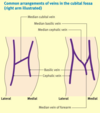
Label this diagram with the tendons and ligaments of the elbow joint.

A
2
Q

Label this diagram of the anterior elbow joint.

A
3
Q

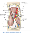
Label this diagram of the cubital fossa.

A
4
Q

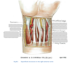
Label this diagram of the anterior forearm muscles.

A
5
Q

Label this diagram of the tendons at the wrist.

A
6
Q

What articulations does the elbow joint consist of?

A

- Humeroulnar: trochlea of humerus and trochlear/sigmoid notch of ulnar

- Humeroradial: capitellum of humerus and raidal head

7
Q

Label this diagram of the elbow joint and what inserts on the medial and lateral epicondyles?

A

Medial: Attachment for flexor-pronator group in anterior forearm

Lateral: Attachment for extensor group in the posterior forearn

8
Q

Label this diagram and state what inserts onto the radial tuberosity?

A

Biceps Brachii

9
Q

Label this diagram of the elbow joint and state what inserts onto the olecranon?

A

Triceps brachii

10
Q

What type of joint is the elbow joint and what ligaments hold the joint together?

A
  • Synovial joint with joint capsule

- Radial collateral: lateral epicondyle and annular ligament of radius, keeping head of the radius and the capitellum in close association during supination and pronation

- Ulnar collateral: triangle from medial epicondyle to coronois and olecranon

- Annular: stabilises proximal radioulnar joint as forms a collar round radial had so can rotate (pivot joint)

11
Q

What are the three bands of the ulnar collateral ligament?

A

- Anterior: Strongest

- Posterior: Weak

  • Oblique/Inferior: deepends the socket for the trochlea of the humerus
12
Q

What movement is the elbow joint capable of and what muscles perform these movements?

A

- Flexion: brachialis, biceps brachii, brachioradialis and weak extensors are those originating from medial epicondyle of humerus

- Extension: triceps brachii, anconeus and muscles from common origin on lateral epicondyle are weak extensors

13
Q

What is the carrying angle and the function of it?

A
  • In full extension ulna makes valgus angle with humerus
  • 5-10 degrees male/ 10-15 degrees female
  • Allows forearm to clear hips in swinging movements of walking
14
Q

What is an excess carrying angle called or a carry angle that deviates towardsthe body?

A

Cubitus Valgus = excessive

Cubitus Varus = towards

15
Q

What provides stability to the elbow?

A
  • Ligaments
  • Surrounding muscles
  • Bones
16
Q

What are the important bursa of the elbow and what is their function?

A

- Subcutaneous Olecranon Bursa: overly the olecranon in subcutaneous CT.

- Subtendinosus Bursa: between triceps tendon and tip of olecranon

Reduce friction between bone and skin or between tendon and bone during movement

17
Q

What joint does pronation and supination occur and what muscles are involved?

A
  • Proximal and distal radio-ulnar joints

- Pronation: pronator teres and pronator quadratus

- Supination: supinator or biceps brachii when there is resistance

18
Q

What is the anatomy of the distal radioulnar joint?

A

- Pivot joint where ulnar notch of radius rotates anteriorly around the head of the ulnar

- Triangular Fibrocartilage Complex binds the radius and the ulnar together but separates DRUJ from wrist

19
Q

What happens within the annular ligament?

A

Radial head rotates within it

20
Q

What are the joints connecting the radius and ulnar and describe the odd one in detail.

A
  • Proximal and distal radioulnar joints

- Interosseous membrane: fibrous joint with fibres running diagonally from radius proximally to ulna more distally. Keeps bones together during pro and supination and prevents proximal displacement of radius when FOOSH

21
Q

What are the muscles of the anterior forearm and what are the layers?

A

- Superficial: PT, FCR, PL, FRU (Pass Fail Pass Fail)

- Intermediate: FDS

- Deep: FDP, FPL, PQ

4 - 1= 3

22
Q

What are the superficial muscles of the anterior forearm and what are they inverted by, and where do they originate from?

A

Median nerve EXCEPT for FCU which is Ulnar

23
Q

What movements can each anterior compartment of the forearm carry out, and what are they all innervated by?

A

All innervated by median apart from FCU and FDP(medial part)

24
Q

What are all the origins, insertions, actions and innervations of the superficial muscles of the anterior forearm?

A
25
Q

What can palmaris longus be used for?

A

Tendon grafting in reconstructive surgery as only a weak flexor

26
Q

What is the easiest way to locate the median nerve in the anterior forearm in the DR?

A
27
Q

What are all the origins, insertions, actions and innervations of the intermediate muscles of the anterior forearm?

A
  • Median nerve and ulnar artery pass between it’s two ‘heads’ to travel deep to muscle belly
  • Can assist flexion of the wrist

- Continous tendious arch at origin not two heads

28
Q

What are all the origins, insertions, actions and innervations of the deep muscles of the anterior forearm?

A

- FDP: assists wrist flexion and flexes metacarpophalangeal joints. also anterior interosseous branch of median nerve

29
Q

What is the origin, insertion, action and innervation of brachioradialis and why is it a paradoxical muscle?

A

Origin: lateral supracondylar ridge of humerus

Insertion: distal radius proximal to radial styloid process

Action: flexes elbow most effectively in mid prone position, in supinated it assists flexion

Innervation: Radial

(paradoxical as it’s origin is from the common extensor origin)

30
Q

What is the origin, insertion, action and innervation of supinator?

A

Origin: humeral head from common extensor orgin and ulnar head from supinator crest on ulna

Insertion: proximal lateral surface of radius up to insertion of pronator teres

Action: supinates when no resistance so when starts pronated. rotation of radial head in annular ligament, rotation of radius externally over ulna at DHUJ

Innervation: deep branch of radial nerve

31
Q

When does the radial nerve split and what does it split into?

A
  • Just before cubital fossa
  • Deep branch exits fossa between two heads of supinator to enter posterior compartment of forearm
  • Superficial branch passes superficial to supinator, deep to brachioradialis
32
Q

What is the origin, insertion, action and innervation of anconeus?

A

- Origin: common extensor origin

- Insertion: lateral side of olecranon and posterior surface of proximal ulna

- Action: Weak extensor and assists triceps. Stabilises elbow joint. When pronated anconeus abducts the ulna and prevents joint capsule being impinged in olecranon fosssa

- Innervation: Radial nerve

33
Q

How would you test the motor function of the main branches of the brachial plexus and what should you do when testing this?

A
  • Stabilise the shoulder and arm to avoid further iatrogenic induced soft tissue injury
  • Only test motor power in more elbow if clavicle fracture, use dermatomes and stabilise arm and shoulder
34
Q

How would you assess the dermatome function of different nerves of the brachial plexus to find out which ones have been damaged?

A

Axillary: lateral arm where deltoid and teres iss

Musculocutaneous: lateral forarm and elbow

Median: wrist, palm of hand, dorsal interphalangeal joints

Ulnar: 4th and 5th digits

Radial: posterior arm, forearm and dorsum of hand

35
Q

What are the median nerves sensory and motor functions?

A

C6 to T1

- Motor: Innervates flexor and pronators in forearm and thenar muscles and lateral two lumbricals in the hand so PRONATION OF FOREARM, FLEXION OF WRIST AND DIGITS

- Sensory: Palmar cutaneous branch innervates lateral palm and Digital cutaneous branch innervate lateral 3.5 fingers on palm and dorsum of respective phalanges

36
Q

What is the anatomical course of the median nerve?

A
  1. Lateral to brachial artery then halfway down arm becomes medial to artery
  2. Enters anterior forearm via cubital fossa
  3. Exits fossa between heads of PT and deep to tendinous arch of FDS
  4. Innervates superficial and intermediate layers directly but gives rise to anterior interosseous nerve to supply deep
  5. Palmar cutaneous branch comes off to innervate lateral palm and it passes superficial to capal tunnel
  6. After these two branches it enters carpal tunnel and terminates in recurrent branch (thenar muscles) and palmar digital branch (cutaneous and lumbrical)
37
Q

What are the sensory and motor functions of the ulnar nerve?

A

C8 - T1

Sensory: Palmar and dorsal surface of medial one and a half fingers and medial palm and dorsum of hand

Motor: intrinsic hand muscles and FCU and FDP

38
Q

What is the anatomical course of the ulnar nerve?

A
  1. Descends medial aspect of arm
  2. At elbow passes posterior to medial epicondyle of humerus in cubital tunnel and gives rises to articular branch to elbow joint
  3. Passes between two heads of FCU and travels deep to FCU next to ulna
  4. Gives off 3 branches in forearm:

- Muscular innervating FCU and FDP

- Palmar Cutaneous innervating medial third of palm

- Dorsal Cutaneous innervating dorsal parts

  1. At wrist travels superficial to flexor retinaculum and medial to ulnar artery to enter hand via ulnar canal
  2. Terminates in hand by superficial (palm skin) and deep (intrinsic muscles) branches
39
Q

How do you test for ulnar nerve palsy?

A

Froment’s Sign

  • Grasp paper as paper is pulled away and they should be able to hold it
  • Positive test is when patient cannot adduct thumb so flex thumb at interphalangeal joint to hold aper
40
Q

What are the sensory and motor functions of the musculocutaneous nerve?

A

C5-C7

Sensory: supplies muscles then contains purely sensory fibres and becomes lateral cutaneous nerve to supply lateral forearm

Motor: anterior cpmpartment of arm

41
Q

What is the anatomical course of the musculocutaneous nerve?

A
  1. Pierces corachobrachialis near insertion then down to flexor compartment of arm superficial to brachialis but deep to biceps to innervate both
  2. Articular branch to elbow joint
  3. Then pierces deep fascia lateral to biceps to emerge lateral to biceps tendon to continous in forearm as lateral cutaneous nerve of forearm close to cephalic vein
  4. Cutaneous distribution entirely in C6
42
Q

How can injury to musculocutaneous nerve occur, and how would you test it’s function?

A
  • Well protected in axilla so have to be traumatic, e.g stabbing, anterior dislocations, surgery
  • Loss of sensation at lateral arm
  • Anterior arm paralysed so flexion of shoulder and elbow weakened as only pec major and brachioradialis. Weak supination as only supinator
43
Q

What is the medial cutaneous nerve of the arm?

A
  • Branch of brachial plexus from medial cord and contains T1
  • Distal medial arm to elbow
  • Can have some T2 dermatome overlap as interacts with intercostobrachial nerve (T2)
44
Q

What is the medial cutaneous nerve of the forearm?

A
  • From medial cord of brachial plexus C8 and T1
  • Supplies distal anterior surface of arm and medial side of forearm to wrist
45
Q

What are the main arteries of the forearm?

A
  • Brachial artery bifurcates to radial and ulnar in cubital fossa

- Radial: Posterolateral aspect of forearm and contributes to anastomotic networks around elbow and carpal bones

- Ulnar: Anteromedial forearm and anastomic network arrround elbow joint. Gives rise to anterior and posterior interosseous arteries for deep forearm structures

  • Both radial and ulnar anastomose in hand to form superficial palmar arch and deep palmar arch
46
Q

Where can you palpate the brachial, radial and ulnar pulse?

A

- Brachial: Cubital fossa immediately medial to tendon of biceps brachii. Used for manual B.P

- Radial: Wrist immediately lateral to FCR prominent tendon

- Ulnar: Wrist immediately lateral to tendon of FCU and proximal to pisiform

47
Q

What are the two common arrangements of the superficial veins in the cubital fossa?

A

They form the roof of the cubitsal fossa and they are important for venepunction as the vein rarely slips

48
Q

What is the course of the deep venous system of the upper limb?

A
  • Deep to deep fascia
  • Formed by venae comitantes beside artery
  • Perforating veins run between deep and superficial veins to connect them
49
Q

What are the borders, roof and floor of the cubital fossa?

A
50
Q

What are the contents of the cubital fossa?

A
51
Q

What is the brachial artery and the median nerve protected by when you are doing venepuncture at the cubital fossa?

A

Bicipital Aponeurosis

52
Q

Label the following diagram.

A
53
Q

What passes through the cubital tunnel and what structures form the boundaries of this tunnel?

A

- Ulnar nerve

- Roof: FCU fascia and Osborne’s ligament

- Floor: Posterior and transverse bands of MCL and elbow joint capsule

- Walls: Medial epicondyle and olecranon