18 - Disorders of the Elbow Flashcards
What does a supracondylar fracture of the distal humerus look like and what is the common mechanism of injury?
- FOOSH e.g falling off monkey bars, or fall onto flexed elbow (elderly and rae)
- Most common under 10 years, mainly boys 5-7 years
- Pain, deformity loss of function

What are the complications of a supracondylar fracture of the distal humerus?
- Malunion: Cubitus varus gunstock deformity
- Damage to median, ulnar or radial nerve
- Volkmann’s Ischaemic Contracture

How does Volkmann’s ischaemic contracture occur with a supracondylar fracture of the humerus?
- Brachial artery damaged or occluded by displaced fracture
- Reflex spasm of collateral circulation around elbow so ischaemia of muscles in anterior forearm
- Oedema and rise in pressure so compartment syndrome leading to more ischaemia
- Muscles undergo infarction and replaced by scar tissue where myofibroblasts contract so flexion contracture

How does Volkmann’s ischaemic contracture typically present?
- Flexed wrist
- Extended fingers at metacarpophalangeal joints
- Flexed at interphalangeal joints
- Forearm pronated and elbow flexed

How should you treat a supracondylar fracture of the humerus?
- Neurovascular examination (radial pulse, OK sign, capillary return)
- Any compromise emergency reduction and fixation with wire stabilisation
- If undisplaced less complicated

How do elbow dislocations tend to occur?
- FOOSH with elbow partially flexed as in midflexion only ligaments stabilising elbow joint not like in flexion where bones holding stability
- 2nd most common dislocation
- Sports injuries

What are the two types of elbow dislocations and what secondary issues can they cause?
Named by displacement of distal arm (ulna and radius)
- Posterior (90%): ulnar collateral ligament torn and associated fracture and/or ulnar nerve damage
- Anterior (10%): direct blow to posterior flexed elbow. fractures of olecranon due to degree of force needed to cause this dislocation

What is pulled elbow and how is it most commonly caused?
- Subluxation of radial head a.k.a Nursemaid’s elbow
- Longitudinal traction with pronated forearm
- Falls or overreaching for an object
- Age 2-5 years as running away

What is subluxation?
Partial disruption of joint with some remaining but abnormal apposition of articular surfaces, i/e an incomplete dislocation

What do children with pulled elbow often present as?
- Reduced movement of elvow
- Pain over lateral proximal forarm
- Parent states not using arm

Why is pulled elbow so common in toddlers?
- In pronation annular ligament is taut in supination and relaxed in pronation so easier for subluxation to occur
- Longitudinal traction tears distal annular ligament on neck of radius so the radial head is displaced through torn ligament
- As child ages anular ligament strengthens making condition less common

How do you treat pulled elbow?

What do radial head/neck fractures occur from and how does the patient present?
- FOOSH where radial head impacts onto capitellum
- Pain in lateral aspect of proximl forearm and loss of range of movement*
- Swelling not as bad compared to supracondylar fracture
- Sail-sign* (haemarthrosis or displacement of anterior fat pad from olecranon fossa)

What do patients with OA of the elbow typically present as?
- Uncommon as strong stabilising ligaments and well matched joint surfaces
- More common in men and manual workers and athletes with throwing
- Crepitus, Locking due to fragments of cartilage, late swelling, paraesthesia/muscle weakness as osteophytes can impinge on ulnar nerve, loss of extension and stiffness of elbow*

What are the x-ray features of rheumatoid arthritis?
- Joint space narrowing
- Periarticular osteopenia
- Juxta-articular (Marginal) bony erosions (in non-cartilage protected bone)
- Subluxation and gross deformity

What are the general principles of pathology of rheumatoid arthritis, and what joints does it mainly affect?
- Autoantibodies called rheumatoid factor attack synovial membranes
- Inflammed synovial cells proliferate to form pannus which penetrates through cartilage and bone leading to joint erosion and deformity
- Affects MCPJ, PIPJ of hands and feet, and cervical spine mainly. Can involve large joints and the autoimmune can damage other organs like eyes, skin, lungs, heart, B.V, kidneys

What are some risk factors of rheumatoid arthritis?
- Women
- Peak age 40-50

How is rheumatoid arthritis managed?
- Medically through prescription of disease-modifying meds like DMARDS and biologics
- Surgery to relieve pain and improve mobility in severe cases
- Total replacement in bad cases of erosion

What is lateral elbow tendinopathy?
- Tennis Elbow
- Pain due to tedinopathy* (chronic overuse disorders in tendons) in common extensor tendon at lateral epicondyle
- ECRB muscles stabilises wrist when elbow is straight so when weakened from overuse microscopic tears form in the tendon where it attaches to the lateral epicondyle leading to inflammation and pain

How will a patient with tennis elbow present and how is it treated?
- Pain over lateral epicondyle during extension of wrist

- Mainly manual workers, sports players, 40-60 years old due to repetitive wrist and elbow movement
- Patients advised to modify activity to allow tendon to heal and will heal in about a year
- If not –> physio, bracing or injections or surgery
What is medial elbow tendinopathy?
- Golfer’s elbow, 10x less common than tennis
- Common flexor origin at medial epicondyle
- Associated with throwing sports that place valgus stress on wrist

Where is the most common site of pathology for medial elbow tendinopathy and how does this condition present and how is it treated?
- Interface between pronator teres and FCR origins
- Pain on resisted flexion or pronation of the wrist and ulnar nerve symptoms in a few cases as close to medial epicondyle
- Activity modification or same as LET

What are the common causes of swelling around the elbow?
- Trauma
- Olecranon bursitis
- Rheumatoid nodules
- Gouty tophi
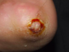
What is olecranon bursitis?
- Student’s elbow
- Inflammation of bursa due to repeated microtrauma
- Soft, cystic, serous filled so transilluminates