MSCT Week 6: Cutaneous Signs of Systemic Disease Flashcards
Skin findings and systemic disease
Question 1

Acanthosis Nigricans
Screen for Diabetes
Acanthosis Nigricans Clinical Features

Acanthosis Nigricans most commonly found where on the body?
- Posterior neck fold
- axillae most common
Acanthosis Nigricans is a sign of
Insulin Resistance
Identify features of histology

acanthosis
IDGF-1 causes thickening and overdevelopment of the epidermis
Acanthosis Nigricans should screen for diabetes mellitus by?
glucose tolerance test
Malignant Acanthosis Nigricans Clinical Features

“Tripe Palms”
Think Malignant Acanthosis Nigricans
Strange manifestations of Malignant Acanthosis Nigricans
thickened velvety plaques

Diabetes and Skin

Diabetes and Skin: Diabetic Dermopathy Clinical Features
deposition of glycosylated proteins in the skin

Identify

Acanthosis Nigricans
Associations with Acanthosis Nigricans
no collagen vascular disease
some hereditary some syndromic
some insulin resistance

Why does this occur?

Diabetic Neuropathy
don’t realize that they are damaging their feet because they have lost sensation
What is this?

- Scleroderma from poorly controlled diabetes or from other disorders but diabetes is most common
- typically asymptomatic, infiltration of the skin (usually in the back) with mucin
- thickened hardened, can be pruritic
What is this?

Bullosis Diabeticorum
- non-infectious
- not-inflammed
- sterile blisters filled with water on the lower limbs and feet
What is this?

Necrobiosis Lipoidica
- typically occurs on the anterior shins
- the atrophic skin where vessels can be seen underneath
- yellowish hue
- if ulcerate are difficult to treat
What is this

Acquired Perforating Disorder
- common for diabetics on renal dialysis
- build up of abnormal collagen and elastin in the skin and the body tries to eliminate it
- epidermis tries to envelope abnormal collagen
- transepidermal elimination
- associated with renal failure
Acquired Perforating Disorder is associated with?
Renal failure
What is this?

Eruptive Xanthomas
- grouped yellow tender papules
- firm
- extensor surfaces and buttocks
- happen when cholesterol and triglycerides are very high
- This is an emergency
- as cholesterol is brought down they resolve
Question

Malignancy
This is called the sign of leser-Trelat
a large number of seborrheic keratoses suddenly appearing responding to same growth factor as does the malignancy

Sign of Leser-Trelat

Identify Histological Features

Pseudocysts
thickened epidermis
well defined basal layer
comedo blackhead like openings
Question

- Abdominal cramping, bloating, especially after consuming bread
- and fatigue from malabsorption
- Gluten-sensitive enteropathy or celiac disease
Dermatitis Herpetiformis
Dermatitis Herpetiformis Clinical Features

Dermatitis Herpetiformis Etiology
IgA Autoantibodies mucosal antibodies are spilling out targeting epidermal
- Tissue transglutaminase
- Epidermal Transglutaminase
- Gliadin

Identify Histological Features

neutrophilic infiltrate in the dermal papillae with some vesiculation separating the dermal papillae from the epidermis
Dermatitis Herpetiformis
Dermatitis Herpetiformis HLA Association
HLA DQ2 and HLA DQ8
Dermatitis Herpetiformis Immunofluorescence Features

Dermatitis Herpetiformis Treatment
5 listed

Question

Autoimmune Disease (Graves Disease, Lupus, Rheumatoid Arthritis)
Dermatitis Herpetiformis Risks for
- Hypothyroidism
- Intestinal lymphoma from chronic intestinal inflammation
Vitiligo Clinical Features

identify Histological Features

Normal skin
Identify Histological Features

- No melanocytes in the basal layer
- when stained for melanin nothing shows up
Vitiligo Treatment
4 listed

Question

Systemic Lupus Erythematosus
Systemic Lupus Erythematosus Clinical Features

Identify Histological Features

SLE
- Perivascular infiltrate (lymphocytes and a few histiocytes
- peradnexal (around hair follicles and eccrine ducts
- vacuolization of the basal layer
- destruction of basal keratinocytes
- thickening of basement membrane
- mucus deposition
Identify Histological Features

SLE
- destruction of keratinocytes
- keratinocytes undergoing apoptosis
- basal layer cuboidal
- vacular dermatitis
- think about erythema multiforme and drug reactions they would look very similar
- do immunofluorescence for antibody deposition to distinguish
Identify Histologic Features

SLE
stained for mucin
thickened basement membrane
Subacute Cutaneous Lupus Erythematosus Clinical Features
10-20% have systemic symptoms as well

Discoid Lupus Erythematosus Clinical Features

Question

Muscle Weakness
Dermatomyositis Clinical Features

Dermatomyositis Associations
proximal muscle weakness
muscle fatigue
muscle tenderness
fever malaise

Dermatomyositis Treatment

Question

Breast Cancer
worry about malignancy with Dermatomyositis
Dermatomyositis & Malignancy

Question

Erythema Nodosum
Erythema Nodosum Clinical Features
can be mistaken with cellulitis

Identify Histological Features

inflammation primarily in the fat
lymphocytes and histiocytes
Erythema Nodosum
Identify Histological Features

Erythema Nodosum
can have giant cells
Erythema Nodosum Associations

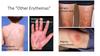
Other Erythemas
4 listed
Question

Pyoderma Gangrenosum
Pyoderma Gangrenosum Clinical Features

Identify Histological Features

Pyoderma Gangrenosum
can look like folliculitis (pustular ingflammation around the hair follicle
in any ulcer see inflammation of blood vessels
biopsy on the edge
Question

Inflammatory Bowel Disease
Pyoderma Gangrenosum Associations

Pyoderma Gangrenosum Associations continued

Question

Cutaneous T-Cell Lymphoma
Identify Histologic Features

atypical epidermotropic lymphoid infiltrate
Cutaneous T-cell Lymphoma
Identify Histologic Features

Cutaneous T-cell Lymphoma Clinical Features

Pruritus

Mediators of Pruritus

Common skin conditions with pruritus

Systemic Disease that cause Pruritus

signs of chronic pruritus


Skin signs of chronic pruritus & workup


Laboratory studies for systemic diseases

Physical Exam for systemic diseases

Hx for systemic diseases

Diagnoses for systemic diseases
