EKGs Flashcards
Basic EKG grid

EKG axis

EKG intervals

Signs of LVH on EKG

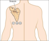
Where to place V7, V8, and V9
WPW on EKG

STEMI evolution

Parkinsonian tremor artifact
The ECG shows classic Parkinsonian tremor artifact (about 5 cycles/sec) simulating atrial flutter or coarse atrial fibrillation. True sinus P waves (obscured in the limb leads) are most evident in leads V2-V4 upon careful inspection. Note also: the variable pseudo-flutter (F) to QRS intervals are not consistent with pure 6:1 conduction. Also, the biphasic (negative/positive) “atrial” waves in V1 are not compatible with typical, clockwise atrial flutter which is associated with upright F waves in that lead.

The prominence of the baseline artifact in lead II (left leg-right arm) and in lead aVR was consistent with the patient’s overt right hand tremor.
The patient’s palpitations were characterized by an intermittent “skipped beat” sensation and were attributed to isolated atrial and ventricular beats, which are not present here but were observed at other times. Compare this record with that in Case #84.
Left bundle branch block
Common in elderly, usually asymptomatic
In patients with ACS, may represent acute anterior wall MI (LAD).

Right bundle branch block
- Asymptomatic and may be the result of degenerative disease or right heart strain
- Characterized by RSR’ pattern (small initial upward deflection followed by a small downward deflection, then a large upward deflection) in lead V1

RBBB vs LBBB

Right heart strain pattern
Called “S1Q3T3”. Sometimes seen in PE, but sensitivity and specificity are both poor.
S present in 1, Q present in 3, and T inverted in 3

Hyperkalemia on ECG
Early: Peaked T waves
Late: Progresses to “sine wave” pattern

Hyper- and hypo-calcemia on ECG
They effect the length of the ST segment inversely
The more calcium, the shorter it gets

Normal p wave axis should align almost perfectly with. . .
. . . Lead II
J waves
aka Osborn waves
Seen in hypothermia

Five mechanisms of bradycardia

Acquired long QT etiologies

Principles of torsades management

Left ventricular aneurysm syndrome and ECG
- Often occurs months post-MI
- EKG shows persistent ST elevations, deep Q waves in associated leads
- Thin and dyskinetic wall on echo
- Presents w/ heart failure, angina, ventricular arrhythmia, arterial embolism
