Abdominal Cavity I Flashcards

(66 cards)

1
Q

Where is the abdominal cavity located?

A

In between the thoracoabdominal diaphragm and the pelvic brim

2
Q

What is the abdominal cavity sometimes referred to as?

A

Abdominopelvic cavity - the abdominal and pelvic cavities are continuous

3
Q

List the abdominal wall layers going from superficial to deep

A
  1. Skin
  2. Superficial fascia
    - Camper’s fascia: outer fatty layer
    - Scarpa’s fascia: innermost membranous layer
  3. Muscles
    - External abdominal oblique
    - Internal abdominal oblique
    - Transversus abdominis
    - Rectus abdominis
  4. Endoabdominal fascia
  5. Parietal peritoneum
4
Q

What is the endoabdominal fascia made up of? And what is it deep to?

A

Transversalis fascia, extraperitoneal fat, psoas and iliacus fascia (“TEPI”)

Deep to: muscle layers of abdomen

5
Q

Parietal peritoneum

A

Serous membrane continuous w/ abdominal visceral peritoneum

6
Q

Small intestine (bowel) segments

A

Duodenum

Jejunum (starts @ duodenojejunal junction)

Ileum (ends @ ileocecal junction)

7
Q

Large intestine (bowel) segments

A

Cecum w/ ileocecal valve & vermiform appendix Ascending colon

Transverse colon

Descending colon

Sigmoid colon

Rectum

8
Q

Abdominal digestive tract components

A

Distal end of esophagus

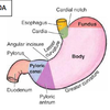
Stomach w/ greater and lesser curvatures & pyloric valve Small intestine

Large intestine

Liver and gall bladder

Pancreas

= DS’S LLP (Diss’ large liver pan)

9
Q

Abdominopelvic organs

A

Spleen, kidneys, adrenal glands, rectum, urinary bladder, uterus, uterine tubes and ovaries (SKAR UR OUUT)

10
Q

Peritoneum “TELL”

A

TELL =

Thin, transparent serous membrane

Enclosed sac - organs develop against it

Lines abdominopelvic cavity, deep to transversalis fascia

Layers are formed - parietal and visceral

11
Q

Parietal peritoneum (“parents LVs”)

A

Lines abdominal wall

Very strong

Sensitive to somatic stimuli (cutting, burning, etc)

12
Q

Visceral peritoneum - CNN

A

Covers viscera directly

Not as strong as parietal

Not sensitive to somatic stimuli

13
Q

Peritoneal cavity

A

Potential space between visceral and parietal

Empty except for thin serous fluid

14
Q

Define ascites - what does it result from?

A

Abnormal accumulation of serous fluid

Results from heart, kidney or liver failure

15
Q

Peritonitis

(-itis = inflammation)

A

Inflammation of peritoneum

16
Q

Subdivisions of peritoneal cavity

A

Greater peritoneal sac

Lesser peritoneal sac

Omental foramen (opening between greater and lesser sacs)

17
Q

Intraperitoneal organs definition and examples

A

Organs protrude completely against the peritoneal sac and become completely covered by the peritoneum (They are NOT within the peritoneal cavity)

Examples: stomach, liver, jejunum

18
Q

Mesentry

A

Double layer of peritoneum suspending the intraperitoneal organs from the posterior abdominal wall

Mesentry allows intraperitoneal organs to move freely in the abdominal cavity

19
Q

What courses to the organ within the mesentry? (between two peritoneal layers)

A

Blood vessels, nerves, lymphatics

20
Q

Omenta

A

Mesentries attached to the stomach

  • Greater omentum
  • Lesser omentum
21
Q

Retroperitoneal (RPT) organs

A

Protrude only slightly against peritoneal sac

Are covered on one side by peritoneum – e.g kidney

22
Q

Retroperitoneal - location, mesentry

A

Organs grow up against posterior abdominal wall - posterior to the parietal peritoneum

RPT organs have no mesentry

23
Q

Secondarily RPT

A

Start out as intraperitoneal but then develop against the posterior abdominal wall to become RPT

  • Duodenum, pancreas, cecum, ascending colon, descending colon
24
Q

Gastrocolic ligament of greater omentum (GGT)

A

Runs from greater curvature of stomach → transverse colon

25
Omental apron of greater omentum
Hangs down inferior to transverse colon Shape and size varies Helps seal off infections by sticking to a site of inflammation
26
Lesser omentum (LLL)
Runs from liver --\> lesser curvature of stomach and duodenum
27
Portal triad
Part of the duodenum (free edge or hepatoduodenal ligament) that contains: Bile duct Hepatic artery proper Portal vein
28
Where does the mesointestine (the mesentery) and root attach at? What courses within it?
Attaches to jejenum and ileum Superior mesenteric vessel and autonomic nerves course within it
29
Mesoappendix -- attachment
Attaches the appendix to the posterior abdominal wall
30
Transverse mesocolon -- attachment
Attaches to inferior margin of transverse colon - has root along pancreas
31
Sigmoid mesocolon -- attachment
Root attaches sigmoid colon to posterior abdominal wall
32
Falciform ligament
Has round ligament of liver (ligamentum teres hepatis) in its free border Round ligament is remnant of the fetal umbilical vein
33
Distal esophagus: entry into abdomen
Enters abdomen via opening in diaphragm = esophageal hiatus EH opens into stomach at the cardiac orifice
34
Lower esophageal sphincter
Prevents reflex of food into esophagus via: - Contraction of voluntary muscles of the diaphragm and smooth muscles Note: this is a physiological sphincter, not an anatomical one -- **no thickening** of smooth muscle
35
What are the stomach's 4 parts it can be divided into?
Cardiac region Fundus Body Pyloric region
36
Fundus
Dilation/bulge that's superior to a horizontal line drawn to the left from region of cardiac notch Rests against left dome of thoracic diaphragm Filled with gas (radiolucent [dark] on an x-ray)
37
Body
Largest part of stomach - Below cardiac region and fundus to a vertical line dropped from angular incisure
38
Pyloric Region
Drop a line from the angular incisure to find this region Has 2 parts: 1. Pyloric Antrum - wider area, adjacent to body 2. Pyloric canal - narrower region, leading to pyloric sphincter
39
Pyloric sphincter (valve) - Location - What type of sphincter is it - Function
L: Junction between stomach and duodenum S: True **anatomical sphincter** - actual thickening of muscle F: Controls rate that stomach empties at * In a state of tonic contraction * Closed unless emitting stomach content
40
Rugae
Stomach is lined by gross folds of mucosa called rugae (Think that rugs line the floor, so rugae lines the stomach)
41
Functions of stomach/pathway of food - DB-CPS
* **D**istensible - acts as blender and reservoir of food * Food enters stomach as lump = **b**olus * Mixes with enzymes to make watery mixture = **c**hyme * **P**eristaltic actions move chyme from stomach --\> intestine * **S**tomach empties 2-3 hours after eating
42
Hiatal Hernia
Part of stomach bulges through esophageal hiatus into thoracic cavity
43
Small intestine: - Attachment - Pilcae circulares - 3 parts
Attachment: proximally to stomach, distally to cecum of large int. Pilcae: Muscosal folds that cover the inner surface 3 parts: duodenum, jejunum, ileum
44
Duodenum - shape - location - parts
Secondarily retroperitoneal - except 1st part that is intraperitoneal Shape: C-shaped, cradles head of pancrease Location: starts to the right of the midline --\> ends to left of midline at the duodenojejunal junction 4 Parts: superior, descending, horizontal, ascending
45
What empties at the 2nd part of duodenum? What is that specific area called?
Common bile duct and main pancreatic duct empty into LT side of 2nd (descending) part of duodenum at the major duodenal papilla Accessory pancreatic duct also opens into 2nd
46
Jejunum
Intraperitoneal Continuous w/ duodenum - proximally Continuous w/ ileum - distally * No clear demarcation from ileum
47
Jejunum characteristics as compared to ileum "OW-RPB"
* Often empty * Wider diameter w/ thicker walls * Redder in living state - greater vascular supply * Prominnet pliae circulares slow passage of food and aid in bsorption * Blood vessels in mesentery * 1-2 layers of arcades * long vasa recta (straight arteries)
48
Ileum
Intraperitoneal Continuous w/ jejunum proximally, cecum distally Most prominent characteristics are at distal end
49
Ileum characterisitics relative to jejunum NP Sib, B Pays Meck.
* Narrower diameter w/ thinner walls * Paler and less vascularized * Small or absent plicae circulares * Increased intermesenteric fat * Blood vessels in mesentry * Payer's patches * Meckel's diverticulum
50
Ileum blood vessels characteristics
4-5 layers of arcades Short vasa recta
51
Peyer's patches Meckel's diverticulum
1. Peyer: submucosal aggregates of lymphatic tissue on antimesenteric border 2. Meckel: common developmental malformation * Fingerlike bind pouch - remnant of embryonic vitelline duct * 2 ft from ileocecal junction * If inflammed, may mimic the symptoms of appendicitis
52
Jejunum and ileum attachment
Attached to posterior abdominal wall by **mesentry** * Mesentry is fan shaped * Root = short, runs obliquely * Long border = 6-7 m, point of attachment of jejunum and ileum
53
Large intestine's 3 distinct surface characteristics "THO"
1. Tenia coli 2. Haustra 3. Omental (epiploic appendices)
54
Large intestine - tenia coli
3 equally spaced bands formed bc: Outer longitudinal muscle layer is incomplete
55
Large Intestine - Haustra (sacculations)
Gut walls that bulge out between tenia coli (bc tenia coli are 1/6 shorter than rest of colon)
56
Large intestine - omental (epiploic) appendices
Fat filled appendices
57
Semilunar fold
* Inner surface of large intestine's mucosal folds * Runs between **tenia coli**
58
LI - Cecum region is "bossy = basi"
* Blind pouch * Appendicular orifice here * Secondarily retroperitoneal * Ileum enters cecum @ ileocecal orifice * Ileocecal valve guards orifice * Most reflux is prevented by contraction of the terminal ileum
59
LI - Vermiform appendix region
* Intraperitoneal * Fixed point = root * Located 2/3 from umbilicus to ant superior iliac spine - where appendectomy incisions are made (McBurney's Point) * Mesentry = mesoappendix * Frequently inflamed = appendicitis
60
Ascending colon of LI
* Secondarily retroperitoneal * From proximal cecum --\> distal right colic (hepatic) flexure * Narrower than cecum
61
Transverse colon - LI
* Intraperitoneal * Largest & most mobile portion of LI * From RT colic flexure --\> LT colic flexure * LT = more superior * 2 mesenteric attachments: * Transverse mesocolon * Gastrocolic ligament
62
Transverse mesocolon - LI
* Transverse colon is posteriorly attached to trans. mesocolon * Courses acrocc post abdominal wall * Across duodenum and pancreas
63
Gastrocolic ligament and transverse colon - LI
* Trans colon is anteriorly attached to GC ligament * Follows curvature of stomach * Can actually hang into pelvis
64
Descending colon - LI
Secondarily retroperitoneal From LT colic --\> sigmoid colon Narrowest portion of colon
65
Sigmoid colon - LI
Intraperitoneal S-shaped loop - from descending colon --\> rectum Attached to: sigmoid mesocolon - v-shaped mesentry
66
Rectum and anal canal
Retroperitoneal W/in true pelvis Continuous w/ sigmoid colon (@ S3 vertebrae) Anal canal - inferior 1.5 in of GI * Continuous w/ rectum @ anorectal junction