Anatomy Eye, retina and colour vision Flashcards

1
Q

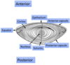
what is the external anatomy of the eye?

A
2
Q

what are the different types of tears?

A

basal

reflex

emotional

3
Q

how are basal teats produced?

A

by lacrimal system at a constant level- even in absence of irritation or stimulation

4
Q

when are reflex tears produced?

A

increased tear production in response to ocular irritation

5
Q

what is the lacrimal reflex pathway made up of?

A
  • afferent pathway, CNS, efferent pathway and lacrimal gland
    • Afferent- cornea, innervation = sensory nerve fibres via cranial nerve V1- ophthalmic trigeminal
      • Trigeminal nerve relays signals to CNS
    • Efferent- parasympathetic
    • Neurotransmitter- acetylcholine
6
Q

what are tears produced by?

A

lacrimal gland

7
Q

where is the lacrimal gland located?

A

within orbit

latero-superior to globe

8
Q

where do tears drain?

A

2 puncta, opening on upper and lower medial lid margins

9
Q

where do puncta drain to?

A
  • Puncta form opening of superior and inferior canaliculi within lower and upper eyelids
  • Both canaliculi converge as single common canaliculus and drain tear into tear sac
  • Tear drained out of tear sac into nasal cavity through tear duct
10
Q

what is the cornea covered with?

A

tear film- thin layer fluid

11
Q

what is the function of the tear film?

A
  • Maintains smooth cornea-to-air surface
  • Important for maintaining clear vision and removing surface debris during blinking
  • bactericide
  • Source of oxygen and nutrient supply to anterior segment
    • Cornea has no blood vessels
12
Q

what are the layers of the tear film?

A

superficial lipid layer

aqueous tear film

mucinous layer

13
Q

what is the superficial lipid layer?

A
  • Responsible protecting tear film from rapid evaporation
  • Secreted by Meibomian glands situated along eyelid margins
14
Q

what is the aqueous tear film?

A
  • Main bullk of tear film
  • Delivers oxygen and nutriet to surrounding tissue
  • Contains factors against potentially harmful bacteria
15
Q

what is the mucinous layer?

A
  • Ensures tear film sticks to eye surface
  • Renders surface eye ‘wettable’
  • Mucin molecules act by binding water molecules to hydrophobic corneal epithelial cell surface
16
Q

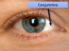
what is the conjunctiva?

A

thin, transparent tissue that covers outer surface of eye

  • Begins at outer edge of cornea, covers visible part of eye and lines inside of eyelids
  • Nourished by tiny blood vessels that are nearly invisible to naked eye
17
Q

what happens in conjunctivitis to the visible conjunctiva?

A

infection of the conjunctiva

blood vessels become visible to naked eye

18
Q

what is the anterio-posterior diameter of eye in adults?

A

24mm

19
Q

where does the eye sit?

A

anatomical space of the oribit

20
Q

what is the eye enclosed by?

A

bone walls

21
Q

what is the posterior coat of the eye

A

sclera

choriod

retina

22
Q

what is the anatomy of the eye in lateral view?

A
23
Q

what is the sclera responsible for?

A
  1. Sclera- hard and opaque
    1. Protection
    2. Shape maintenance of eye
    3. High water content
24
Q

what is the choroid responsible for?

A
  1. Choroid- pigmented and vascular
    1. Providing circulation to eye
    2. Shielding out unwanted scattered light
25
Q

what is the retina responsible for?

A
  1. Retina- neurosensory tissue
    1. Converting light into neurological impulses
    2. Transmitted to brain via optic nerve
26
Q

what is the cornea?

A
  • Transparent, dome shaped window covering front of eye
  • Low water content
  • Refracting surface providing 2/3 eye focusing power
  • Clear window to look through
27
Q

what are the layers of the cornea?

A
  1. Epithelium
  2. Bowman’s membrane
  3. Stroma- contributes towards transparency
  4. Descement’s membrane
  5. Endothelium-pumps fluid out corneal and prevents corneal oedema
28
Q

what is the uvea?

A
  • Vascular coat of eyeball and lies between sclera and retina
  • 3 parts
  1. Iris
  2. Ciliary body
  3. Choroid
29
Q

what can disease of uvea cause?

A

connected intimately

disease of one part affects other portions but not always to same degree

30
Q

where is the choroid?

A

between retina and sclera

31
Q

what does the choroid do?

A

composed of layers of blood vessels that nourish back of eye

32
Q

what does the iris do?

A

controls light levels inside eye

rebound opening in center-pupil

embedded with tiny muscles that dilate and constrict pupil size

33
Q

what is the structure of the lens?

A
  • Outer acellular capsule
  • core of Regular inner elongated cell fibres- transparency
34
Q

what can happen to the lens with age?

A

loss of transparency- cararacts

35
Q

what are the functions of the lens?

A
  • Transparency
  • Regular structure
  • Refractive power
  • 1/3 eye focusing power- higher refractive index than aqueous fluid and vitreous
  • Accommodation
  • Elasticity
36
Q

what is the retina?

A
  • Thin layer of tissue
  • Lines inner part of eye
  • Capture light rays that enter eye
  • Light impulses sent to brain for processing via optic nerve
37
Q

what does the optic nerve do?

A

transport electrical impulses from retina to brain

connects near back of eye near macula

visible portion= optic disc

38
Q

what is the blind spot caused by?

A

where optic disc meets retina= no light cells= blind spot= optic disc

39
Q

where is the macula?

A

located in centre of retina. temporal to optic nerve

40
Q

what is the macula responsible for?

A
  • Small and highly sensitive
  • Responsible for detailed coloured vision
  • Macula allows detail and to perform tasks that require central vision such as reading
41
Q

what is the centre of the macula?

A

fovea

42
Q

what is the make up of the fovea?

A
  • Highest concentration of cones, low concentration rods
  • Only fovea has concentration of cones to perceive in detail
43
Q

what is central vision? (macula vision)

A
  • Detail day vision
  • Colour vision- highest conc cone photoreceptors on fovea
  • Reading, facial recognitions
  • Assessed by visual acuity assessment
  • Loss foveal vision- poor visual acuity
44
Q

what is peripheral vision?

A
  • Shape, movement, night vision
  • Navigation vision
  • Assessed by visual field assessment
45
Q

what is the effects of extensive loss visual field?

A
  • = unable navigate in an environment – may need white stick even with perfect acuity
46
Q

what is the structure of the retina?

A

outlayer- retinal pigment epithelium

middle layer- bipolar cells (2nd order neuron)

inner layer- neuroretina

47
Q

what is the retinal pigment epithelium?

A
  • Situated in front choroid
  • Photoreceptors (1st order neuron)
  • Detection light
  • Transport nutrient from choroid to photoreceptors and remove metabolic waste from retina
48
Q

what do the bipolar cells do?

A

Local signal processing to improve contrast sensitivity, regulate sensitivity

49
Q

what does neuroretina do?

A
  • Retinal ganglial cells (3rd order neuron)
  • Transmission signal from eye to brain
50
Q

what is neuroretina made up of?

A

photo-receptors and neurons

51
Q

what are the classes of photoreceptors?

A

rods

cones

(overall more rods than cones)

52
Q

what are the features of rods?

A
  • Longer outer segment with more photosensitive pigment
  • More sensitive to light than cones
  • Main photoreceptors in night vision (scotopic vision)
  • Slow response to light
53
Q

what are the features of cones?

A
  • Less sensitive to light
  • Shorter outer segment
  • Day fine vision and colour vision
  • Faster response
54
Q

where are photoreceptors synthesised?

A
  • in inner segment and transported to outer segment
55
Q

what is the outer segment made up of?

A

Stacks of discs

56
Q

what happens to distal discs?

A

distal discs with deactivated photo-pigments are shedded from tips

phagocytosed by retinal epithelial cells

57
Q

what happens to deacitvated photopigments in retinal epithelial cells?

A

regenerated and transported to photo-receptors

58
Q

what is the distribution of photoreceptors?

A
  • Central eye- highest cone cells (only cone cells in macula for photopic (day vision)
  • Peripheral= highest rod- highest density outside macula and tail off towards periphery (20-40deg away from fovea)
  • No photoreceptors in blind spot
  • Rod cells absent in macula
59
Q

what is the frequency spectrum of rod cells?

A

only has single peak light sensitivity so not sensitive to any particular colour

60
Q

what is the frequency spectrum of cone cells?

A
  • S-cones- short wavelength sensitivity= blue
  • M-cones- medium wavelength sensitivity = green
  • L-cones- long wavelength sensitivity= red
  • Yellow light has wavelength between M cones and L cones
    • Stimulates both equally
61
Q

what is the commonest vision deficiency in frequency spectrum?

A

Deuteranomoly

shifting M-cone sensitivity peak towards that of L-cone curve

red- green confusion

62
Q

what are the different colour vision deficiencies?

A
  • Deuteranomaly= daltonism and most frequent form colour blindness
    • Not completely colour blind but don’t perceive colour red due to anomalous trichromatism
  • Full colour blindness only in very small % population= ACHROMATOPSIA= monochromatism
63
Q

what can achromatopsia be caused by?

A
  • Can be caused by blue cone monochromatism with only presence blue L-cones
    • Normal day light visual acuity
  • Can be cause by rod monochromatism
    • Total absence of all cone photoreceptors
    • No functional day vision
64
Q

what is the test for colour blindness?

A

Ishaihara test

65
Q
A