BL Session 7 - Skeletal System, Cartilage and Bone Flashcards
(46 cards)
What is cartilage?
Cartilage: is an avascular tissue that consists of an extensive extracellular matrix in which lie chondrocytes.
Describe the structure and function of chondrocytes.
- The chondrocytes produce and maintain the extracellular matrix.
- Each chondrocyte lies in a lacuna (pl. lacunae). There is artefactual shrinkage of the cells away from the lacunae walls in this preparation
- Pressure loads applied to the cartilage create mechanical, electrical, and chemical signals that direct the synthetic activity of the chondrocytes.

Describe the structure and function of cartilage.
- The large ratio of glycosaminoglycans (GAGs) to type II collagen in the cartilage matrix permits ready diffusion of substances between the chondrocytes and the blood vessels surrounding the cartilage.
- The extracellular matrix is solid and firm, but also rather pliable and therefore resilient to the repeated application of pressure.
- The large amount of hyaluronic acid in the extracellular matrix assists this resilience to the repeated application of pressure.
Describe the proteoglycan structure in cartilage ground substance.
- The proteoglycan monomer consists of a core protein to which 100 GAG units are joined.
- Hyaluronic acid molecule, forming linear aggregates, each with many proteoglycan monomers, are interwoven with a network of collagen fibrils (pink).
- The high density of negative charges on the GAGs attracts water, forming a hydrated gel.

Identify and outline the three types of cartilage.
- Hyaline cartilage which has a matrix containing proteoglycan, hyaluronic acid and type II collagen. The hyaluronate proteoglycan aggregates are bound to the fine collagen matrix fibres.
- Elastic cartilage which has a matrix like that of hyaline cartilage but with the addition of many elastic fibres and elastic lamellae.
- Fibrocartilage which has abundant type I collagen fibres in addition to the matrix material of hyaline cartilage.
Outline the structure and function of hyaline cartilage, as well as areas where it can be found.
- Cell type: chondrocyte (no other cell types are present)
- Chondrocytes are present singly or, if recently divided, as small clusters called isogenous groups. The chondrocytes within the isogenous groups separate as they elaborate (lay down) extracellular matrix.
- In early foetal development hyaline cartilage is the precursor model of those bones which develop by endochondral ossification.
- As long bones develop some hyaline cartilage remains at the articulating surface, (and also at the epiphyseal plate until growth ceases).
- Hyaline cartilage is sited at articulating surfaces, in parts of the rib cage, nose, and in the trachea, bronchi and larynx.

Describe the main features of hyaline cartilage as observed in an H&E stained micrograph.
- One observes dense connective tissue at the top
- Perichondrium (dense connective tissue) covers the margin of hyaline cartilage. It contains many elongate, fibroblast-like cells which can develop into chondroblasts and thereafter into chondrocytes.
- Chondrocytes lying in cartilage extracellular matrix, which they produce and maintain. The matrix is highly hydrated.
- One observes the territorial and inter-territorial matrices as well.

Outline what is meant by appositional and interstitial growth.
- Fibroblast-like cells of the perichondrium give rise to flat, newly formed chondroblasts, which secrete matrix components and round up to develop into chondrocytes.
- The cartilage grows as a result. This growth from the periphery is known as appositional growth.
- Chondrocytes deeper in the cartilage may divide and give rise to isogenous groups. Deposition of further matrix by these isogenous groups results in interstitial growth. The cells of the isogenous group separate as they lay down further matrix.
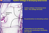
Describe the hyaline cartilage of developing tarsal bones.
- Hyaline cartilage of developing tarsal ‘bone’. This cartilage will ossify.
- No perichondrium at articulating surfaces.
- Perichondrium at non-articulating surfaces and contributing to developing joint capsule.
Describe the positioning of cartilage at the end of the long bone.
- Hyaline cartilage lines the articulating surface of the bone. In this position it is not lined by perichondrium.
- There is spongy or cancellous bone.
- Hyaline cartilage also forms the epiphyseal growth plate (no perichondrium here either)
- Compact bone

Describe the epiphyseal edge of a long bone.
- Chondrocytes lying in lacuna
- The articular surface of the bone is very smooth and composed of hyaline cartilage, without a perichondrium
- There is an irregular boundary between the articular cartilage and the underlying bone
- Osteocytes lying in lacuna.

Describe the structure and function of elastic cartilage, as well as the areas where it can be found.
- The presence of many elastic fibres in the extracellular matrix confers elasticity upon the cartilage, in addition to the resilience characteristic of hyaline cartilage.
- Unlike hyaline cartilage which calcifies with ageing, elastic cartilage does not calcify.
- Elastic cartilage is found in the external ear (pinna), in the external acoustic meatus, in the epiglottis, and in the Eustachian tube.

Describe the appearance of elastic cartilage as seen from the pinna of the ear.

Describe the structure and function of fibrocartilage, as well as the areas where it can be found.
- Cell types: Chondrocytes and fibroblasts
- Fibrocartilage is a combination of dense regular connective tissue and hyaline cartilage.
- The cells are often seen to be distributed in rows.
- There is no surrounding perichondrium.
- Fibrocartilage is present in intervertebral discs, articular discs of the sternoclavicular and temporomandibular joints, the menisci of the knee joint and in the pubic symphysis.
- The fibrocartilage has the resilience to act as a shock absorber and to resist shearing forces.

Describe the appearance of fibrocartilage in a light micrograph.
- Collagen fibres are stained green
- The rounded chondrocytes tend to be arranged in rows, or as isogenous groups.
- There are a relatively small number of elongated fibroblast nuclei evident

What is endochondral ossification?
Endochondral ossification involves the replacement of a pre-existing hyaline cartilage template by bone and is the way in which most of the bones of the body develop.

The bone increases in length by endochondral ossification. Outline the steps of long bone development by endochondral ossification.
- Initial cartilage model (miniature version of adult bone)
- Collar of periosteal bone appears in the shaft
- Central cartilage calcifies. Nutrient artery penetrates supplying bone-depositing osteogenic cells. Primary ossification centre formed.
- Medulla becomes cancellous bone. Cartilage forms epiphyseal growth plates. Epiphyses develop decondary centres of ossification.
- Epiphyses ossify and growth plates continue to move apart, lengthening the bone.
- Epiphyseal growth plates replaced by bone. Hyaline articular cartilage persists.

What is a synovial joint?
A moveable joint in which the juxtaposed (opposed) bone ends are:
- Covered by hyaline cartilage or fibrocartilage, and
- Lie within lubricating synovial fluid bounded by an articular capsule (joint cavity) which is lined by synovial membrane, and reinforced with fibrous tissue and ligaments.

Describe the zones observed in a micrograph of an LS through an epiphyseal growth plate.
- Zone of reserve cartilage: no cellular proliferation or active matrix production
- Zone of proliferation: cells actively dividing to form columns; cells enlarge and secrete matrix
- Zone of hypertrophy: cells enlarge greatly. Matrix compressed into linear bands between cell columns
- Zone of calcified cartilage: enlarged cells begin to degenerate and matrix calcifies
- Zone of resorption: in which the calcified matrix is in direct contact with the marrow cavity. Small blood vessels and connective tissue invade the region occupied by the dying chondrocytes, leaving the calcified cartilage as spicules between them. Bone is laid down on these cartilage spicules.

The bone increases in length by intramembranous ossification. What is intramembranous ossification?
- Intramembranous ossification takes place within condensations of mesenchymal tissue and not by replacement of a bone pre-existing hyaline cartilage template.
- The process also contributes to the thickening (not the lengthening) of long bones, at their periosteal surfaces (appositional growth).
Where can intramembranous ossification be observed?
Flat and skull bones develop by intramembranous ossification, examples being:
- Skull
- Clavicle
- Scapula
- Pelvic bones

Outline the process of intramembranous ossification.
- A small cluster of mesenchymal stem cells (MSCs) form a tight cluster of cells (a nidus).
- The MSCs become osteoprogenitor cells (each developing more Golgi apparatus and rough endoplasmic reticulum).
- The osteoprogenitor cells become osteoblasts and lay down an extracellular matrix containing Type I collagen (osteoid).
- The osteoid mineralises to form rudimentary bone tissue spicules, which are surrounded by osteoblasts, and contain osteocytes.
- The spicules join to form trabeculae, which merge to form woven bone, which is finally replaced by the lamellae of mature compact bone.
Distinguish between cancellous and compact bone.
- Cancellous bone forms a network of fine bony columns or plates to combine strength with lightness. The spaces are filled by bone marrow.
- Compact bone forms external surfaces of the bones and comprises approx. 80% of the body’s skeletal mass.

What can be observed when viewing a section of the shaft of long bone (compact bone)?
- Osteonal artery
- Osteon
- Periosteum
- Outer circumferential lamellae
- Inner circumferential lamellae
- Haversian canal
- Lamellae of bone
- Volkmann’s canal
- Endosteum
- Interstitial lamellae


