Blood Supply to the Central Nervous System Flashcards
(35 cards)
What percentage of cardiac output goes to the brain?
10-20%
What percentage of liver glucose does the brain use?
66%
State the two main sources of blood supply to the brain?
Vertebral arteries (posteriorly) Internal carotid arteries (anteriorly)

State the major artery that the vertebral arteries branch off and describe the path of the vertebral arteries to the brain.
Subclavian artery The vertebral arteries pass through the transverse foramina of the cervical vertebrae and through the foramen magnum into the brain

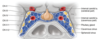
Draw the circle of Willis.
DO NOT FORGET the superior cerebellar artery and the anterior inferior cerebellar arteries

State the names of the venous sinuses that are at the top and bottom of the falx cerebri.
Superior sagittal sinus Inferior sagittal sinus

What is the name given to the place where all the sinuses meet?
Confluence of sinuses
What connects the inferior sagittal sinus to the confluence of sinuses?
Straight sinus

What vessel does the inferior sagittal sinus join with to form thestraight sinus?
Great cerebral vein
Which sinus ascends to join the confluence of sinuses?
Occipital sinus

Which two sinuses run along the temporal bone?
Superior petrosal sinus Inferior petrosal sinus

Which main sinus drains into the internal jugular vein through the jugular foramen?
Sigmoid sinus
Which sinus connects the confluence of sinuses to the sigmoid and superior petrosal sinuses?
Transverse sinus
Which sinuses run on either side of the pituitary stalk?
Anterior and posterior intercavernous sinuses
Which extension of dura mater separates the cerebellum from the inferior portion of the occipital lobe?
Tentorium cerebelli
Define Stroke.
A rapidly developing focal disturbance of brain function of presumed vascular origin that lasts more than 24 hours
Define Transient Ischaemic Attack (TIA).
A rapidly developing focal disturbance of brain function of presumedvascular origin that resolves completely within 24 hours
What percentage of strokes are caused by infarction and what percentage are caused by haemorrhage?
85% infarction 15% haemorrhage
State two causes of occlusions.
Thrombus Embolus
Describe the location of the leg in the motor and sensory homunculus compared to the arm.
Leg is more MEDIAL
Describe the features of a disturbance in the anterior cerebral artery.
Contralateral hemiplegia in the LEG more than the arm Disturbance of intellect and executive function Loss of appropriate social behaviour
Describe the features of a disturbance in the middle cerebral artery.
This is a CLASSIC STROKE Contralateral hemiplegia in the ARM more than the leg Contralateral hemisensory deficits Hemianopia Aphasia (can’t speak) – left-sided lesion of the middle cerebral artery will result in aphasia because the language centres are more on the left side than the right
Describe the features of a disturbance in the posterior cerebral artery.
The posterior cerebral artery supplies the occipital lobe, which is where the primary visual cortex is located This causes visual defects such as homonymous hemianopia and visual agnosia (unable to recognise what you are seeing)
Which parts of the brain are involved in speech and understanding language?
Broca’s area – speech Wernicke’s area – understanding language


